Ringworm Information for Healthcare Professionals
Definition and sources of infection
Ringworm, also called “tinea” or “dermatophytosis,” is a common infection of the epidermis (skin, hair, or nails) caused by dermatophyte molds. People can acquire ringworm through direct skin contact with people and animals who are infected. People can also acquire ringworm by sharing personal items (e.g., towels, clothing, bedding) or through contact with surfaces found in moist areas (e.g., shower stalls, locker room floors, pool areas). Ringworm can also spread from one part of the body to another.
Learn more about how steroid creams can make ringworm worse.
Learn more about emerging antimicrobial-resistant ringworm infections.
Physical examination
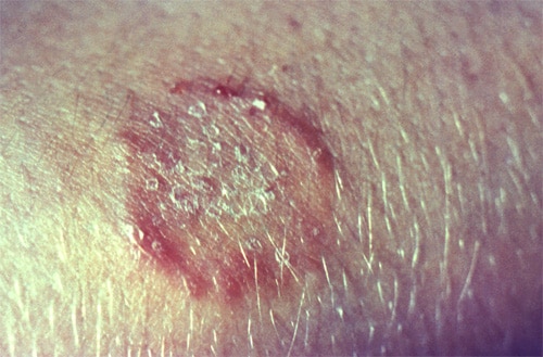
The classic ringworm lesion is an erythematous, raised, scaly ring with central clearing. Multiple lesions might be present. The severity of the infection can range from mild, scaly lesions to erythematous, exudative lesions if a bacterial superinfection has developed.
Ringworm may be difficult to distinguish from other skin conditions.1 Physical examination and clinical history alone may not be enough to diagnose ringworm. Clinicians should generally use a diagnostic test to confirm suspected ringworm, especially before prescribing antifungal treatment.
Diagnostic testing
Potassium hydroxide preparation
Clinicians can use a potassium hydroxide (KOH) preparation of skin scrapings or nail clippings to confirm a diagnosis of ringworm. This test can provide rapid results, but the test’s accuracy depends on clinician experience and technique.2
Fungal culture
Fungal culture can be used to diagnose ringworm. Fungal culture is more specific than KOH stain, but results may take several weeks.3,4
Histopathologic examination with a periodic acid-Schiff stain
Histopathologic examination of nail clippings with a periodic acid-Schiff (PAS) stain is a method for confirming the diagnosis for patients with suspected onychomycosis, a fungal nail infection most often caused by dermatophytes.
Polymerase chain reaction
Polymerase chain reaction (PCR) is a quick and increasingly used method for diagnosing ringworm.
Ultraviolet light (Wood’s lamp)
Ultraviolet light can be useful for diagnosing ringworm caused by Microsporum canis and Microsporum audouinii. Although both species fluoresce blue-green under a Wood’s lamp, these species are uncommon causes of ringworm infections in people.
Treatment
Tinea pedis: Athlete’s foot can usually be treated with over-the-counter topical antifungal products. Chronic or extensive tinea pedis may require treatment with systemic antifungal agents such as terbinafine, itraconazole, or fluconazole.6 In addition, chronic tinea pedis may require adjunctive therapy such as foot powder or talcum powder to prevent skin maceration.
Tinea capitis: Treatment with systemic antifungal medication is required, as topical antifungal products are ineffective for treatment of tinea capitis. Many experts consider griseofulvin to be the drug of choice.6 Terbinafine is also FDA-approved for the treatment of tinea capitis in patients four years of age and older. Itraconazole and fluconazole have been shown to be safe and effective, but are not FDA-approved for this indication.6 Selenium sulfide shampoos can be used as adjunctive therapy.6-7 Clinicians should generally confirm the diagnosis of tinea capitis using a laboratory test.8
Tinea corporis/cruris: Tinea corporis and tinea cruris can usually be treated with topical antifungal products.6 Patients who have tinea cruris should be advised to keep the groin area clean and dry and to wear cotton underwear. Patients who have extensive or recurrent infections may require systemic antifungal therapy.6
Get information about the treatment of tinea unguium.
Treatment failure
Most ringworm infections improve with antifungal treatment, but treatment failure may occur. Common reasons for treatment failure may include incorrect diagnosis and inadequate treatment. Clinicians should counsel patients on the importance of taking antifungal medications as prescribed.
Emerging resistance is another potential cause of treatment failure. Antifungal resistant Trichophyton rubrum and Tinea indotineae are emerging global public health concerns.
- Yadgar RJ, Bhatia N, Friedman A. Cutaneous fungal infections are commonly misdiagnosed: A survey-based study. J Amer Acad Dermatol. 2017 March; 76(3): 562-563.
- Levitt JO, Levitt BH, Akhavan A, Yanofsky H. The Sensitivity and Specificity of Potassium Hydroxide Smear and Fungal Culture Relative to Clinical Assessment in the Evaluation of Tinea Pedis: A Pooled Analysis. Derma Res and Prac. 2010 Jun; 2010.
- Hainer BL. Dermatophyte infections. Am Fam P 2003 Jan 1;67(1):101-8.
- Crawford F, Hollis S. Topical treatments for fungal infections of the skin and nails of the foot. Cochrane Database Syst. Rev. 2007(3):CD001434.
- Gupta AK, Cooper EA. Update in antifungal therapy of dermatophytosis. Mycopathologia. 2008 Nov-Dec;166(5-6):353-67.
- Michaels BD, Del Rosso JQ. Tinea capitis in infants: recognition, evaluation, and management suggestions. J Clin Aesthet Dermatol. 2012 Feb;5(2):49-59.
- Allen HB, Honig PJ, Leyden JJ, McGinley KJ. Selenium sulfide: adjunctive therapy for tinea capitis. Pediatrics. 1982 Jan;69(1):81-3.
- Ely JW, Rosenfeld S, Seabury Stone M. Diagnosis and Management of Tinea Infections. Am Fam Physician. 2014;90(10):702-711
- Gold JAW, Wu K, Jackson BR, Benedict K. Opportunities to improve guideline adherence for the diagnosis and treatment of onychomycosis: Analysis of commercial insurance claims data, United States. J Amer Acad of Derm. 2022 July.
- Levitt JO, Levitt BH, Akhavan A, Yanofsky H. The sensitivity and specificity of potassium hydroxide smear and fungal culture relative to clinical assessment in the evaluation of tinea pedis: a pooled analysis. Derm Res Pract. 2010;2010:764843.
- Noble SL, Forbes RC, Stamm PL. Diagnosis and management of common tinea infections. Am Fam Physician. 1998 Jul;58(1):163-74, 77-8.