 An official website of the United States government



Notes from the Field: Molecular Detections of Cladophialophora bantiana at a Clinical Reference Laboratory — United States, 2009–2026
Weekly / October 15, 2026 / 75(40);609-611
On October 2, 2026, this report was posted online as an MMWR Early Release.
Maria T. Nelson, MD, PhD1; Thao T. Truong, PhD1; Lori A. Bourassa, PhD1; Dallas J. Smith, PharmD2; Joshua A. Lieberman, MD, PhD1 (View author affiliations)
View suggested citationSummary
What is already known about this topic?
Cladophialophora bantiana is a neurotropic fungus; infection is associated with a 60%–70% case-fatality rate. National surveillance does not exist, and neither the environmental reservoir nor the route of infection is fully understood.
What is added by this report?
During January 2009–July 2026, 48 confirmed cases and one probable case of C. bantiana were detected in clinical samples (predominantly central nervous system tissue [88%]) originating in 22 jurisdictions and tested at a single clinical reference laboratory; detections of the fungus increased approximately fourfold from 2023 to 2025.
What are the implications for public health practice?
Reasons for the apparent increase are unknown. Further research into epidemiologic risk factors and potentially evolving virulence are warranted. Systematic reporting of C. bantiana cases is needed and C. bantiana should be considered in patients with a brain abscess. Patients with infection require prompt antifungal therapy, and surgical excision might improve patient outcomes.
Altmetric:
During 2017–2023, the number of cases of Cladophialophora bantiana detected by a clinical reference laboratory at the University of Washington (UW) increased substantially. C. bantiana is a highly virulent, neurotropic dematiaceous mold*; infection is associated with a 60%–70% case-fatality rate, even with aggressive therapy (1). Approximately one half of patients with C. bantiana infections have no preceding immunocompromising condition (1). Despite its virulence, relatively little is known about the risk factors for or epidemiology of this fungus. The presumed route of infection is inhalation or direct inoculation after cutaneous injury, followed by hematogenous spread to the brain. Rarely, pulmonary (2) and cutaneous (3) infections have been reported; most cases occur as brain abscesses, often with no extracranial disease and no clear source of inoculation. Furthermore, C. bantiana has only rarely been isolated from environmental sources, and many patients do not have similar exposure histories or risk factors (1). No routine surveillance is available; data consist primarily of case reports and retrospective literature reviews, limiting assessment of evolving epidemiologic characteristics and patterns. This report describes a substantial increase in the rate of detection of C. bantiana at a molecular microbiology clinical reference laboratory, with cases originating from a wide geographic range in the United States.
Investigations and Outcomes
Methods
All cases of C. bantiana detected by broad-range fungal polymerase chain reaction (PCR)† at the UW laboratory during January 2009–July 2026 were compiled. Cases were categorized by test order year, tissue site, and state of origin, determined both by referring laboratory location and surgical pathology report, when available.§ To control for increased testing over time, the number of C. bantiana cases was normalized to that of all intracranial central nervous system (CNS) specimens submitted for broad-range fungal PCR during the same period. The DNA extraction method was changed in February 2021; the remainder of the assay has not changed substantially over time. Data were analyzed in R (version 4.5.2; R Foundation). This activity was approved by the institutional review boards at UW and was reviewed by CDC, deemed not research, and conducted consistent with applicable federal law and CDC policy.¶
Characteristics of Detected C. bantiana Cases
During January 2009–July 1, 2026, a total of 48 confirmed cases of C. bantiana infection and one probable case** were identified by broad-range fungal PCR; the median patient age was 68 years (IQR = 63–74 years). Among these 49 cases, 43 (88%) were detected in intracranial CNS tissue specimens and six (12%) in lung specimens. No extracranial CNS infections were identified. During 2009–2023, the number of cases per year ranged from one to five. Case counts increased to eight in 2024 and 14 in 2025 (Figure). During 2017–2023, an average of 8.4 cases of C. bantiana were detected per 1,000 CNS specimens submitted for broad-range fungal PCR at UW. By 2025, this increased by 210%, to 26 per 1,000, suggesting that the increase in C. bantiana cases is not attributable to an increase in laboratory referral volume alone. Cases originated from 21 U.S. states and the District of Columbia (Supplementary Figure); the largest number of cases originated from California (six), Florida (five), Texas (five), and Pennsylvania (four); state of origin was not available for three cases. Broad-range fungal PCR testing of cerebrospinal fluid was performed antemortem for the single, fatal case managed at the UW Medical Center and was negative for C. bantiana in samples collected concurrently and 1 month before biopsy. Outcome information for other cases was not available.
Preliminary Conclusions and Actions
C. bantiana is a highly virulent mold that predominantly causes brain abscesses in both immunocompetent and immunocompromised persons. The frequency of C. bantiana cases detected by broad-range fungal PCR at UW per 1,000 total submitted CNS specimens increased approximately fourfold in 2 years (2023–2025). Whereas most cases were detected in CNS specimens, detection of 12% of cases in pulmonary samples supports the presumed respiratory route of infection. Because of the nature of reference laboratory testing, limited demographic, occupational, and clinical data were available, and the causes of this recent increase in cases are unclear, highlighting the need for more systematic reporting mechanisms for this mold and for clinical laboratories to publish numbers of detected cases. This approach could help in collecting epidemiologic data to identify risk factors and transmission patterns more effectively. Clinicians should be aware of this increase in incidence and consider C. bantiana in patients who have signs and symptoms of a brain abscess. Testing can be ordered through clinical reference laboratories that offer molecular testing for fungi in tissue or CSF specimens. In a 2024 study of 23 patients in France (4), surgical excision or drainage of brain abscesses was associated with more favorable outcomes; thus, when possible, surgical exision should be considered, concurrently with administration of combination antifungal therapy. Although the most effective antifungal regimen is not known, triple therapy (treatment with lipsosomal amphotericin B, posaconazole or voriconazole, and flucytosine) for at least 2 weeks has demonstrated some effectiveness (4).
Corresponding author: Joshua A. Lieberman, joshuaal@uw.edu.
1Department of Laboratory Medicine and Pathology, University of Washington School of Medicine, Seattle, Washington; 2Mycotic Diseases Branch, Division of Foodborne, Waterborne, and Environmental Diseases, CDC.
All authors have completed and submitted the International Committee of Medical Journal Editors form for disclosure of potential conflicts of interest. No potential conflicts of interest were disclosed.
* A large, diverse group of molds and black yeasts defined by the presence of melanin or melanin-like pigments in their cell walls.
† This fungal organism–agnostic method amplifies conserved ribosomal RNA loci (28S D1/D2, ITS1, and ITS2) directly from fresh and formalin-fixed paraffin-embedded tissue samples or body fluids and performs sequencing to assign taxonomic identification of detected fungal DNA.
§ State of origin was unavailable for three cases.
¶ 45 C.F.R. part 46, 21 C.F.R. part 56; 42 U.S.C. Sect. 241(d); 5 U.S.C. Sect. 552a; 44 U.S.C. Sect. 3501 et seq.
** One case of intracranial CNS infection was reported in 2024 as a Cladophialophora species infection with a significant sequence homology to C. bantiana.
References
- Kantarcioglu AS, Guarro J, De Hoog S, Apaydin H, Kiraz N. An updated comprehensive systematic review of Cladophialophora bantiana and analysis of epidemiology, clinical characteristics, and outcome of cerebral cases. Med Mycol 2017;55:579–604. https://doi.org/10.1093/mmy/myw124 PMID:28007938
- Hernandez C, Lawal F. Cerebral and pulmonary phaeohyphomycosis due Cladophialophora bantiana in an immunocompromised patient. IDCases 2021;25:e01240. https://doi.org/10.1016/j.idcr.2021.e01240 PMID:34381691
- Mackrill SW, Schramm DC, Amini A, et al. A case of disseminated autochthonous Cladophialophora bantiana infection in a renal transplant recipient in the UK. Lancet Infect Dis 2025;25:e104–12. https://doi.org/10.1016/S1473-3099(24)00579-6 PMID:39515363
- Lortholary O, Garcia-Hermoso D, Sturny-Leclère A, et al. Reappraising Cladophialophora bantiana phaeohyphomycosis in France: retrospective nation-based study. Lancet Microbe 2024;5:100907. https://doi.org/10.1016/S2666-5247(24)00139-3 PMID:39395429
 FIGURE. Number of proven Cladophialophora bantiana cases detected by broad-range fungal polymerase chain reaction (A)* and number of C. bantiana cases per 1,000 intracranial central nervous system specimens submitted for the same assay (B),† by year — United States, January 2009–July 2026

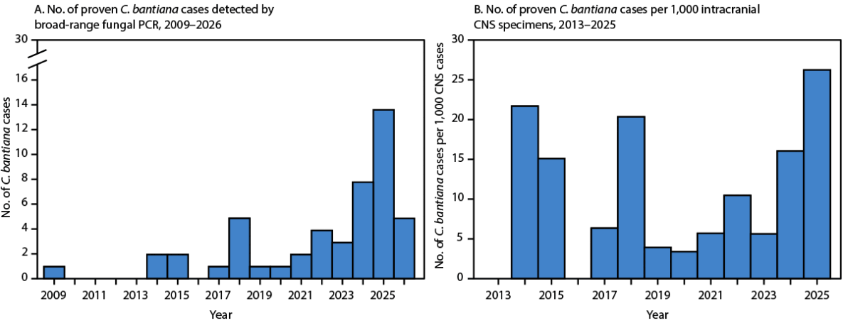
Abbreviations: CNS = central nervous system; PCR = polymerase chain reaction.
* Data for 2026 are through July 1.
† Baseline CNS cases were available beginning in 2013.
Suggested citation for this article: Nelson MT, Truong TT, Bourassa LA, Smith DJ, Lieberman JA. Notes from the Field: Molecular Detections of Cladophialophora bantiana at a Clinical Reference Laboratory — United States, 2009–2026. MMWR Morb Mortal Wkly Rep 2026;75:609-611. DOI: http://dx.doi.org/10.15585/mmwr.mm7540e1.
MMWR and Morbidity and Mortality Weekly Report are service marks of the U.S. Department of Health and Human Services.
Use of trade names and commercial sources is for identification only and does not imply endorsement by the U.S. Department of
Health and Human Services.
References to non-CDC sites on the Internet are
provided as a service to MMWR readers and do not constitute or imply
endorsement of these organizations or their programs by CDC or the U.S.
Department of Health and Human Services. CDC is not responsible for the content
of pages found at these sites. URL addresses listed in MMWR were current as of
the date of publication.
All HTML versions of MMWR articles are generated from final proofs through an automated process. This conversion might result in character translation or format errors in the HTML version. Users are referred to the electronic PDF version (https://www.cdc.gov/mmwr) and/or the original MMWR paper copy for printable versions of official text, figures, and tables.
Questions or messages regarding errors in formatting should be addressed to mmwrq@cdc.gov.