 An official website of the United States government



Notes from the Field: Fatal Pneumonic Plague — Coconino County, Arizona, July 2025
Weekly / October 1, 2026 / 75(38);588–589
Christine A. Wang, DVM, PhD1,2; Jacob Hojnacki, MPH3; Sierra Jaramillo3; Saba Qasmieh, PhD1,2; Irene Ruberto, PhD2; Laura Platt, MD4; Schante Patton3; Guillermo Adame, MPH2; Drew Francis2; Grace Marx, MD4; Shane Brady, MPH2; Kenneth K. Komatsu, MPH2; Kathryn Burr, DVM2,5; Joel Terriquez, MD2 (View author affiliations)
View suggested citationSummary
What is already known about this topic?
Pneumonic plague is a rare and often fatal disease that can be transmitted by respiratory droplets from animals or persons infected with Yersinia pestis bacteria.
What is added by this report?
In July 2025, Arizona reported its first pneumonic plague death since 2007, in a man with occupational exposure to ill cats. Two days after the patient’s death, preliminary diagnostic test results supported a suspected pneumonic plague diagnosis, prompting immediate identification of potentially exposed persons for postexposure prophylaxis.
What are the implications for public health practice?
In areas with endemic plague, preliminary diagnostic test results combined with clinical suspicion for pneumonic plague can result in timely public health interventions.
Altmetric:
Pneumonic plague is transmitted through inhalation of respiratory droplets containing the bacterium Yersinia pestis and has a nearly 100% case-fatality rate when untreated (1). Although Y. pestis is endemic among rodents in northern Arizona, only eight human plague cases were reported in Arizona during 2000–2024. In July 2025, Arizona reported its first pneumonic plague death since 2007 (2).
Investigation and Outcomes
In July, a young adult male was evaluated in a Coconino County, Arizona emergency department after 2 days of abdominal pain, chest pain, and dry cough (Figure). He was afebrile, tachycardic, and tachypneic, and had leukocytosis (28.3 ×10³ white blood cells/µL). Chest radiographs and computed tomographic angiography of the chest, abdomen, and pelvis identified pulmonary infiltrates consistent with pneumonia; no buboes (i.e., painful, swollen lymph nodes) were observed. Clinicians initially suspected hantavirus disease or bacterial pneumonia. The patient was admitted and received empiric antibiotic treatment with piperacillin-tazobactam, vancomycin, and azithromycin. Despite progressively intensive oxygen therapy, his hypoxemia worsened, and he died <8 hours after admission.
One day later, bacterial growth was detected in the patient’s blood cultures. The following day, matrix-assisted laser desorption/ionization time-of-flight (MALDI-TOF) mass spectrometry of bacterial growth from that blood culture identified Yersinia. pseudotuberculosis, and bipolar-staining gram-negative bacilli were identified (Supplementary Figure). Together, both results raised suspicion for pneumonic plague, and the local public health department was immediately notified.
Two days after the patient’s death, 46 potentially exposed persons were identified, and all commenced a 7-day course of oral ciprofloxacin postexposure prophylaxis (PEP) (3). Potentially exposed persons included household members, coworkers, and hospital staff members; no contact became ill. Six days after the patient’s death, the Arizona State Public Health Laboratory (ASPHL) detected Y. pestis from initial blood cultures using quantitative (real-time) polymerase chain reaction testing. Four days later (10 days after the patient’s death), ASPHL isolated Y. pestis from blood and lung tissue cultures.
The public health investigation focused on patient exposures during the pneumonic plague incubation period of 1–5 days preceding illness onset. Family members reported that the patient did not engage in outdoor activities and only spent time at his workplace or residence. Five and 4 days before his illness onset, he had occupational exposure to two indoor domestic cats with respiratory symptoms and other signs of systemic illness consistent with pneumonic plague. Inhalation transmission from either cat was possible with the patient’s occupational exposure. Feline specimens were unavailable for Y. pestis testing because both cats had been euthanized before the patient’s illness onset. No other persons exposed to these cats reported illness.
CDC was consulted after the patient’s death and primarily focused on supporting the interpretation of clinical signs for each animal to which the patient was exposed. This activity was reviewed by CDC, deemed not research, and conducted consistent with applicable federal law and CDC policy.*
Preliminary Conclusions and Actions
Y. pestis misidentification can occur with automated pathogen detection systems, including MALDI-TOF (4). The combination of Gram stain test results, MALDI-TOF results, the patient’s clinical signs and symptoms, and known endemicity of Y. pestis in northern Arizona sufficiently raised suspicion for pneumonic plague. These circumstances supported prompt identification of persons potentially exposed to the patient for PEP initiation 8 days before confirmation of Y. pestis.
Health care providers in areas where plague is endemic should consider plague in patients with severe respiratory and systemic disease who have been exposed to ill animals. Appropriate laboratory safety practices must also be considered for clinical laboratory personnel handling specimens from these patients. Pneumonic plague risk increases with occupational exposure to animals infected with Y. pestis, especially cats, because of their susceptibility to plague (5). During evaluation of ill animals and persons suspected of having plague or handling their clinical specimens, consistent use of personal protective equipment, especially appropriate respiratory protection, can reduce occupational exposures to zoonotic pathogens such as plague among veterinary, medical, and clinical microbiology personnel.
Corresponding author: Christine A. Wang, cwang4@cdc.gov, christine.wang@azdhs.gov.
1Epidemic Intelligence Service, CDC; 2Arizona Department of Health Services; 3Coconino Department of Health and Human Services, Flagstaff, Arizona; 4Division of Vector-borne Diseases, National Center for Emerging and Zoonotic Infectious Diseases, CDC; 5Career Epidemiology Field Officer Program, CDC.
All authors have completed and submitted the International Committee of Medical Journal Editors form for disclosure of potential conflicts of interest. Drew Francis reports a leadership role on the American Public Health Laboratory’s Infectious Diseases Committee. Kenneth K. Komatsu reports travel and meeting support to the Council of State and Territorial Epidemiologists, participation in the Arizona Department of Health Services Human Subjects Review Board and the Phoenix BioWatch Advisory Committee. No other potential conflicts of interest were disclosed.
* 45 C.F.R. part 46.102(l)(2), 21 C.F.R. part 56; 42 U.S.C. Sect. 241(d); 5 U.S.C. Sect. 552a; 44 U.S.C. Sect. 3501 et seq.
References
- Pechous RD, Sivaraman V, Stasulli NM, Goldman WE. Pneumonic plague: the darker side of Yersinia pestis. Trends Microbiol 2016;24:190–7. https://doi.org/10.1016/j.tim.2015.11.008 PMID:26698952
- Wong D, Wild MA, Walburger MA, et al. Primary pneumonic plague contracted from a mountain lion carcass. Clin Infect Dis 2009;49:e33–8. https://doi.org/10.1086/600818 PMID:19555287
- Nelson CA, Meaney-Delman D, Fleck-Derderian S, Cooley KM, Yu PA, Mead PS. Antimicrobial treatment and prophylaxis of plague: recommendations for naturally acquired infections and bioterrorism response. MMWR Recomm Rep 2021;70:1–27. https://doi.org/10.15585/mmwr.rr7003a1 PMID:34264565
- Dale AP, Kretschmer M, Ruberto I, et al. Notes from the field: delays in identification and treatment of a case of septicemic plague—Navajo county, Arizona, 2020. MMWR Morb Mortal Wkly Rep 2021;70:1063–4. https://doi.org/10.15585/mmwr.mm7031a1 PMID:34351879
- Kugeler KJ, Staples JE, Hinckley AF, Gage KL, Mead PS. Epidemiology of human plague in the United States, 1900-2012. Emerg Infect Dis 2015;21:16–22. https://doi.org/10.3201/eid2101.140564 PMID:25529546
 FIGURE. Timeline of events associated with a fatal case of pneumonic plague — Coconino County, Arizona, July 2025

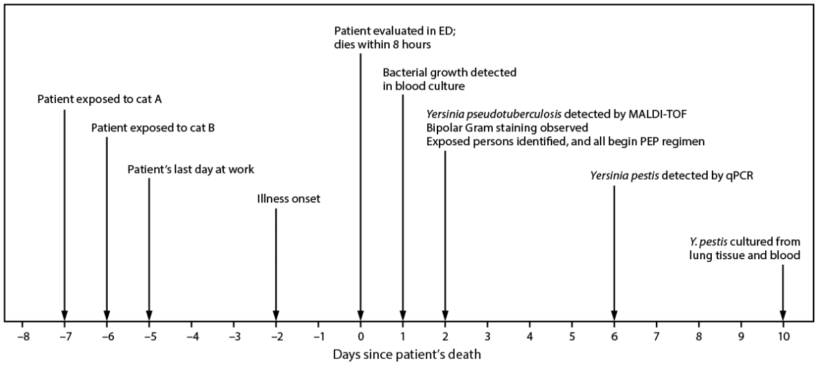
Abbreviations: ED = emergency department; MALDI-TOF = matrix-assisted laser desorption/ionization time-of-flight; PEP = postexposure prophylaxis; qPCR = quantitative polymerase chain reaction.
Suggested citation for this article: Wang CA, Hojnacki J, Jaramillo S, et al. Notes from the Field: Fatal Pneumonic Plague — Coconino County, Arizona, July 2025. MMWR Morb Mortal Wkly Rep 2026;75:588–589. DOI: http://dx.doi.org/10.15585/mmwr.mm7538a2.
MMWR and Morbidity and Mortality Weekly Report are service marks of the U.S. Department of Health and Human Services.
Use of trade names and commercial sources is for identification only and does not imply endorsement by the U.S. Department of
Health and Human Services.
References to non-CDC sites on the Internet are
provided as a service to MMWR readers and do not constitute or imply
endorsement of these organizations or their programs by CDC or the U.S.
Department of Health and Human Services. CDC is not responsible for the content
of pages found at these sites. URL addresses listed in MMWR were current as of
the date of publication.
All HTML versions of MMWR articles are generated from final proofs through an automated process. This conversion might result in character translation or format errors in the HTML version. Users are referred to the electronic PDF version (https://www.cdc.gov/mmwr) and/or the original MMWR paper copy for printable versions of official text, figures, and tables.
Questions or messages regarding errors in formatting should be addressed to mmwrq@cdc.gov.