Radiation in Healthcare: Nuclear Medicine
Nuclear medicine uses radioactive material inside the body to see how organs or tissue are functioning (for diagnosis) or to target and destroy damaged or diseased organs or tissue (for treatment).
Nuclear medicine vs common imaging procedures using x-rays: how they work
| Nuclear medicine | x-rays |
| Radioactive material (tracer) is injected, ingested, or inhaled | Beams of radiation pass through the body |
| Images of the body show where and how the tracer is absorbed. | Images of the structure in the body are produced |
| Shows function | Shows structure |
| Used in diagnosis or treatment | Used in diagnoses |
Although we all are exposed to ionizing radiation every day from the natural environment, added exposures like those from nuclear medicine procedures can slightly increase the risk of developing cancer later in life.
Talk to your healthcare provider to decide on the best procedure for your health needs and discuss any concerns you have.
What You Should Know
Your healthcare provider may recommend a nuclear medicine procedure to diagnose or treat a health problem.
When It’s Used for Diagnosis
Nuclear medicine can show how the organs or tissues are functioning. For most diagnostic procedures, a tracer, which contains the radioactive material, is injected, swallowed, or inhaled. Then the healthcare provider or radiologist (a healthcare professional with special training to use radiation in healthcare) uses a radiation detector to see how much of the tracer is absorbed or how it reacts in the organ or tissue. This will give the provider information about how well it is functioning.
Common uses of nuclear medicine for diagnosis include:
- Scans of the heart, lung, kidneys, gallbladder, and thyroid
In a type of nuclear medicine called positron emission tomography (PET), the tracer is used to show the natural activity of cells, providing more detailed information on how organs are working and if there is damage to the cells. PET scans are often combined with computed tomography (CT) scans or magnetic resonance imaging (MRI) which provide three-dimensional images of the organ.
Common uses of PET scans include:
- Diagnosing heart disease, Alzheimer’s disease, and brain disorders
- Getting detailed information about cancerous tumors to decide the best treatment option
When it’s used for treatment
When used in treatment, the tracer targets a harmful organ or tissue and radioactivity damages or stops the growth of its cells.
Two common uses of nuclear medicine for treatment include radioactive iodine therapy and brachytherapy (a form of radiation treatment where a sealed radiation source is placed inside or next to the area requiring treatment).
What To Expect
Find information on special considerations pregnant women and children.
Before the procedure
- You will receive a tracer either through an injection, inhalation (breathing it in), or through a pill or substance to swallow.
- You may need to wait a certain amount of time for the tracer to travel through your body to the tissue or organ being diagnosed or treated.
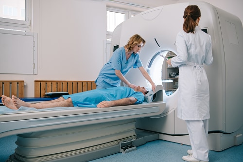
During the procedure
- You may be asked to lie down on a table or to walk on a treadmill.
- A camera that detects radiation will be placed over your body to collect information on how the tracer is acting in an organ or tissue.
After the procedure
- The radiologist and your healthcare provider use this information to see how an organ or tissue is functioning.
- The radioactive material from the tracer will pass out of your body in a few hours to a few days, depending on the type of tracer and test you receive.
When You Go Home
Right after your procedure, your body is very slightly radioactive (giving off radiation) but this wears off with time and is not directly harmful to others. Your healthcare provider may give you special instructions to help reduce any small amounts of radiation you give off from exposing others such as washing your hands frequently. Drinking a lot of water may help the radioactive material leave your body quicker.
- Before you leave, ask your healthcare provider if there are steps you should take to protect others or if you have any concerns or questions about information you were given.
- Talk to your healthcare provider if you are currently breastfeeding.
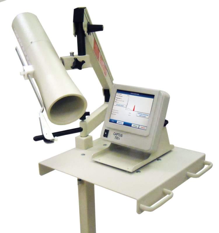
Photo of a machine used for a thyroid scan
The ionizing radiation dose for these procedures is typically higher than the dose received from a common x-ray procedure. There are always some possible risks from exposure to ionizing radiation in healthcare, but these procedures should be used when the health benefits outweigh these risks.
Benefits and Risks of Nuclear Medicine
Benefits
- Provides information on how organs, tissues, and cells are working. (Other common imaging procedures only show the structures.)
- Can be used also in targeted treatments to kill or damage harmful or cancerous cells, reduce the size of tumors, or reduce pain.
Risks
- Radiation doses are usually higher than in common imaging like x-rays. This means these procedures are slightly more likely to increase the possibility you may get cancer later in life.
- Some nuclear medicine procedures are longer and use more radiation than others. These could cause skin reddening and hair loss.
- You may give off small amounts of radiation right after your procedure and need to take steps to protect others from exposure.
What are nuclear medicine procedures?
Nuclear medicine procedures are used in diagnosing and treating certain illnesses. These procedures use radioactive materials called radiopharmaceuticals. Examples of diseases treated with nuclear medicine procedures are hyperthyroidism, thyroid cancer, lymphomas, and bone pain from some types of cancer.
The amount of radioactive materials used in diagnosing illnesses depends on the needs of the person and range from a small amount to a large amount. These materials flow through different body organs and in some cases are taken up by specific organs or tissue. The radiation that comes from the radiopharmaceutical is used for treatment or is detected by a camera to take pictures of the corresponding body organ, region or tissue.
What happens during a nuclear medicine imaging procedure?
- During a nuclear medicine imaging procedure, doctors give patients radiopharmaceuticals. Depending on the type of medical examination they can be breathed in (inhaled), injected, or swallowed.
- Once the radiopharmaceutical is given, the patient is usually asked to lie down on a table. A special camera that detects radiation is placed over the patient’s body to take pictures. A computer is used to show where the body concentrates the radioactive material. This allows doctors to check if organs are working properly and diagnose diseases.
- The radioactive materials usually leave the body within hours to months.
 image icon[JPG - 31 KB]
Photo of a machine used for a thyroid scan (click to enlarge)
What are radioactive materials?
Radioactive materials are chemicals that release radiation (energy). Radioactive materials can be natural or they can be man-made. They can be solids (like some rocks on earth) or liquids or they can also be gases that people can breathe (like radon). Each radioactive material has a unique half-life, which tells how quickly it stops being radioactive.
Are any health effects associated with nuclear medicine procedures?
The Nuclear Regulatory Commission (NRC), the U.S. Food and Drug Administration (FDA) and states regulate the use of radioactive materials for nuclear medicine to make sure patients, medical personnel, and the public are safe. Before any type of nuclear medicine procedure is used, it must be justified to ensure the benefits of the procedures outweigh risks to the patient. However, exposure to too much radiation can quickly damage organs or tissues, while exposure to any amount of radiation might lead to an increase in the risk of cancer years after the exposure occurs. Image Gentlyexternal icon is a campaign that encourages medical facilities to use a “child size” amount of radioactive material when a child has a nuclear medicine procedure.
What are some common nuclear medicine procedures?
There are several nuclear medicine procedures for diagnosing illnesses and treating diseases.
Some common procedures are as follows:
Diagnostic Procedures
- Heart disease can be diagnosed with a stress test using Sestamibi that contains technetium-99m or through the use of positron emission tomography (PET) scans. See more information about how PET scans are used in nuclear medicine in the section below.
- Gallbladder problems can be diagnosed using hepatobiliary iminodiacetic acid (HIDA) scans that contain a radioactive material tracer, usually technetium-99m.
- Thyroid disease can be diagnosed with a radioactive iodine thyroid scan that utilizes sodium iodine which contains iodine-131.
Therapeutic Procedures
- Radioimmunotherapy in which a radioactive element like iodine-133 90 is tagged to an antibody that targets a specific cancerous cell.
- Thyroid Ablation that uses sodium iodide which contains iodide-131.
- Brachytherapy in which a radioactive source like iridium-192 is inserted inside or near a tumor to kill cancer cells while sparing neighboring cells.
How are PET scans used?
Doctors use PET scans to get more data about how body organs are functioning. PET scans may be performed together with a computerized axial tomography (CAT) scan that provides an image of the organ. PET scans provide a clear view of how the organs are working at the cellular level and if they have been damaged. The scan helps doctors determine effective treatment options. PET scans are commonly used to diagnose heart conditions, help doctors determine appropriate cancer treatment, help in diagnosing Alzheimer’s disease and brain disorders. They also can provide data for medical research.
Cdc-pdfpdf iconexternal iconExternalpdf iconexternal icon
- Nuclear Medicine-Diagnostic ProceduresExternalexternal icon
- Image Gently and SNMExternalexternal icon
- What is Nuclear Medicine?Externalexternal icon
- What is Molecular Imaging?Externalexternal icon
- PET Professional Resources and Outreach SourceCdc-pdfExternalpdf iconexternal icon
- Nuclear Medicine: What it is and Isn’tExternalexternal icon