Key points
- Pulmonary atresia (pronounced PULL-mun-airy ah-TREE-sha) is a congenital heart defect. Congenital means present at birth.
- It occurs when the valve that controls blood flow from the heart to the lungs doesn't form at all.
- People with pulmonary atresia should schedule routine checkups with a heart doctor throughout their lives to stay as healthy as possible.

What it is
Pulmonary atresia is a birth defect of the pulmonary valve. This valve controls blood flow from the right ventricle to the main pulmonary artery. The right ventricle is the lower right chamber of the heart. The pulmonary artery is the blood vessel that carries blood from the heart to the lungs.
Pulmonary atresia is when this valve doesn't form at all. This means that no blood can go from the right ventricle of the heart out to the lungs. With pulmonary atresia, blood must use other routes to bypass the unformed pulmonary valve.
In babies with pulmonary atresia, the foramen ovale often remains open to allow blood flow to the lungs. The foramen ovale is a natural opening between the upper chambers of the heart that usually closes after birth.
A baby with pulmonary atresia may need surgery or other procedures soon after birth. Therefore, pulmonary atresia is considered a critical congenital heart defect (critical CHD).
Occurrence
About 1 in 8,333 (about 416) babies in the United States are born with pulmonary atresia each year.
Types
There are typically two types of pulmonary atresia. The types are determined by whether or not a baby also has a ventricular septal defect (VSD). A VSD is a hole in the wall that separates the two lower chambers, or ventricles, of the heart.
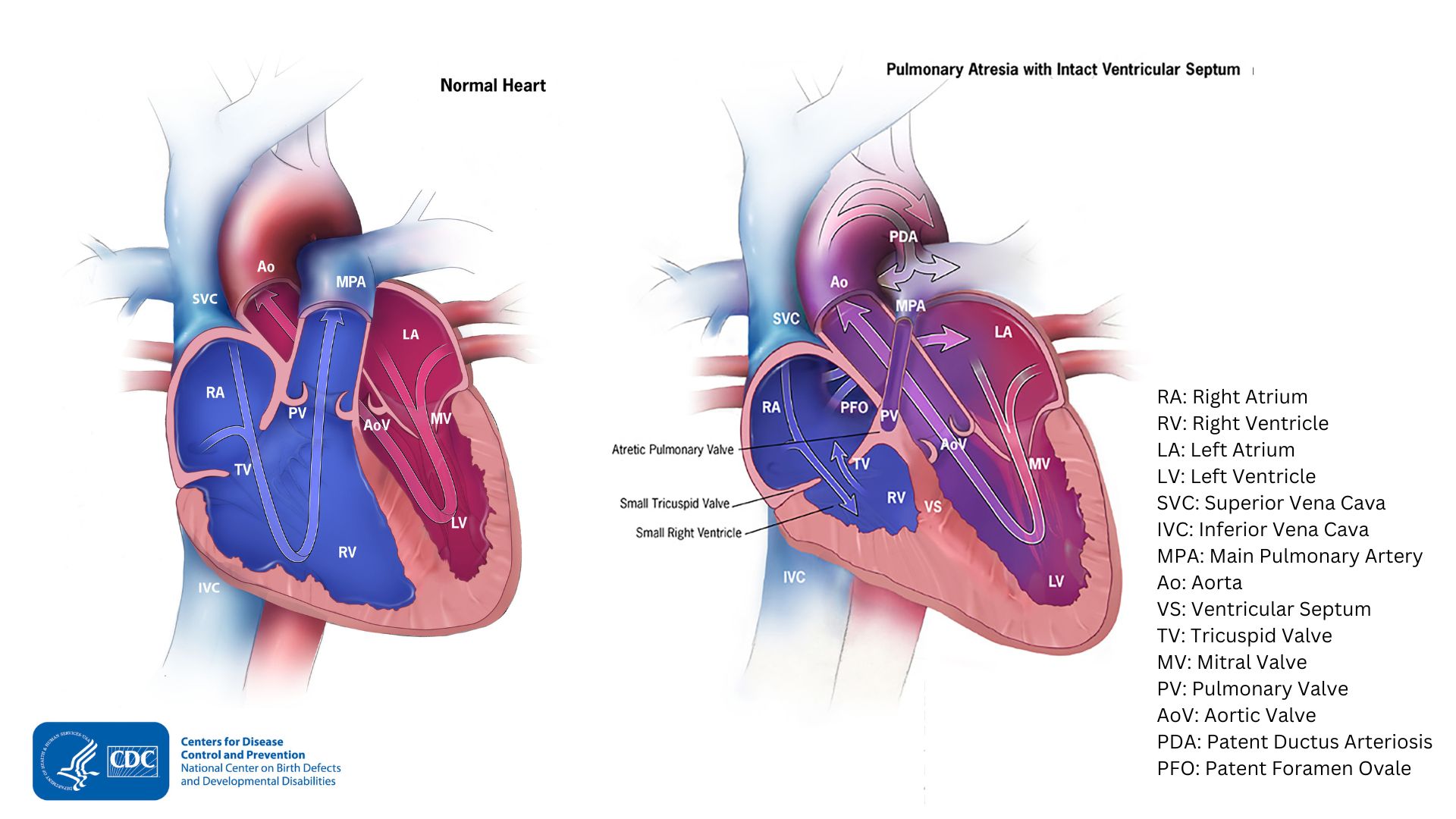
Pulmonary atresia with an intact ventricular septum
In this form, the wall, or septum, between the ventricles remains complete and intact. During pregnancy when the heart is developing, very little blood flows into or out of the right ventricle (RV). Therefore, the RV doesn't fully develop and remains very small. If the RV is under-developed, the heart can have problems pumping blood to the lungs and the body. The artery that carries blood out of the RV is the main pulmonary artery (MPA). The MPA also remains very small, because the pulmonary valve (PV) didn't form.
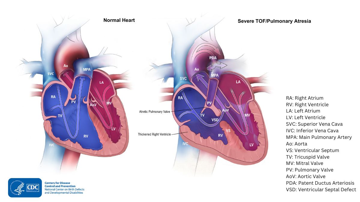
Pulmonary atresia with a ventricular septal defect (VSD)
In this form, a VSD allows blood to flow into and out of the RV. Therefore, blood flowing into the RV can help the ventricle develop during pregnancy. In this form, the RV is typically not as small as in pulmonary atresia with an intact ventricular septum. Pulmonary atresia with a VSD is similar to another condition called tetralogy of Fallot. However, in tetralogy of Fallot, the pulmonary valve (PV) forms, but it is small (called pulmonary valve stenosis). Thus, pulmonary atresia with a VSD is like a very severe form of tetralogy of Fallot.
Signs and symptoms
Babies born with pulmonary atresia will show symptoms at birth or very soon afterwards. They may have a bluish looking skin color, called cyanosis, because their blood doesn't carry enough oxygen. Infants with pulmonary atresia can have additional symptoms such as:
- Problems breathing
- Ashen or bluish skin color
- Poor feeding
- Extreme sleepiness
Risk factors
The causes of pulmonary atresia among most babies are unknown. Some babies have heart defects because of changes in their genes or chromosomes. A combination of genes and other risk factors may increase the risk for pulmonary atresia. These factors can include things in a mother's environment, what she eats or drinks, or the medicines she uses.
Diagnosis
Pulmonary atresia may be diagnosed during pregnancy or soon after the baby is born.
During pregnancy
An ultrasound, a tool that creates pictures of the baby, may detect pulmonary atresia during pregnancy. If the health care provider suspects pulmonary atresia, they can request a fetal echocardiogram to confirm the diagnosis. A fetal echocardiogram is a more detailed ultrasound of the baby's heart. This test can show problems with the structure of the heart and how the heart is working.
After the baby is born
During a physical examination, a doctor can see the symptoms, such as bluish skin or problems breathing. Using a stethoscope, a doctor will check for a heart murmur (an abnormal "whooshing" sound caused by blood not flowing properly). However, it is not uncommon for a heart murmur to be absent right at birth.
If a doctor suspects a problem, the health care provider might request an echocardiogram, which is an ultrasound of the heart. Cardiac catheterization can also confirm the diagnosis by looking inside the heart and measuring the blood pressure and oxygen levels. An electrocardiogram (EKG) may also be conducted to confirm the diagnosis.
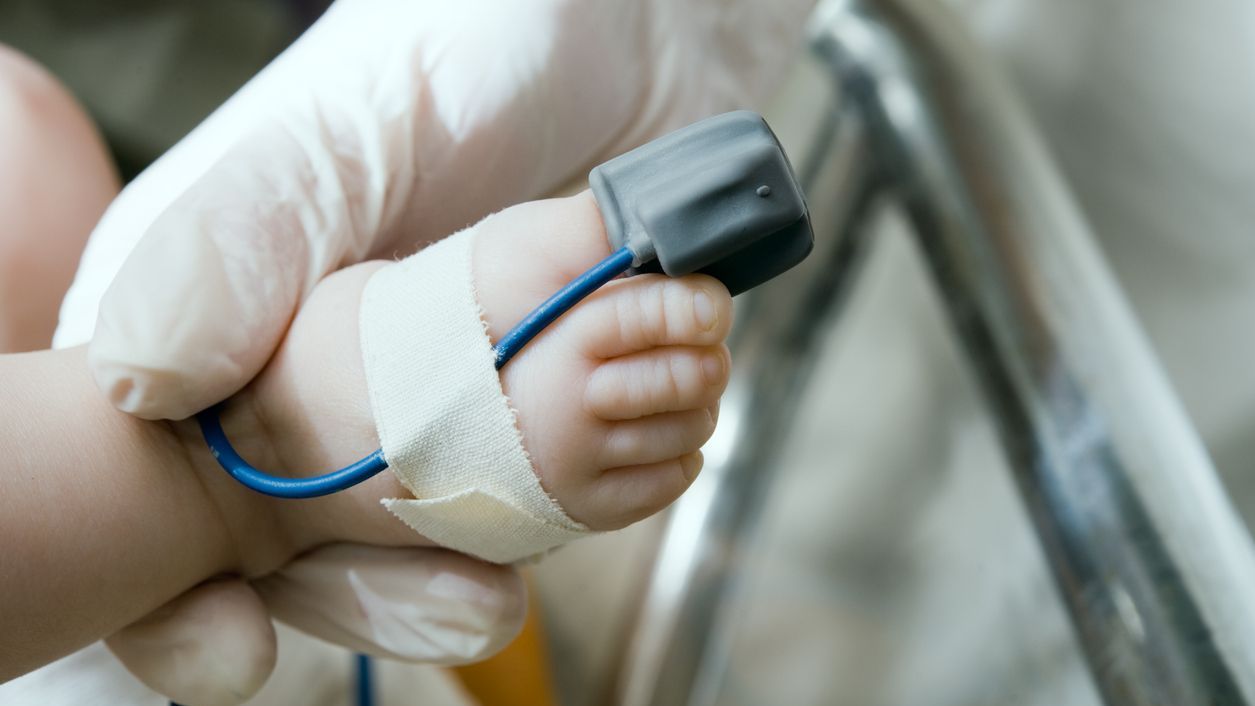
Pulmonary atresia can also be detected with newborn pulse oximetry screening. Low levels of oxygen in the blood can be a sign of a critical CHD like pulmonary atresia. Newborn screening using pulse oximetry can identify some infants with pulmonary atresia before they show any symptoms.
Treatments
Most babies will need medication to keep their ductus arteriosus open after birth. Keeping this blood vessel open will help with blood flow to the lungs until the pulmonary valve can be repaired.
Treatment for pulmonary atresia depends on its severity. In some cases, blood flow can be improved by using cardiac catheterization. During this procedure, doctors can expand the valve using a balloon to keep the ductus arteriosus open. In some cases, they may need to place a stent (a small tube) to keep it open.
In most cases of pulmonary atresia, a baby may need surgery soon after birth. During surgery, doctors widen or replace the pulmonary valve and enlarge the passage to the pulmonary artery. If a baby has a VSD, the doctor will use a patch. The patch closes the hole between the lower chambers of the heart. These actions will improve blood flow to the lungs and the rest of the body. If a baby has an underdeveloped right ventricle, staged surgical procedures might be needed. The staged surgical procedures are similar to surgical repairs for hypoplastic left heart syndrome (HLHS)
What to expect long-term
Most babies with pulmonary atresia will need routine checkups with a heart doctor. A heart doctor will monitor a baby's progress and check for other health conditions that might develop as they get older. As adults, they may need more surgery or medical care for other possible problems.
- Stallings EB, Isenburg JL, Rutkowski RE, et al. National population-based estimates for major birth defects, 2016–2020. Birth Defects Res. 2024;116(1):e2301. https://doi.org/10.1002/bdr2.2301