
Information on group A strep bacteria and the health problems they can cause.

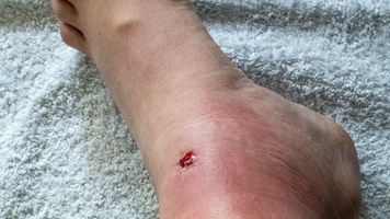
Diagnosis, testing, and treatment guidance on pyoderma caused by group A strep.

Clinical guidance on diagnosis, testing, and treatment of streptococcal pharyngitis.
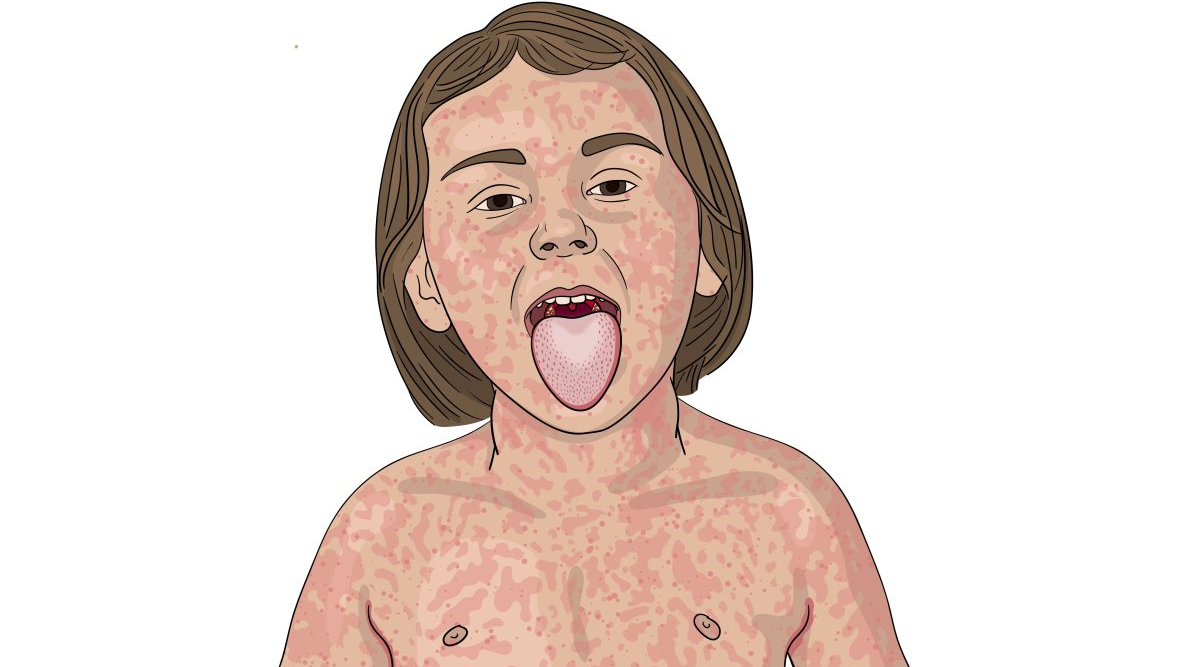
Guidance on differentiating scarlet fever from viral exanthems.
Invasive diseases
Guidance on differentiating streptococcal cellulitis from other skin infections and treating it.

STSS diagnosis, testing, and treatment guidance.

Diagnosis and treatment guidance for streptococcal necrotizing fasciitis.
Immune-mediated sequelae

Clinical guidance on diagnosis, testing, and treatment of post-streptococcal glomerulonephritis.

Summary of clinical guidance on diagnosis, testing, and treatment of acute rheumatic fever.