Photos and Videos of Naegleria fowleri

On This Page

Clinicians: For 24/7 diagnostic assistance, specimen collection guidance, shipping instructions, and treatment recommendations, please contact the CDC Emergency Operations Center at 770-488-7100. More detailed guidance is under Information for Public Health & Medical Professionals.

Photos of Naegleria fowleri

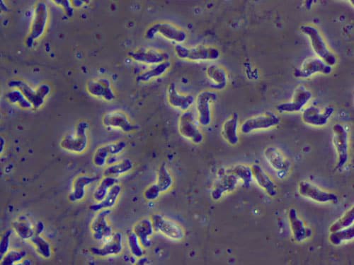
A wet mount of Naegleria fowleri trophozoites cultured from the CSF of a patient with primary amebic meningoencephalitis (PAM) viewed using phase contrast microscopy. Magnification: 600x.
Phase Contrast Microscopy

A wet mount of Naegleria fowleri trophozoites cultured from the cerebrospinal fluid (CSF) of a patient with primary amebic meningoencephalitis (PAM) viewed using phase contrast microscopy. Magnification: 600x.

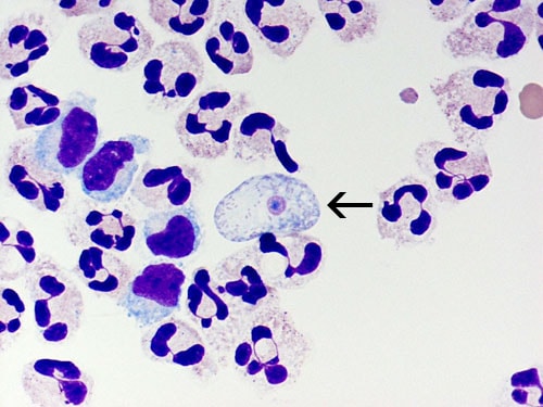
A cytospin of fixed CSF showing a Naegleria fowleri trophozoite (arrow) stained with Giemsa-Wright amidst polymorphonuclear leukocytes and a few lymphocytes. Within the trophozoite, the nucleus and nucleolus can be seen. Magnification: 1000x.
Giemsa-Wright Stain

A cytospin of fixed CSF showing a Naegleria fowleri trophozoite (arrow) stained with Giemsa-Wright amidst polymorphonuclear leukocytes and a few lymphocytes. Within the trophozoite, the nucleus and nucleolus can be seen. Magnification: 1000x.

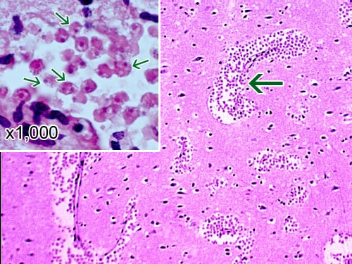
A section of the cerebral portion of the brain from a PAM patient, stained with hematoxylin and eosin, showing large clusters of Naegleria fowleri trophozoites and the destruction of the normal brain tissue architecture. Cysts are not seen. Magnification: 100x. Inset: Higher magnification (1000x) of Naegleria fowleri trophozoites (arrows) with the characteristic nuclear morphology.
Brain Tissue Stained with Hematoxylin and Eosin

A section of the cerebral portion of the brain from a PAM patient, stained with hematoxylin and eosin, showing large clusters of Naegleria fowleri trophozoites and the destruction of the normal brain tissue architecture. Cysts are not seen. Magnification: 100x. Inset: Higher magnification (1000x) of Naegleria fowleri trophozoites (arrows) with the characteristic nuclear morphology.

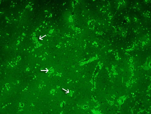
A section of the cerebral portion of the brain from a PAM patient reacted with the specific anti-Naegleria fowleri antibody which has been conjugated to a fluorescent antibody (immunofluorescent staining) viewed using microscopy with an exciter filter. Note the large numbers of Naegleria fowleri trophozoites staining bright green. No cysts are seen. Magnification: 100x.
Immunofluorescent Stain

A section of the cerebral portion of the brain from a PAM patient reacted with the specific anti-Naegleria fowleri antibody which has been conjugated to a fluorescent antibody (immunofluorescent staining) viewed using microscopy with an exciter filter. Note the large numbers of Naegleria fowleri trophozoites staining bright green. No cysts are seen. Magnification: 100x.

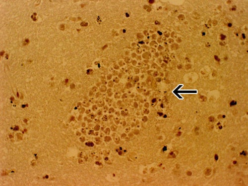
A section of the brain from a PAM patient stained with hemotoxylin and eosin showing a large cluster of Naegleria fowleri trophozoites surrounding capillaries. Magnification: 400x.
Brain Tissue Stained with Hematoxylin and Eosin

A section of the brain from a PAM patient stained with hemotoxylin and eosin showing a large cluster of Naegleria fowleri trophozoites surrounding capillaries. Magnification: 400x.

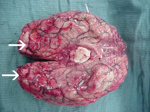
Extensive hemorrhage and necrosis is present in the brain, mainly in the frontal cortex.
Whole Brain 1

Extensive hemorrhage and necrosis are present in the brain, mainly in the frontal cortex.

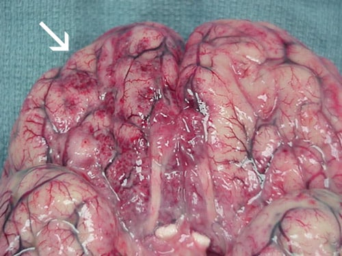
Extensive hemorrhage and necrosis is present in the brain, mainly in the frontal cortex.
Whole Brain 2

Extensive hemorrhage and necrosis are present in the brain, mainly in the frontal cortex.

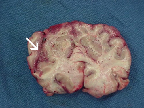
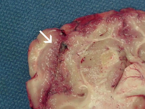
Focal hemorrhage and necrosis in frontal cortex due to Naegleria fowleri
Brain Section 1

Focal hemorrhage and necrosis in frontal cortex due to Naegleria fowleri.

Focal hemorrhage and necrosis in frontal cortex due to Naegleria fowleri
Brain Section 2

Focal hemorrhage and necrosis in frontal cortex due to Naegleria fowleri.

Videos of Naegleria fowleri

Naegleria fowleri under a microscope in real-time.

Naegleria fowleri under a microscope with increased speed.