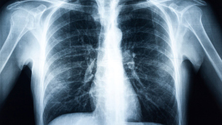
Study Syllabus for Classification of Radiographs of Pneumoconioses

Clinical Overview

Major Occupational Lung Diseases

Pneumoconioses

Coal Mine Dust Lung Disease (Part 1)

Inhalation of coal mine dust can lead to a variety of respiratory conditions that are included under the broad category of coal mine dust lung disease (CMDLD) [Petsonk et al. 2013]. Coal mine dust is a complex and variable mixture that may contain coal, silica and silicates, limestone used for rock dusting, diesel exhaust and, depending on specific conditions, other particulates and volatile chemicals. Mining methods and the coal miner’s job duties (including proximity to the coal face) determine the concentration and type of exposure, with underground miners in general experiencing higher disease risks than surface miners.

The spectrum of CMDLD includes coal workers’ pneumoconiosis (CWP), silicosis, mixed dust pneumoconiosis, dust-related diffuse fibrosis, and emphysema and chronic bronchitis. CWP typically requires at least 10 years’ exposure to manifest chest radiographic findings, classified as either simple or complicated. Simple CWP classically manifests as small rounded opacities, often more profuse in the upper zones (Fig. 4). The q type of small rounded opacities are associated with macules and micronodules on pathology, while r type opacities are associated with macronodules [Laney and Petsonk 2012]. Although upper lung predominant rounded opacities are more common, lower lung predominant irregular opacities may also be seen [Laney and Petsonk 2012; Blackley et al. 2015; Young et al. 1992]. Though many patients with early simple CWP have normal lung function, decrements in FEV1, FVC and FEV1/FVC ratio are greater with increasing profusion of small opacities [Blackley et al. 2015]. Complicated CWP (PMF) has radiograph and CT appearances similar to complicated silicosis (Fig. 5), with opacities of at least 1 cm.

Large opacities typically start in the lung periphery, have a round or lentiform shape paralleling the pleura and may enlarge over time. As a large opacity evolves, it may migrate toward the hilum, with a concomitant decrease in background small nodule profusion, and may cavitate and/or calcify. The large opacities of CWP may be unilateral, or may involve the lower lungs. Complicated CWP is associated with substantial impairment in lung function, often with mixed restrictive and obstructive changes and with decreased diffusion capacity.