Images of Influenza Viruses
This image illustrates the very beginning stages of an influenza (flu) infection. Most experts think that influenza viruses spread mainly through small droplets containing influenza virus. These droplets are expelled into the air when people infected with the flu cough, sneeze or talk. Once in the air, these small infectious droplets can land in the mouths or noses of people who are nearby. This image shows what happens after these influenza viruses enter the human body. The viruses attach to cells within the nasal passages and throat (i.e., the respiratory tract). The influenza virus’s hemagglutinin (HA) surface proteins then bind to the sialic acid receptors on the surface of a human respiratory tract cell. The structure of the influenza virus’s HA surface proteins is designed to fit the sialic acid receptors of the human cell, like a key to a lock. Once the key enters the lock, the influenza virus is then able to enter and infect the cell. This marks the beginning of a flu infection.

![Image without Labels or Text [JPG, 1.3 MB] Image without Labels or Text](/flu/images/freeresources/flu-virus-images/influenza-virus-nolabels-600px.jpg?_=99144)
![Image with Labels only [JPG, 1.4 MB] Image with Labels only - JPG, 1.4 MB](/flu/images/freeresources/flu-virus-images/influenza-virus-labels-600px.jpg?_=98939)

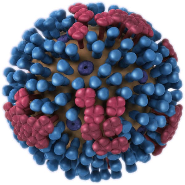
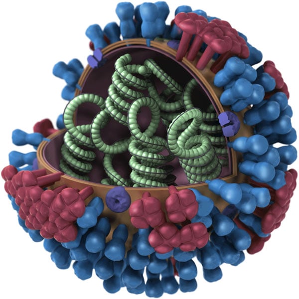
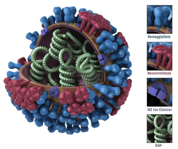
The above image shows the different features of an influenza virus, including the surface proteins hemagglutinin (HA) and neuraminidase (NA). Following influenza infection or receipt of the influenza vaccine, the body’s immune system develops antibodies that recognize and bind to “antigenic sites,” which are regions found on an influenza virus’ surface proteins. By binding to these antigenic sites, antibodies neutralize flu viruses, which prevents them from causing further infection.

The above image shows the different features of an influenza virus, including the surface proteins hemagglutinin (HA) and neuraminidase (NA). Following influenza infection or receipt of the influenza vaccine, the body’s immune system develops antibodies that recognize and bind to “antigenic sites,” which are regions found on an influenza virus’ surface proteins. By binding to these antigenic sites, antibodies neutralize flu viruses, which prevents them from causing further infection.

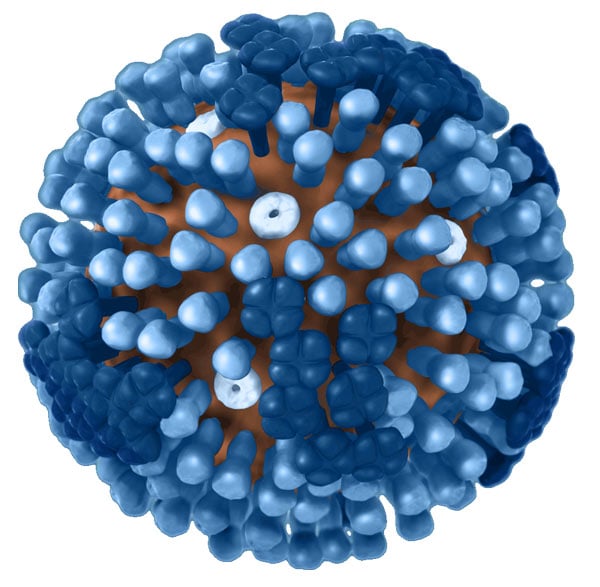
A 3D computer-generated rendering of a whole influenza (flu) virus in semi-transparent blue with a clear background. The virus’ hemagglutinin (HA) and neuraminidase (NA) surface proteins are displayed in semi-transparent blue sticking out of the surface of the virus. HA is a trimer (which is comprised of three subunits), while NA is a tetramer (which is comprised of four subunits and its head region resembles a 4-leaf clover).
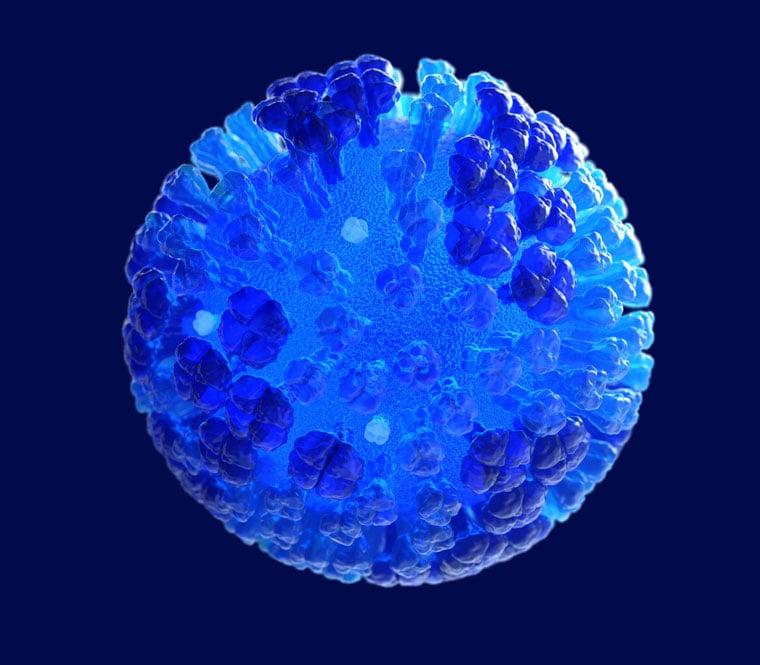
A 3D computer-generated rendering of a whole influenza (flu) virus in semi-transparent blue with a navy-blue background. The virus’ hemagglutinin (HA) and neuraminidase (NA) surface proteins are displayed in semi-transparent blue sticking out of the surface of the virus. HA is a trimer (which is comprised of three subunits), while NA is a tetramer (which is comprised of four subunits and its head region resembles a 4-leaf clover)
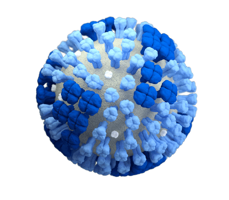
A 3D computer-generated rendering of a whole influenza (flu) virus with a light grey surface membrane set against a clear background. The virus’ surface proteins – hemagglutinin (HA) and neuraminidase (NA) – are depicted in light and dark blue, respectively. HA is a trimer (which is comprised of three subunits), while NA is a tetramer (which is comprised of four subunits and its head region resembles a 4-leaf clover)
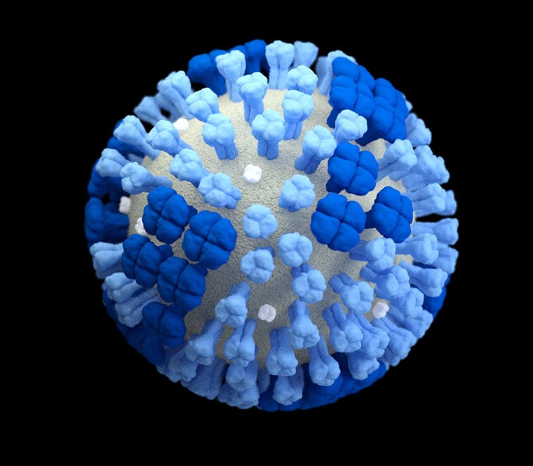
A 3D computer-generated rendering of a whole influenza (flu) virus with a light grey surface membrane set against a black background. The virus’ surface proteins – hemagglutinin (HA) and neuraminidase (NA) – are depicted in light and dark blue, respectively. HA is a trimer (which is comprised of three subunits), while NA is a tetramer (which is comprised of four subunits and its head region resembles a 4-leaf clover)
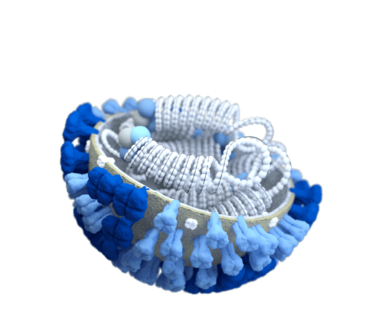
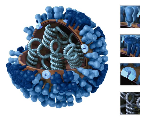
A 3D computer-generated rendering of a half-sliced influenza (flu) virus with a grey surface membrane set against a clear background. The virus’ surface proteins – hemagglutinin (HA) and neuraminidase (NA) – are depicted in light and dark blue, respectively. Inside of the virus, its ribonucleoproteins (RNPs) are shown with their coiled structures and three-bulbed polymerase complex on the ends. An influenza virus’ RNP is composed of both RNA and protein. Every influenza virus has eight RNP segments that correspond to the virus’ eight total gene segments. Three of these RNP segments encode the virus’ surface proteins (i.e., the HA, NA and M proteins).
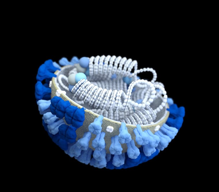
A 3D computer-generated rendering of a half-sliced influenza (flu) virus with a grey surface membrane set against a black background. The virus’ surface proteins – hemagglutinin (HA) and neuraminidase (NA) – are depicted in light and dark blue, respectively. HA is a trimer (which is comprised of three subunits), while NA is a tetramer (which is comprised of four subunits and its head region resembles a 4-leaf clover). Inside of the virus, its ribonucleoproteins (RNPs) are shown with their coiled structures and three-bulbed polymerase complex on the ends. An influenza virus’ RNP is composed of both RNA and protein. Every influenza virus has eight RNP segments that correspond to the virus’ eight total gene segments. Three of these RNP segments encode the virus’ surface proteins (i.e., the HA, NA and M proteins).
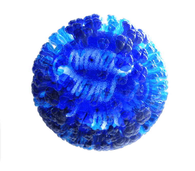
3D computer-generated rendering of a whole influenza (flu) virus in semi-transparent blue with a clear background. On the inside of the virus, its ribonucleoproteins (RNPs) are shown in white with their coiled structures and three-bulbed polymerase complex on the ends. An influenza virus’ RNP is composed of both RNA and protein. Every influenza virus has eight RNP segments that correspond to the virus’ eight total gene segments. Three of these RNP segments encode the virus’ surface proteins (i.e., the HA, NA and M proteins).
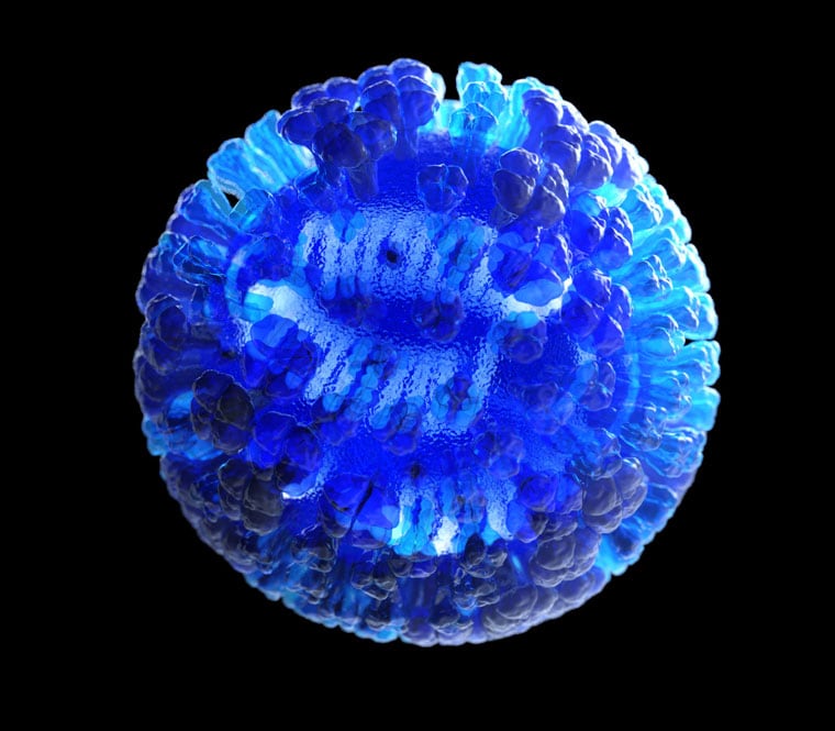
3D computer-generated rendering of a whole influenza (flu) virus in semi-transparent blue with a black background. On the inside of the virus, its ribonucleoproteins (RNPs) are shown in white with their coiled structures and three-bulbed polymerase complex on the ends. An influenza virus’ RNP is composed of both RNA and protein. Every influenza virus has eight RNP segments that correspond to the virus’ eight total gene segments. Three of these RNP segments encode the virus’ surface proteins (i.e., the HA, NA and M proteins).
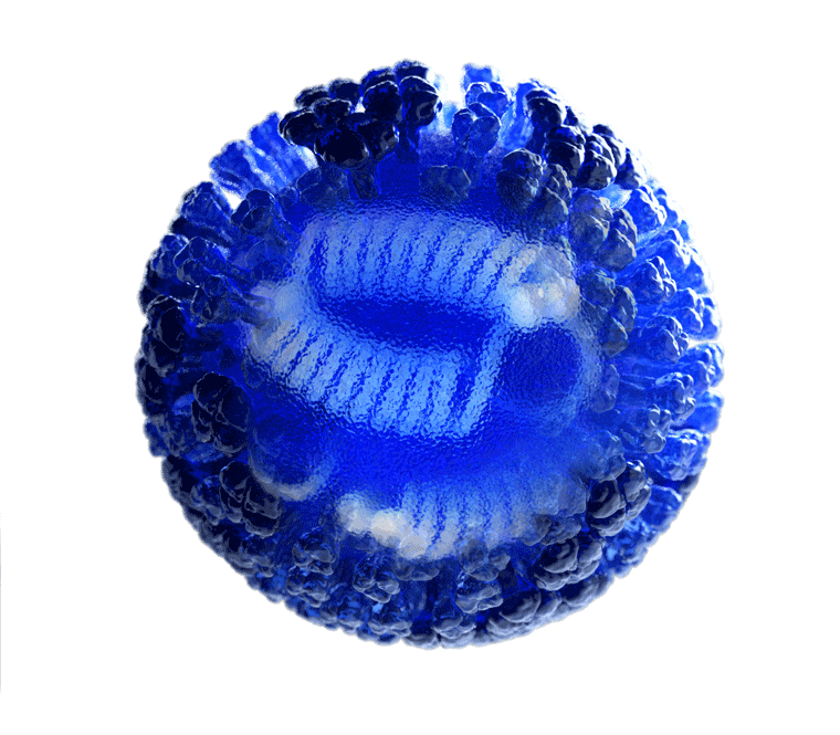
3D computer-generated rendering of a whole influenza (flu) virus in semi-transparent blue with a clear background. A transparent area in the center of the image allows the viewer to see inside of the influenza virus to see its ribonucleoproteins (RNPs). The RNPS are shown in white with their coiled structures and three-bulbed polymerase complex on the ends. An influenza virus’ RNP is composed of both RNA and protein. Every influenza virus has eight RNP segments that correspond to the virus’ eight total gene segments. Three of these RNP segments encode the virus’ surface proteins (i.e., the HA, NA and M proteins). The virus’ hemagglutinin (HA) and neuraminidase (NA) surface proteins are displayed in semi-transparent blue sticking out of the surface of the virus. HA is a trimer (which is comprised of three subunits), while NA is a tetramer (which is comprised of four subunits and its head region resembles a 4-leaf clover)

3D computer-generated rendering of a whole influenza (flu) virus in semi-transparent blue with a black background. A transparent area in the center of the image allows the viewer to see inside of the influenza virus to see its ribonucleoproteins (RNPs). The RNPS are shown in white with their coiled structures and three-bulbed polymerase complex on the ends. An influenza virus’ RNP is composed of both RNA and protein. Every influenza virus has eight RNP segments that correspond to the virus’ eight total gene segments. Three of these RNP segments encode the virus’ surface proteins (i.e., the HA, NA and M proteins). The virus’ hemagglutinin (HA) and neuraminidase (NA) surface proteins are displayed in semi-transparent blue sticking out of the surface of the virus. HA is a trimer (which is comprised of three subunits), while NA is a tetramer (which is comprised of four subunits and its head region resembles a 4-leaf clover)
