
About Cyclosporiasis
Cyclosporiasis is an intestinal illness caused by the microscopic parasite Cyclospora cayetanensis ,...

Preventing Cyclosporiasis
Based on currently available information, the best way to prevent cyclosporiasis is to avoid food or...
Symptoms of Cyclosporiasis
People experience symptoms of cyclosporiasis about one week (ranging as soon as two days to two week...
Countries or Regions at Risk for Cyclosporiasis
Cyclosporiasis occurs in many countries but is most common in tropical and subtropical regions and a...
For Health Care and Public Health Professionals
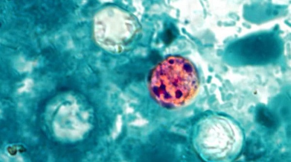
Clinical Overview of Cyclosporiasis
Cyclospora cayetanensis is a unicellular parasite that causes an intestinal infection called cyclosp...
Clinical Care of Cyclosporiasis
Most healthy people will eventually recover from cyclosporiasis without treatment although their ill...
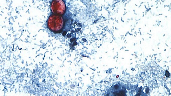
Clinical Overview of Cyclosporiasis
Cyclospora cayetanensis is a unicellular parasite that causes an intestinal infection called cyclosp...
Surveillance of Cyclosporiasis
Cyclosporiasis is a nationally notifiable disease and is reportable in 43 states, the District of Co...
Cyclosporiasis Publications
CDC regularly documents all publications regarding outbreaks, surveillance, and the epidemiology of ...