Photographs
The images below depict symptoms of STDs and are intended for educational use only.
Secondary stage syphilis sores (lesions) on the palms of the hands. Referred to as "palmar lesions."
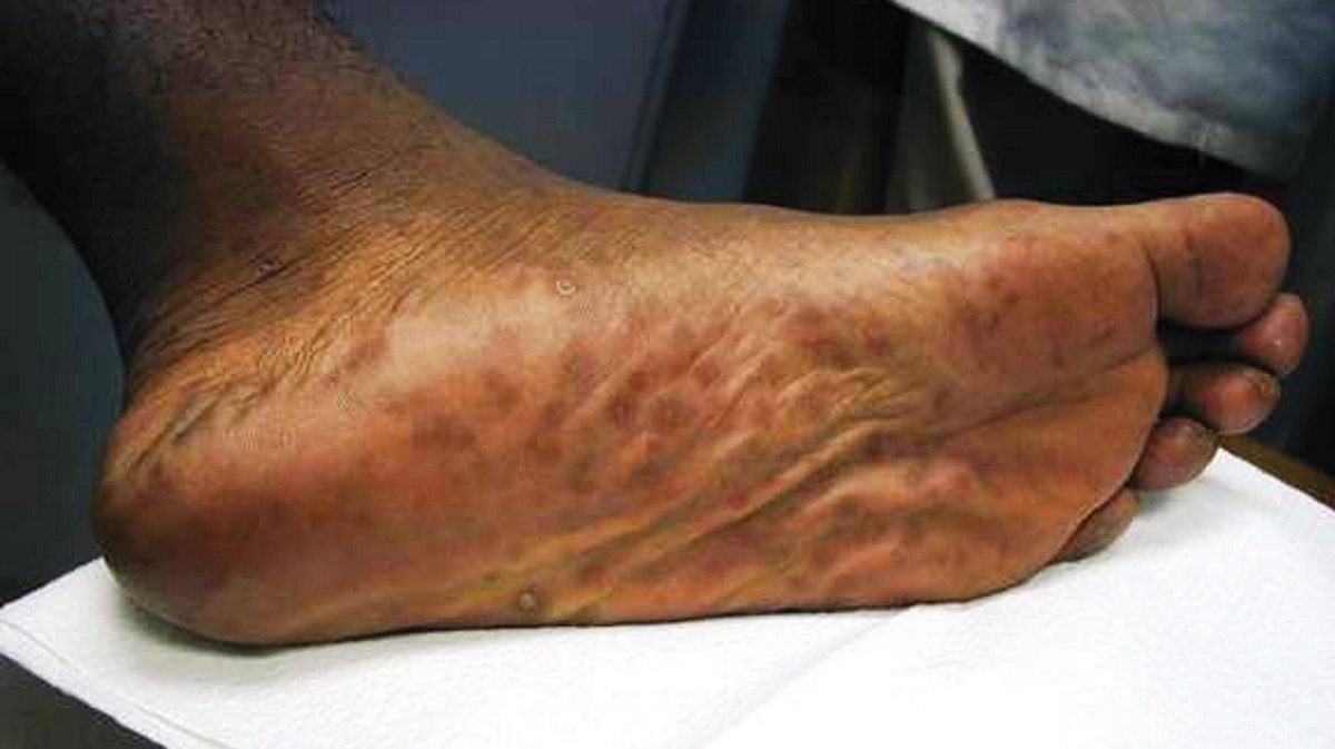
Secondary stage syphilis sores (lesions) on the bottoms of the feet. Referred to as “plantar lesions.”
Darkfield micrograph of Treponema pallidum.
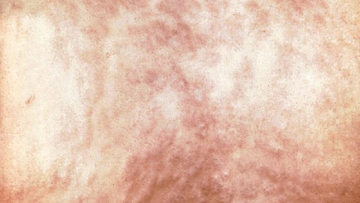
Secondary syphilis rash on the back.
Lesions of secondary syphilis.
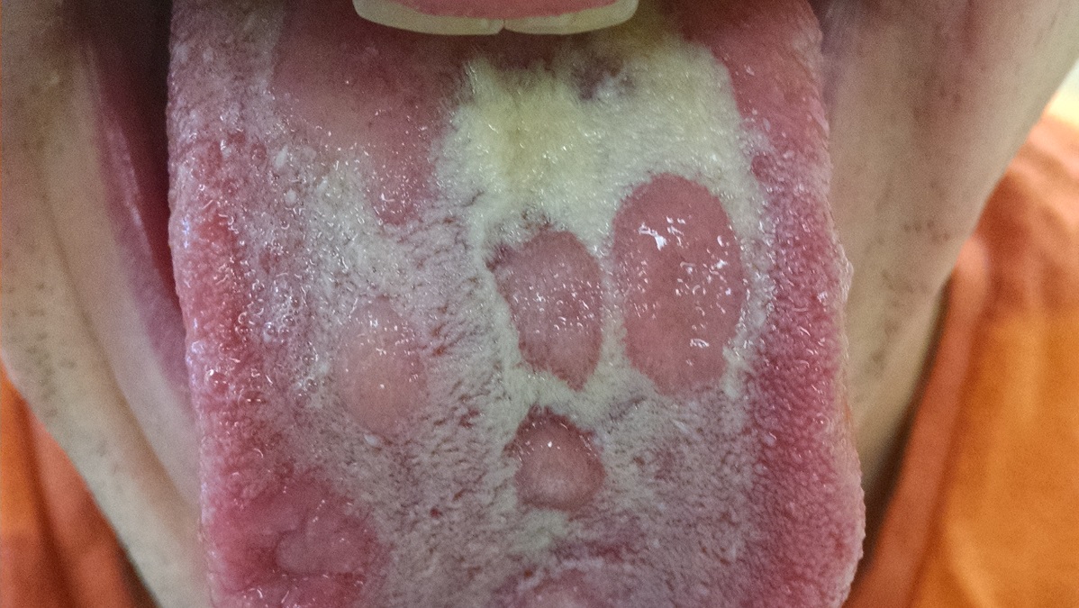
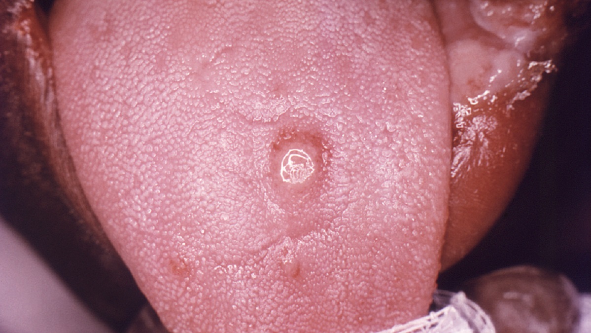
Primary stage syphilis sore (chancre) on the surface of a tongue.
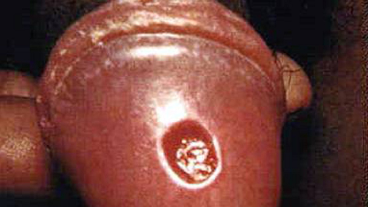
Primary stage syphilis sore (chancre) on glans (head) of the penis.
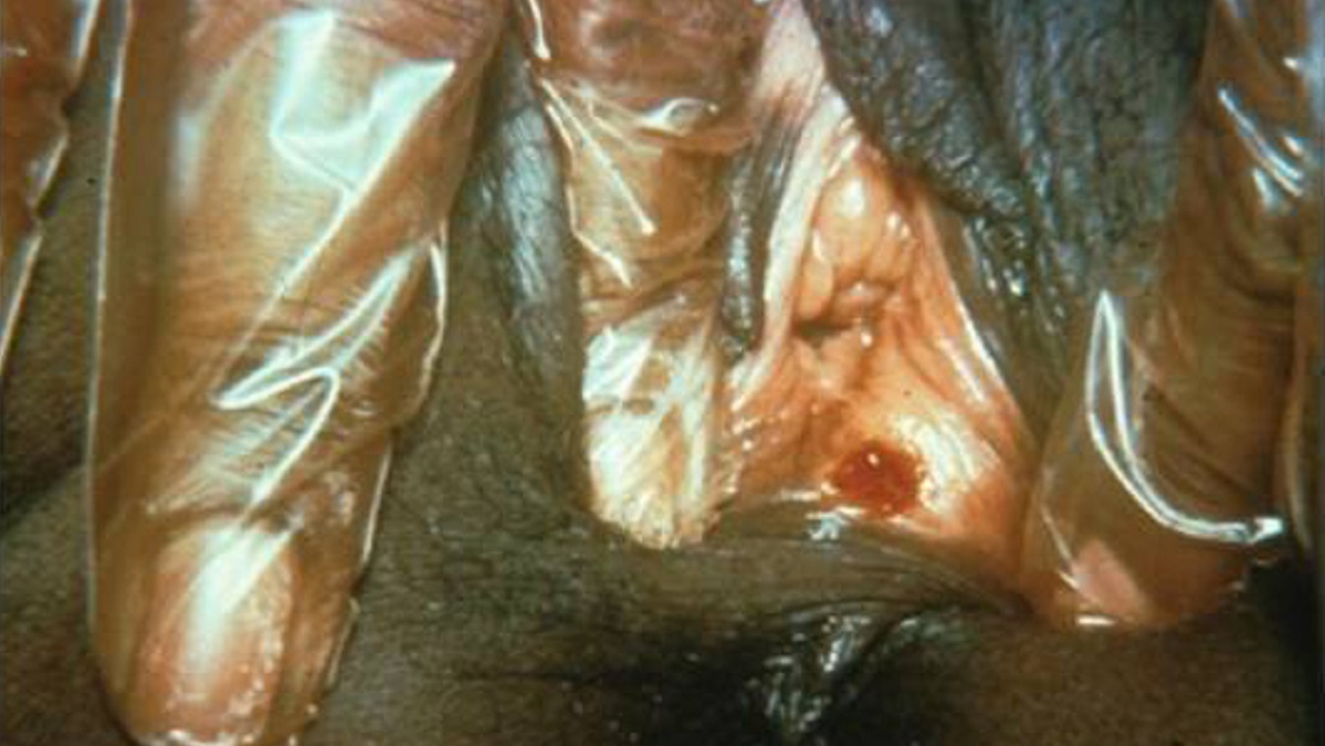
Primary stage syphilis sore (chancre) inside the vaginal opening.