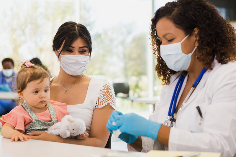
Learn about polio in the United States and when to get a vaccine for yourself or your child.
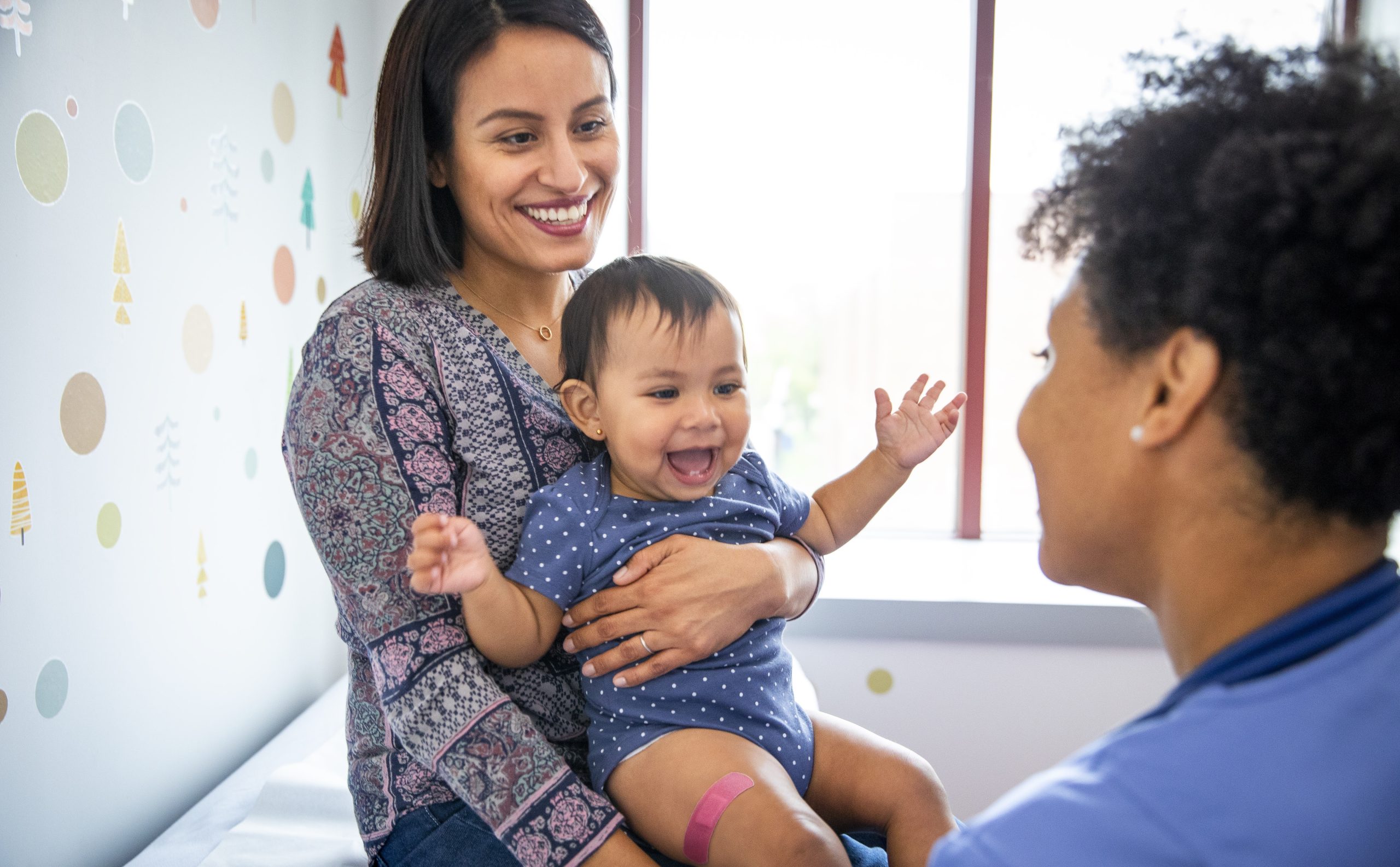
Learn about polio vaccine basics, who should get it, when to get it, and why it's important.

Find polio vaccination recommendations for people who travel internationally.
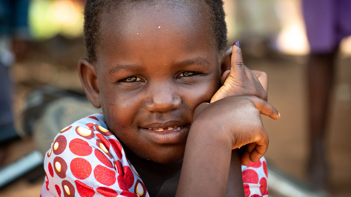
CDC works with partners to eradicate polio worldwide through sound science and effective programming
For Professionals

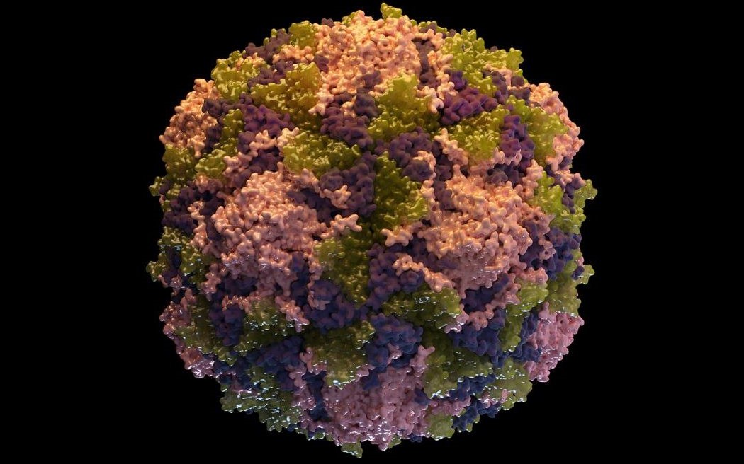
Learn about clinical signs of polio disease, transmission, diagnosis, and case definition.
Find routine recommendations, accelerated schedules, other considerations for polio vaccine.
Learn about what to do if you suspect your patient may have polio.
Learn about laboratory testing and diagnostic methods for poliovirus.
Learn how to report suspected poliovirus and the rules for reporting.