Nocardiosis Information for Healthcare Workers
Clinical Feature
Overall, 80% of nocardiosis cases present as invasive pulmonary infection, disseminated infection, or brain abscess; 20% present as cellulitis.
Pulmonary infection commonly presents with fever, cough, or chest pain. It can also present as pneumonia, lung abscesses, or cavitary lesions.
Central nervous system (CNS) symptoms include:
- Headache
- Lethargy
- Confusion
- Seizures
- Sudden onset of neurologic deficit
Contiguous spread within the thoracic cavity and hematogenous dissemination, particularly to the CNS, are possible.
Although incidence data are extremely limited, the number of cases is likely rising as a result of the increase in the number of severely immunocompromised people.
Approximately 10% of cases with uncomplicated pneumonia are fatal.
The case-fatality rate increases with overwhelming infection, disseminated disease, or brain abscess. Surgical drainage may be indicated and may improve patient outcome.
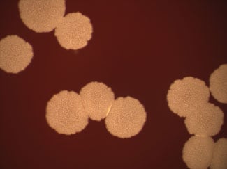
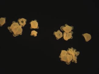
Etiologic Agent of Nocardiosis
The most commonly reported species from clinical sources are:
- Nocardia nova
- Nocardia farcinica
- Nocardia cyriacigeorgica
- Nocardia brasiliensis
- Nocardia abscessus
More than 40 valid Nocardia species are considered clinically relevant.
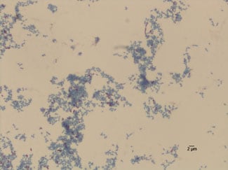
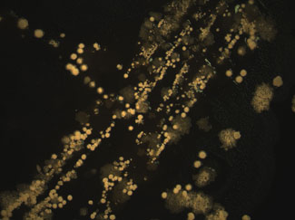
Laboratory Diagnostics
Infections due to Nocardia species are often overlooked due to the extended incubation time needed to isolate the organism from primary clinical specimens. Routine cultures must be held for at least 14 days. Accurate identification of Nocardia species recovered requires molecular methods. Referral of isolates to a reference laboratory, such as CDC’s Special Bacteriology Reference Laboratory, may be needed for identification and Antimicrobial Susceptibility Testing.
For more information on laboratory diagnostics for Nocardia species and other aerobic actinomycetes, see the references in the Treatment section below.
For information on laboratory submissions, visit Special Bacteriology Reference Laboratory (SBRL). All submissions from within the United States must be approved by the state’s department of health.
For faster and more accurate pathogen identification, go to MicrobeNET [Access Request Required].
Treatment
Some nocardiae are reported to have species-specific susceptibility profiles, but multidrug-resistant strains are common. Because of this, antimicrobial susceptibility testing (AST) should be performed on every isolate of clinical significance.
N. farcinica is often resistant to multiple antimicrobial agents, including trimethoprim-sulfamethoxazole (TMP-SMX), and has been shown to be more virulent in an animal model. TMP-SMX therapy for HIV-infected patients may be complicated by frequent occurrence of adverse events and drug resistance.
CDC’s Special Bacteriology Reference Laboratory performs AST on all isolates received. See the Laboratory Diagnostics section above for information on diagnostics, including laboratory submission to CDC.
For more information on treatment of Nocardia species and other aerobic actinomycetes infections, see the following references:
- Bell M, McNeil MM, and Brown JM. Nocardia species (Nocardiosis). 2014. http://www.antimicrobe.org/b117.aspExternalExternal. Accessed April 9, 2015.
- McNeil MM, Brown JM. 2002. Nocardia species (Nocardiosis). p.481-500. ln Yu VL, Merigan TC, Barriere SL (eds.), Antimicrobial Therapy and Vaccines, 2nd ed, vol 1, Apple Trees Productions, New York, NY.
- McNeil MM, and Brown JM. 1994. The medically important aerobic actinomycetes: epidemiology and microbiology. Clin Microbiol Rev 7:357-417.
- Conville P S, and Witebsky FG. 2011. Nocardia, Rhodococcus, Gordonia, Actinomadura, Streptomyces, and Other Aerobic Actinomycetes. p. 443-471. In Versalovic J,
- Carroll KC, Funke G, Jorgensen JH, Landry ML, and Warnock DW (eds.), Manual of Clinical Microbiology, 10th ed. ASM Press, Washington, DC.