Key points
- High-quality laboratory science keeps laboratory professionals safe and protects community health.
- CDC provides clinical and public health laboratories with training and technical assistance.
- Learn about the laboratory quality management systems used in clinical testing.
Purpose
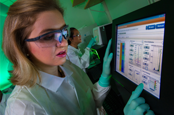
Laboratories are on the frontline for protecting our communities’ health. CDC provides clinical and public health laboratories with training and technical assistance to achieve the highest quality in laboratory science. This support also ensures the safety of laboratory professionals and the communities where they work.
CDC’s Division of Laboratory Systems (DLS) helps laboratories achieve high-quality testing and accurate, timely results by supporting top standards. This approach positively influences how the results are used by a patient’s care team.
Our approach
DLS is responsible for:
- Advancing laboratory quality management systems (QMS).
- Technical guidance.
- Training materials based on the Clinical Laboratory Improvement Amendments (CLIA) requirements.
This regulation requires laboratories to have quality control (QC) and quality assurance (QA) measures in place. This ensures the accuracy and precision of the complete testing process. In addition, DLS works to enhance quality resources throughout clinical and public health laboratories across the country.
DLS works with other CDC programs to develop and deliver QMS trainings for laboratory professionals within the agency. They also provide training for the broader U.S. clinical and public health laboratory community.
Program priorities
Molecular diagnostic testing combines laboratory testing with the precision of molecular biology. It has revolutionized the way clinical and public health laboratories investigate human, viral, and microbial genomes. It has also transformed how they examine genes and the products they encode.
Molecular diagnostic tests are increasingly being used, and have supplanted numerous conventional tests, in many areas of laboratory medicine including:
- Oncology.
- Infectious diseases.
- Clinical chemistry.
- Clinical genetics.
Advancements in molecular diagnostic testing continue to improve the accuracy and speed of detecting microbial pathogens and analyzing a patient's genes. This progress is becoming an essential aspect of patient-tailored interventions and treatments. DLS continues to advance the quality of clinical laboratory testing, and the molecular tools used in clinical practice.