Key points
- Fasciola is a type of flat, leaf-shaped parasitic worm, also known as a "liver fluke."
- A parasite is an organism (a living thing) that lives on or inside another organism.
- Fasciola can occur in over 70 countries, especially where there are sheep or cattle.
- People can get Fasciola by eating raw watercress or other freshwater plants contaminated by larvae (young worms).
Overview
Fasciola is a liver fluke (a type of parasitic worm) that can infect the liver and bile duct of exposed people and animals, such as sheep, cattle, goats, and other plant-eating domestic and wild animals.
- Fasciola parasites can cause an infectious disease called fascioliasis. Generally, fascioliasis is more common in people who live or work around livestock (e.g., sheep, cattle, goats) in areas where animal infections are common.
Two Fasciola species (types) can infect people:
- Fasciola hepatica: the main species that infects people. Other names for it include "the common liver fluke" and "the sheep liver fluke."
- Fasciola gigantica: a related species that primarily affects domestic and wild animals but can also infect people.
Fascioliasis occurs in all continents except Antarctica, in over 70 countries, especially where there are sheep, cattle, or goats.
- In most cases, Fasciola infections can occur when people eat raw watercress or freshwater plants contaminated by larvae.
- People also can get infected by consuming contaminated water, such as by drinking it or by eating vegetables that people washed or irrigated with contaminated water.
- You cannot get Fasciola from another person.
Resource
Signs and symptoms
Not all people with Fasciola infection have symptoms.
There are two phases: the acute (migratory) phase and the chronic phase.
Acute (migratory) phase
- Some people feel sick during the acute phase, when flukes that have not fully developed are migrating (passing) from the intestine through the abdominal cavity and liver.
- Symptoms can start four to seven days after exposure and can last for several weeks or months.
Chronic phase
- Some people feel sick during the chronic phase, when fully developed flukes are in the bile ducts (the duct system of the liver).
- Symptoms can start months after exposure.
Signs and symptoms from both phases of the infection can include:
- Fever
- Malaise
- Nausea and/or vomiting
- Abdominal pain
- Diarrhea or change in bowel habits
- Eosinophilia (when your blood has too many eosinophils, which are a type of white blood cell that fights infections/ allergies)
- Hepatomegaly (enlarged liver)
- Abnormal liver tests
Risk factors
Eating raw watercress or other water plants from areas where the disease occurs or consuming contaminated water are the key risk factors for Fasciola infection.
At-risk populations
Fascioliasis occurs in more than 70 countries, especially where there are sheep, cattle, or goats.
Fasciola hepatica occurs in all continents except Antarctica. In contrast, Fasciola gigantica only occurs in parts of Africa and Asia.
Although rare, people can become locally infected with Fasciola in the United States. There have been a few reported cases in Hawaii, California, and Florida. Most reported cases in the US occurred in immigrants that became infected in countries where fascioliasis occurs.
Causes
A person can get Fasciola infection when they consume raw watercress or other freshwater plants contaminated by larvae. A person can also get infected by consuming contaminated water when drinking it or eating vegetables that people washed or irrigated with contaminated water. A person cannot get Fasciola from another person.
Prevention
To avoid Fasciola infection, do not eat raw watercress and other freshwater plants, especially from areas where livestock feed on land covered by grass (grazing areas) in places where Fasciola infection occurs. It is important to avoid areas with poor sanitation that can have contaminated food and water.
There is no vaccine available to protect people against Fasciola.
Diagnosis
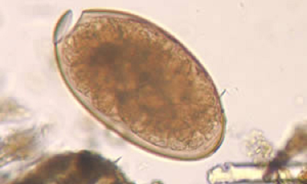
Talk to a healthcare provider if you think you might have Fasciola infection. Healthcare providers can diagnose Fasciola infection by seeing if you have Fasciola eggs in stool (poop) specimens (samples) via microscopy.
Stool specimen
- Stool specimen is the main way to diagnose Fasciola infection.
- Microscopy can identify Fasciola eggs.
- A healthcare provider may need more than one stool sample to identify the eggs.
Ultrasound, CT, or MRI
- At times, ultrasound, CT, or MRI can detect dilated bile ducts containing the fluke and help make the diagnosis.
Blood test
- A blood test for detecting Fasciola is available in the US at CDC.
- A blood test is less useful than a stool sample for diagnosis and patient management because it cannot tell the difference between a past or current infection.