Persons using assistive technology might not be able to fully access information in this file. For assistance, please send e-mail to: mmwrq@cdc.gov. Type 508 Accommodation and the title of the report in the subject line of e-mail.
Guidelines for Prevention and Treatment of
Opportunistic Infections in HIV-Infected Adults and Adolescents
Recommendations from CDC, the National Institutes of Health, and
the HIV Medicine Association of the Infectious Diseases Society of America
Prepared by
Jonathan E. Kaplan, MD1
Constance Benson, MD2
King K. Holmes, MD, PhD3
John T. Brooks, MD1
Alice Pau, PharmD4
Henry Masur, MD4
1CDC, Atlanta, Georgia
2 University of California San Diego, San Diego, California
3University of Washington, Seattle, Washington
4National Institutes of Health, Bethesda, Maryland
Disclosure of Relationship
CDC, our planners, and our content specialists wish to disclose they have no financial interests or other relationships
with the manufacturers of commercial products, suppliers of commercial services, or commercial supporters, with the exception
of Constance Benson and King K. Holmes. Dr. Benson discloses being on the Advisory Board for Merck, GlaxoSmithKline,
and Boehringer Ingelheim; being a grant recipient for Gilead; and being a Data Safety Monitoring Board (DSMB) member
for Achillion and JJR Australia. Her spouse also is a consultant for Merck, Gilead, Achillion, Monogram, and Vertex. Dr.
Holmes discloses being a DSMB member of Merck, receiving an honorarium at the 2005 Infectious Diseases Society of
America Conference, and serving on the Mycology Research Laboratories scientific advisory board. Presentations will not include
any discussion of the unlabeled use of a product or a product under investigational use.
The material in this report originated in the National Center for HIV/AIDS, Viral Hepatitis, STD, and TB
Prevention, Kevin Fenton, MD, Director.
Corresponding preparer: John T. Brooks, M.D., Division of HIV/AIDS Prevention, NCHHSTP, CDC, 1600
Clifton Road NE, MS E-45, Atlanta, GA 30333, Telephone: 404-639-3894, Fax: 404-639-6127, Email: zud4@cdc.gov.
Summary
This report updates and combines earlier versions of guidelines for the prevention and treatment of opportunistic infections (OIs)
in HIV-infected adults (i.e., persons aged
>18 years) and adolescents (i.e., persons aged 13--17 years), last published in 2002 and
2004, respectively. It has been prepared by the Centers for Disease Control and Prevention (CDC), the National Institutes of Health
(NIH), and the HIV Medicine Association (HIVMA) of the Infectious Diseases Society of America (IDSA). The guidelines are intended
for use by clinicians and other health-care providers, HIV-infected patients, and policy makers in the United States. These
guidelines address several OIs that occur in the United States and five OIs that might be acquired during international travel. Topic
areas covered for each OI include epidemiology, clinical manifestations, diagnosis, prevention of exposure; prevention of disease
by chemoprophylaxis and vaccination; discontinuation of primary prophylaxis after immune reconstitution; treatment of
disease; monitoring for adverse effects during treatment; management of treatment failure; prevention of disease recurrence; discontinuation
of secondary prophylaxis after immune reconstitution; and special considerations during pregnancy.
These guidelines were developed by a panel of specialists from the United States government and academic institutions. For
each OI, a small group of specialists with content-matter expertise reviewed the literature for new information since the guidelines were
last published; they then proposed revised recommendations at a meeting held at NIH in June 2007. After these presentations
and
discussion, the revised guidelines were further reviewed by the co-editors; by the Office of AIDS Research, NIH; by specialists at
CDC; and by HIVMA of IDSA before final approval and publication.
The recommendations are rated by a letter that indicates the strength of the recommendation and a Roman numeral that
indicates the quality of evidence supporting the recommendation, so that readers can ascertain how best to apply the recommendations in
their practice environments.
Major changes in the guidelines include 1) greater emphasis on the importance of antiretroviral therapy for the prevention
and treatment of OIs, especially those OIs for which no specific therapy exists; 2) information regarding the diagnosis and management
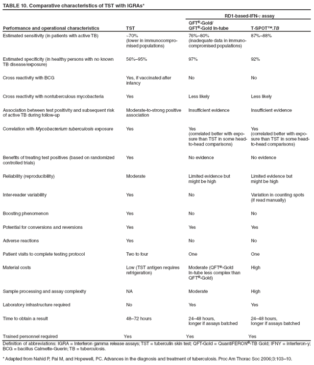
of immune reconstitution inflammatory syndromes; 3) information regarding the use of interferon-gamma release assays for the
diagnosis of latent Mycobacterium tuberculosis (TB) infection; 4) updated information concerning drug interactions that affect the use
of rifamycin drugs for prevention and treatment of TB; 5) the addition of a section on hepatitis B virus infection; and 6) the addition
of malaria to the list of OIs that might be acquired during international travel.
This report includes eleven tables pertinent to the prevention and treatment of OIs, a figure that pertains to the diagnois
of tuberculosis, a figure that describes immunization recommendations, and an appendix that summarizes recommendations
for prevention of exposure to opportunistic pathogens.
Introduction
Before the widespread use of potent combination antiretroviral therapy (ART), opportunistic infections (OIs), which
have been defined as infections that are more frequent or more severe because of immunosuppression in HIV-infected persons,
were the principal cause of morbidity and mortality in this population. In the early 1990s, the use of
chemoprophylaxis, immunization, and better strategies for managing acute OIs contributed to improved quality of life and improved survival
(1). However, the widespread use of ART starting in the mid-1990s has had the most profound influence on reducing
OI-related mortality in HIV-infected persons in those countries in which these therapies are accessible and affordable
(1--8).
Despite the availability of ART in the United States and other industrialized countries, OIs continue to cause
considerable morbidity and mortality for three primary reasons: 1) many patients are unaware of their HIV infection and seek medical
care when an OI becomes the initial indicator of their disease; 2) certain patients are aware of their HIV infection, but do not
take ART because of psychosocial or economic factors; and 3) certain patients are prescribed ART, but fail to attain
adequate virologic and immunologic response because of factors related to adherence, pharmacokinetics, or unexplained biologic
factors (4,9,10). Thus, although hospitalizations and deaths have decreased since the implementation of ART, OIs remain a
leading cause of morbidity and mortality in HIV-infected persons
(11--19). Clinicians must be knowledgeable about
optimal strategies for prevention and management of OIs to provide comprehensive high-quality care for these patients.
Recognizing that the relation between OIs and HIV infection is bidirectional is important. HIV leads
to immunosuppression that allows opportunistic pathogens to cause disease in HIV-infected persons. OIs and other
coinfections that might be common in HIV-infected persons, such as sexually transmitted infections, can also have adverse effects on
the natural history of HIV infection. Certain OIs are associated with reversible increases in circulating viral load
(20--25), and these increases could lead to accelerated HIV progression or increased transmission of HIV
(26). Thus, although chemoprophylaxis and vaccination directly prevent pathogen-specific morbidity and mortality, they might also contribute
to reduced rate of progression of HIV disease. For instance, randomized trials using trimethoprim-sulfamethoxazole
(TMP-SMX) have documented that chemoprophylaxis can both decrease OI-related morbidity and improve survival. The
survival benefit is likely to be partially attributable to reduced progression of HIV
infection(27--31). Reduced progression of
HIV infection would also indirectly delay or reduce the occurrence of subsequent OIs.
History of the Guidelines
In 1989, the Guidelines for Prophylaxis against
Pneumocystis carinii Pneumonia for persons infected with the
human immunodeficiency virus became the first HIV-related treatment guideline published by the U.S. Public Health Service
(32). This report was followed by a guideline on prevention of
Mycobacterium avium complex (MAC) disease in 1993
(33). In 1995, these guidelines were expanded to include the prevention of all HIV-related OIs and the Infectious Diseases Society
of America (IDSA) joined as a cosponsor (34). These prevention guidelines were revised in 1997, 1999, and 2002 and have
been
published in MMWR (35--37), Clinical Infectious Diseases
(38--40), the Annals of Internal Medicine
(41,42), American Family Physician
(43,44), and Pediatrics (45); accompanying editorials have appeared in
the Journal of the American Medical Association
(46,47).
In 2004, CDC, the National Institutes of Health (NIH), and the HIV Medicine Association (HIVMA) of the
IDSA published a new guideline including recommendations for treating HIV-infected adults and adolescents with OIs
(48). Companion guidelines were published for HIV-infected children
(49).
Responses to these guidelines (e.g., numbers of requests for reprints, website contacts, and observations from
health-care providers) have demonstrated that these guidelines have served as valuable references for HIV health-care providers.
Because the guidelines include ratings indicating the strength of each recommendation and the quality of supporting evidence,
readers have been able to assess the importance of each recommendation. The present report includes recommendations for
both prevention and treatment of OIs in HIV-infected adults and adolescents; an accompanying report includes
recommendations for HIV-exposed and -infected children.
These guidelines are intended for clinicians, other health-care providers, HIV-infected patients, and policy makers
residing in the United States; guidelines pertinent to other regions of the world, especially countries with limited resources,
might differ regarding the spectrum of OIs of interest and their diagnostic and therapeutic capacity.
Guidelines Process
These guidelines were prepared by the Opportunistic Infections Working Group under the auspices of the Office of
AIDS Research Advisory Council (OARAC) of NIH. Group leaders and team members with expertise in specific OIs were
selected from the membership of the Working Group; each group reviewed the literature since the last publication of the
prevention and treatment guidelines, conferred for several months, and produced draft revised guidelines. Recommendations
were reviewed and discussed by the Working Group at a meeting in Bethesda, Maryland, on June 25--26, 2007. A draft version
of these recommendations was posted at AIDSInfo (http://aidsinfo.nih.gov/contentfiles/Adult_OI.pdf) on June 18, 2008.
Since the June 18, 2008 posting, the draft recommendations were reviewed and updated for this report by Working Group
members and subject matter experts. Suggested updates were reviewed by the co-editors, who amended the report, as warranted.
The final version of the report was further reviewed by the co-editors, the Office of AIDS Research, NIH; experts at CDC; and
the HIVMA of IDSA before final approval and publication.
The current guidelines share key features with prior versions. They are labeled as guidelines, indicating that
the recommendations should be considered in the context of the individual patient situation and the community where
the patient is being managed. They are evidence based. For each recommendation, the strength and quality of the
evidence supporting it are indicated using a revised version of the rating system of the IDSA. As noted above, they have been
developed by a broadly based panel that included representatives from academic medical centers, federal governmental
agencies, community-based practices, and consumer advocates; representatives from Europe, Latin America, and Asia also took part
in the process. The guidelines are available in print media and on the Internet. They are written for physicians and other
health-care providers who care for HIV-infected persons in the United States and Western Europe where access is available to a
full range of up-to-date medical services; however, these recommended strategies might not be feasible or appropriate in all
settings where the spectrum of HIV-related complications and diagnostic capacity differ from those observed in the United States
and Western Europe. Final versions of the guidelines were reviewed by respective members of each panel to ensure
the recommendations were complete and appropriate. They are endorsed by CDC, the NIH, and
the HIVMA of the IDSA. They are intended to complement more comprehensive textbooks, journals, and other relevant informational materials. Information
is summarized in 11 tables.
Major Changes in Guidelines Since Last Publication
Major changes include 1) additional emphasis on the importance of ART for prevention and treatment of OIs,
especially those for which specific chemoprophylaxis and treatment do not exist; 2) information on diagnosis and management
of immune reconstitution inflammatory syndromes (IRIS); 3) information on interferon-gamma release assays (IGRAs) for
the detection of latent Mycobacterium tuberculosis
infection; 4) updated information on drug interactions affecting use of
rifamycin
drugs for prevention and treatment of tuberculosis (TB); 5) addition of a section on hepatitis B virus (HBV) infection; and
6) addition of a section on malaria to the OIs of geographic interest.
How to Use the Information in this Report
For each of the OIs discussed in this report, recommendations are provided that address 1) preventing exposure
to opportunistic pathogens, 2) preventing disease, 3) discontinuing primary prophylaxis after immune reconstitution, 4)
treating patients with disease, 5) monitoring for adverse effects (including IRIS), 6) managing treatment failure, 7) preventing
disease recurrence ("secondary prophylaxis" or chronic maintenance therapy), 8) discontinuing secondary prophylaxis after
immune reconstitution, and 9) special considerations during pregnancy. Recommendations are rated by a revised version of the
IDSA rating system (Box). In this system, the letters A--E signify the strength of the recommendation for or against a preventive
or therapeutic measure, and Roman numerals I--III indicate the quality of evidence supporting the recommendation.
The guidelines include eleven tables pertinent to the prevention and treatment of OIs (Tables 1--11), a figure that pertains to
the diagnosis of tuberculosis (Figure 1), a figure that describes immunization recommendations (Figure 2), and an appendix
that summarizes recommendations pertinent to prevention of exposure to opportunistic pathogens.
Effect of ART on the Management of OIs
Clinicians treating HIV-infected patients often have to consider two questions related to OIs and ART: 1) when to
initiate ART in ART-naïve persons who experience an acute OI, and 2) how ART should be managed for persons who are on
ART but who experience an acute OI.
Initiation of ART in the Setting of an Acute OI (Treatment-Naïve Patients)
When an acute OI is present, initiation of ART is usually expected to improve immune function and contribute to
faster resolution of the OI. Initiation of ART has been documented to be effective for OIs for which effective therapy does not
exist; cryptosporidiosis, microsporidiosis, and progressive multifocal leukoencephalopathy (PML) might resolve or at least
stabilize after the institution of effective ART
(50--52). For Kaposi's sarcoma (KS), initiation of ART has been documented to lead
to resolution of lesions in the absence of specific therapy for the sarcoma
(53). The initiation of ART in the setting of an
acute OI also has preventive benefit; a second OI is less likely to occur if ART is started promptly rather than delaying the
initiation of ART.
Starting ART in the setting of an acute OI has several potential disadvantages. Severely ill patients might not absorb
ART drugs, leading to subtherapeutic serum levels and the development of antiretroviral drug resistance. ART toxicities might
be confused with disease manifestations or toxicities associated with drugs used for treating patients with an OI.
Drug-drug interactions among ART and anti-OI drugs might be difficult to manage. Renal or hepatic dysfunction during acute
OIs might make dosing of ART drugs difficult to estimate. Finally, IRIS events can occur and cause manifestations that
are difficult to distinguish from other clinical conditions.
The term IRIS has been used to describe a group of clinical syndromes associated with immune reconstitution that
have been observed most commonly for mycobacterial infections (TB and disseminated MAC disease), but also for other
OIs, including Pneumocystis jirovecii pneumonia (PCP), toxoplasmosis, hepatitis B and hepatitis C viruses, cytomegalovirus
(CMV) infection, varicella-zoster virus (VZV) infection, cryptococcal infection, histoplasmosis, and PML
(54--65). IRIS manifestations are diverse and have not been defined precisely; they are usually characterized by fever and worsening of
the clinical manifestations of the underlying OI. These clinical manifestations might be at the site of previously
recognized opportunistic disease or might "unmask" disease at new sites not previously known to be infected by the pathogen. They
also might represent a response to a previously unrecognized additional pathogen. The majority of patients who manifest IRIS
do so within the first 4--8 weeks after starting ART, and have had high viral loads and low
CD4+ T-lymphocyte (CD4+)
counts. However, IRIS has occurred weeks after ART was started and in sequestered sites such as bone.
Diagnosis of IRIS is clinically challenging and involves differentiation from progression of the initial OI (including
the possibility of antimicrobial resistance and treatment failure), development of a new OI, unrelated organ dysfunction, or
drug toxicity. Therapy for IRIS has been empiric. No well-controlled trials exist to help decide when nonsteroidal drugs
or
corticosteroids are needed or when ART should be suspended. The inflammation might take weeks or months to
subside. IRIS does not appear to have favorable or unfavorable implications about patient survival, with the possible exception of
IRIS associated with cryptococcal meningitis (66,
67).
For these reasons, no consensus has been reached concerning the optimal time to start ART in the setting of a
recently diagnosed OI. However, one recently completed randomized clinical trial has demonstrated a clinical and survival benefit
of starting ART early, within the first 2 weeks, of initiation of treatment for an acute OI, excluding TB
(68). The majority of OIs represented in this study were PCP and invasive bacterial infections, although cryptococcal, other fungal diseases,
and disseminated MAC disease occurred in substantial numbers; the results suggest that unless other individual
compelling contraindications are present, early initiation of ART near the time of initiating OI treatment should be considered for
most patients with an acute OI, excluding TB. Other elements that should be considered when making this decision are degree
of immunosuppression, availability of effective therapy for the OI, risk for drug interactions, overlapping drug toxicities, risk
for the consequences of the development of IRIS, and willingness of the patients to adhere to their drug regimens. In cases
of cryptosporidiosis, microsporidiosis, PML, KS, PCP, and invasive bacterial infections, the early benefits of ART
outweigh increased risk related to these other factors and ART should be started as soon as possible. In the setting of TB
disease, awaiting a response to OI therapy might be warranted before initiating ART.
Management of Acute OIs in Patients Receiving ART
OIs that occur after patients have been started on ART can be categorized into three groups. The first group includes
OIs that occur shortly after initiating ART (within 12 weeks). These cases might be subclinical infections that have been
unmasked by early immune reconstitution or simply OIs that occurred because of advanced immunosuppression and are not
considered to represent early failure of ART. Many of these cases represent
IRIS (54,56,69--72).
The second group includes OIs that occur >12 weeks after initiation of ART among patients with suppressed
HIV ribonucleic acid (RNA) levels and sustained
CD4+ counts >200 cells/µL
(73,74). Determining whether these represent a
form of IRIS rather than incomplete immunity with the occurrence of a new OI is difficult. The third group includes OIs
that occur among patients who are experiencing virologic and immunologic failure while on ART. These represent clinical
failure of ART.
When an OI occurs within 12 weeks of starting ART, treatment for the OI should be started and ART should be
continued. When an OI occurs despite complete virologic suppression (i.e., late OI), therapy for the OI should be initiated and
ART should be continued. If the CD4+ response to ART has been suboptimal, modification of the ART regimen may
be considered, although no evidence exists to indicate that changing the ART regimen in this setting will improve the
CD4+ response. When an OI occurs in the setting of virologic failure, OI therapy should be started, antiretroviral resistance
testing should be performed, and the ART regimen should be modified, if possible, to achieve better virologic control.
Special Considerations During Pregnancy
No large studies have been conducted concerning the epidemiology or manifestations of HIV-associated OIs
among pregnant women. No data demonstrate that the spectrum of OIs differs from that among nonpregnant women
with comparable CD4+ counts.
Physiologic changes during pregnancy can complicate the recognition of OIs and complicate pharmacokinetics. Factors
to consider include the following (75):
Increased cardiac output by 30%--50% with concomitant increase in glomerular filtration rate and renal clearance.
Increased plasma volume by 45%--50% while red cell mass increases only by 20%--30%, leading to dilutional anemia.
Tidal volume and pulmonary blood flow increase, possibly leading to increased absorption of aerosolized
medications. The tidal volume increase of 30%--40% should be considered if ventilatory assistance is required.
Placental transfer of drugs, increased renal clearance, altered gastrointestinal absorption, and metabolism by the
fetus might affect maternal drug levels.
Limited pharmacokinetic data are available; use usual adult doses based on current weight, monitor levels if available,
and consider the need to increase doses if the patient is not responding as expected.
Fetal risk is not increased with cumulative radiation doses below 5 rads; the majority of imaging studies result in
radiation exposure to the fetus that is lower than the 5-rad recommended limit. In humans, the primary risks associated with
high-dose radiation exposure are growth restriction, microcephaly, and developmental disabilities. The most vulnerable period is
8--15 menstrual weeks of gestation with minimal risk before 8 weeks and after 25 weeks. The apparent threshold for development
of mental retardation is 20--40 rads, with risk of more serious mental retardation increasing linearly with increasing
exposures above this level. Among children, risk for carcinogenesis might be increased approximately 1 per 1,000 or less per rad of
in utero radiation exposure (76). Therefore, pregnancy should not preclude usual diagnostic evaluation when an OI is
suspected (76--78). Abdominal shielding should be used when feasible to further limit radiation exposure to the fetus. Experience
with use of magnetic resonance imaging (MRI) in pregnancy is limited, but no adverse fetal effects have been noted
(76).
Other procedures necessary for diagnosis of suspected OIs should be performed in pregnancy as indicated for
nonpregnant patients. A pregnant women who is >20 weeks of gestation should not lie flat on her back but should have her left hip
elevated with a wedge to displace the uterus off the great vessels and prevent supine hypotension. Oxygenation should be
monitored when pregnant patients are positioned such that ventilation or perfusion might be compromised.
For pregnant women who have had an OI diagnosed and are not on ART, immediate initiation of ART with OI
therapy should be encouraged to minimize the risk for perinatal transmission of HIV
(79). Decisions about immediate versus
delayed initiation of ART in pregnancy should include consideration of gestational age, maternal HIV RNA levels and
clinical condition, and potential toxicities and interactions between ART and OI drugs.
After first-trimester exposure to agents of uncertain teratogenic potential, a detailed ultrasound examination at 18--20
weeks should be conducted to detect possible major anomalies. For women who receive drugs that have not been
extensively evaluated during pregnancy, an ultrasound should be conducted every 4--6 weeks to assess fetal growth and fluid volume,
with antepartum testing if growth lag or decreased fluid are noted. Women in the third trimester of pregnancy should be
instructed in daily fetal movement counting to detect decreased activity that might indicate fetal compromise
(80).
Disease Specific Recommendations
Pneumocystis Pneumonia
Epidemiology
Pneumocystis pneumonia (PCP) is caused by
Pneumocystis jirovecii, a ubiquitous organism that is classified as a fungus
but that also shares biologic characteristics with protozoa. The taxonomy of the organism has been changed;
Pneumocystis carinii now refers only to the pneumocystis that infects rodents, and
Pneumocystis jirovecii refers to the distinct species that
infects humans. The abbreviation PCP is still used to designate
Pneumocystis pneumonia. Initial infection with
P. jirovecii usually occurs in early childhood; two thirds of healthy children have antibody to
P. jirovecii by age 2--4 years (81). Rodent
studies and case clusters among immunosuppressed patients suggest that
Pneumocystis spreads by the airborne route. Disease
probably occurs by new acquisition of infection and by reactivation of latent infection
(82--84). Before the widespread use of
primary PCP prophylaxis and ART, PCP occurred in 70%--80% of patients with AIDS
(85); the course of treated PCP was
associated with a mortality of 20%--40% in persons with profound immunosuppression. Approximately 90% of cases occurred
among patients with CD4+ counts of <200
cells/µL. Other factors associated with a higher risk for PCP included
CD4+ cell percentage <14%, previous episodes of PCP, oral thrush, recurrent bacterial pneumonia, unintentional weight loss, and
higher plasma HIV RNA (86,87).
Incidence of PCP has declined substantially with widespread use of prophylaxis and ART; recent incidence among
patients with AIDS in Western Europe and the United States is 2--3 cases per 100 person-years
(88). The majority of cases occur among patients who are unaware of their HIV infection or are not receiving ongoing HIV care
(89) or among those with advanced immunosuppression
(CD4+ counts <100
cells/µL) (90).
Clinical Manifestations
The most common manifestations of PCP among HIV-infected persons are the subacute onset of progressive dyspnea,
fever, nonproductive cough, and chest discomfort that worsens within days to weeks. The fulminant pneumonia observed
among non-HIV-infected patients is less common
(91,92).
In mild cases,pulmonary examination is usually normal at rest. With exertion, tachypnea, tachycardia, and diffuse
dry ("cellophane") rales might be observed
(92). Oral thrush is a common coinfection. Fever is apparent in the majority of
cases and might be the predominant symptom among some patients. Extrapulmonary disease is rare but can occur in any organ
and has been associated with use of aerosolized pentamidine prophylaxis.
Hypoxemia, the most characteristic laboratory abnormality, might range from mild (room air arterial oxygen [pO2] of
>70 mm Hg or alveolar-arterial O2 difference, [A-a] DO2 <35 mm Hg) to moderate ([A-a] DO2 >35 and <45 mm Hg) to
severe levels ([A-a] DO2 >45 mm Hg). Oxygen desaturation with exercise is indicative of an abnormal A-a gradient but
is nonspecific (93). Elevation of lactate dehydrogenase levels to >500 mg/dL is common but nonspecific
(94). The chest radiograph typically demonstrates diffuse, bilateral, symmetrical interstitial infiltrates emanating from the hila in a
butterfly pattern (92); however, patients with early disease might have a normal chest radiograph
(95). In addition, atypical presentations with nodules, blebs and cysts, asymmetric disease, upper lobe localization, and pneumothorax
occur. Pneumothorax in a patient with HIV infection should raise the suspicion of PCP
(96,97). Cavitation, intrathoracic adenopathy, and pleural effusion are uncommon in the absence of other pulmonary pathogens or malignancy, and
their presence might indicate an alternative diagnosis. Approximately 13%--18% of patients with documented PCP have
another concurrent cause of pulmonary dysfunction (e.g., TB, KS, or bacterial pneumonia)
(98,99).
Thin-section computerized tomography (CT) demonstrating patchy ground-glass attenuation
(100,101) or a gallium scan indicating increased pulmonary
uptake (102) increases the likelihood that a diagnostic study such as bronchoscopy
would demonstrate PCP in patients with mild-to-moderate symptoms and a normal chest radiograph and might be useful
as adjunctive studies.
Diagnosis
Because the clinical presentation, blood tests, or chest radiographs are not pathognomonic for PCP and the organism
cannot be cultivated routinely, histopathologic demonstration of organisms in tissue, bronchoalveolar lavage fluid, or induced
sputum samples (98,99,103,104) are required for a definitive diagnosis. Spontaneously expectorated sputum has low sensitivity
and should not be submitted to the laboratory to diagnose PCP. Giemsa, Diff-Quik, and Wright stains detect both the cyst
and trophozoite forms but do not stain the cyst wall; Gomori methenamine silver, Gram-Weigert, cresyl violet, and toluidine
blue stain the cyst wall. Certain laboratories prefer direct immunofluorescent staining. Nucleic acid tests have greater sensitivity
but less specificity than colorimetric or immunologic stains and can be combined with noninvasive samples such as
induced sputum or oral wash samples; however, their availability is limited
(105--107). (1→3)ß-D-glucan (a component of fungal
cell walls) might be elevated in patients with PCP, but the sensitivity and specificity of this assay to establish a diagnosis of
PCP has not been adequately evaluated. (108).
Previous studies of stained respiratory tract samples obtained by various methods indicate the following relative
diagnostic sensitivities: induced sputum <50%-->90% (the sensitivity and specificity depend on the quality of the specimens and
the experience of the microbiologist or pathologist), bronchoscopy with bronchoalveolar lavage 90%--99%, transbronchial
biopsy 95%--100%, and open lung biopsy 95%--100%.
Because of the potential for certain processes to have similar clinical manifestations, a specific diagnosis of PCP should
be sought rather than relying on a presumptive diagnosis, especially in patients with moderate-to-severe disease. Treatment can
be initiated before making a definitive diagnosis because organisms persist in clinical specimens for days or weeks after
effective therapy is initiated (104).
Preventing Exposure
Certain authorities might recommend that persons who are at risk for PCP not share a hospital room with a patient who
has PCP, a recommendations based on animal studies and anecdotal human experience. Data are insufficient to support
this recommendation as standard practice
(CIII).
Preventing Disease
Initiating Primary Prophylaxis
HIV-infected adults and adolescents, including pregnant women and those on ART, should receive
chemoprophylaxis against PCP if they have a
CD4+ count of <200
cells/µL (AI) or a history of oropharyngeal
candidiasis (AII) (32,85,86). Persons who have a
CD4+ cell percentage of <14% or a history of an AIDS-defining illness, but do not otherwise
qualify, should be considered for prophylaxis (BII)
(32,85,86).When monitoring
CD4+ counts frequently (e.g., every 1--3 months)
is not possible, initiating chemoprophylaxis at a
CD4+ count of >200, but <250
cells/µL, also should be considered (BII)
(86).
TMP-SMX is the recommended prophylactic
agent (AI) (32,109--111). One double-strength tablet daily is the
preferred regimen (AI). However, one single-strength tablet
daily (111)also is effective and might be better tolerated than one
double-strength tablet daily (AI).One double-strength tablet three times weekly also is
effective (BI) (112).TMP-SMX at a dose
of one double-strength tablet daily confers cross-protection against toxoplasmosis
(113) and selected common respiratory bacterial infections
(109,114). Lower doses of TMP-SMX also likely confer such protection. For patients who have an
adverse reaction that is not life threatening, chemoprophylaxis with TMP-SMX should be continued if clinically feasible; for
those who have discontinued such therapy because of an adverse reaction, reinstituting TMP-SMX should be strongly
considered after the adverse event has resolved
(AII).Patients who have experienced adverse events, including fever and rash, might
better tolerate reintroduction of the drug with a gradual increase in dose (i.e., desensitization), according to published
regimens (BI) (115,116) or reintroduction of TMP-SMX at a reduced dose or frequency
(CIII);as many as 70% of patients can
tolerate such reinstitution of therapy
(114).
If TMP-SMX cannot be tolerated, alternative prophylactic regimens include
dapsone (BI) (109), dapsone
plus pyrimethamine plus leucovorin (BI)
(117--119), aerosolized pentamidine administered by the Respirgard II
nebulizer (manufactured by Marquest, Englewood,
Colorado) (BI) (110), and
atovaquone (BI) (120,121). Atovaquone is as effective
as aerosolized pentamidine (120)or dapsone (BI) (121)but is substantially more expensive than the other regimens. For
patients seropositive for Toxoplasma gondii who cannot tolerate TMP-SMX, recommended alternatives to TMP-SMX for
prophylaxis against both PCP and toxoplasmosis include dapsone plus pyrimethamine plus
leucovorin (BI) (117--119)or
atovaquone with or without pyrimethamine plus
leucovorin (CIII).
Oral pyrimethamine plus sulfadoxine also has activity in preventing
PCP (CIII) (122--124). This combination should
not be used in patients with hypersensitivity to sulfonamides. Pyrimethamine plus sulfadoxine has an increased risk for
severe cutaneous reactions, including Stevens-Johnson syndrome
(125), and the long half-life of both pyrimethamine
and sulfadoxine will result in a delayed clearance when the drug is stopped. Largely because TMP-SMX has superior
safety, widespread availability, and is low cost, oral pyrimethamine plus sulfadoxine should be used rarely in the United
States (CIII).
The following regimens cannot be recommended as alternatives because data regarding their efficacy for PCP
prophylaxis are insufficient:
Aerosolized pentamidine administered by other nebulization devices
However, clinicians might consider using these agents in unusual situations in which the recommended agents cannot
be administered (CIII).
Discontinuing Primary Prophylaxis
Primary pneumocystis prophylaxis should be discontinued for adult and adolescent patients who have responded to
ART with an increase in CD4+ counts to >200
cells/µL for >3 months (AI). In observational and randomized studies
supporting this recommendation, the majority of patients were taking antiretroviral regimens that included a protease inhibitor (PI),
and
the majority had a CD4+ count of >200
cells/µL for >3 months before discontinuing PCP prophylaxis (88,126--134).
The median CD4+ count at the time prophylaxis was discontinued was >300
cells/µL, most had a CD4+ cell percentage of
>14 %, and many patients had a sustained suppression of HIV plasma RNA levels below detection limits of the assay
employed. Median follow-up was 6-- -- 19 months.
Discontinuing primary prophylaxis among these patients is recommended because prophylaxis adds limited
disease prevention (i.e., for PCP, toxoplasmosis, or bacterial infections)
(127,133) and because discontinuing drugs reduces
pill burden, potential for drug toxicity, drug interactions, selection of drug-resistant pathogens, and cost.
Prophylaxis should be reintroduced if the
CD4+ count decreases to <200
cells/µL (AIII).
Treatment of Disease
TMP-SMX is the treatment of choice (AI)
(135,136). The dose must be adjusted for abnormal renal function.
Multiple randomized clinical trials indicate that TMP-SMX is as effective as parenteral pentamidine and more effective than
other regimens. Adding leucovorin to prevent myelosuppression
during acute treatment is not recommended because of
questionable efficacy and some evidence for a higher failure
rate (DII) (137). Oral outpatient therapy of TMP-SMX is highly effective
among patients with mild-to-moderate disease (AI)
(136).
Mutations associated with resistance to sulfa drugs have been documented, but their effect on clinical outcome is
uncertain (138--140). Patients who have PCP despite TMP-SMX prophylaxis are usually effectively treated with standard doses
of TMP-SMX (BIII).
Patients with documented or suspected PCP and moderate-to-severe disease, as defined by room air
pO2 <70 mm Hg or arterial-alveolar
O2 gradient >35 mm Hg, should receive adjunctive corticosteroids as early as possible, and certainly within
72 hours after starting specific PCP therapy
(AI) (141--146). If steroids are started at a later time, their benefits are
unclear, although the majority of clinicians would use them in such circumstances for patients with moderate-to-severe disease
(BIII). Methylprednisolone at 75% of the respective prednisone dose can be used if parenteral administration is necessary.
Alternative therapeutic regimens for mild-to-moderate disease include 1) dapsone and TMP
(BI) (136,147) (this regimen might have similar efficacy and fewer side effects than TMP-SMX but is less convenient because of the number of pills),
2) primaquine plus clindamycin (BI)
(148--150) (the clindamycin component can be administered intravenously for more
severe cases; however, primaquine is only available orally), and 3) atovaquone
suspension (BI) (135,151) (this is less effective
than TMP-SMX for mild-to-moderate disease but has fewer side
effects).Patients should be tested for G6PD deficiency
whenever possible before administration of
primaquine.Alternative therapeutic regimens for patients with moderate-to-severe
disease include clindamycin-primaquine or intravenous (IV)
pentamidine (AI) (150,152,153) (usually the drug of second choice
for severe disease). Certain clinicians prefer IV pentamidine because of convincing data regarding its high degree of efficacy.
Other clinicians prefer clindamycin-primaquine because this combination is better tolerated than pentamidine, although
data regarding efficacy are not as robust as the data supporting
pentamidine.Aerosolized pentamidine should not be used for
the treatment of PCP because of limited efficacy and more frequent
relapse (DI) (152,154,155). Trimetrexate is no
longer available commercially.
The recommended duration of therapy for PCP is 21
days (AII) (91). The probability and rate of response to
therapy depend on the agent used, number of previous PCP episodes, severity of illness, degree of immunodeficiency, and timing
of initiation of therapy.
Although the overall prognosis of patients whose degree of hypoxemia requires intensive care unit (ICU) admission
or mechanical ventilation remains poor, survival in up to 50% of patients requiring ventilatory support has been reported
in recent years (156--158). Because long-term survival is possible for patients in whom ART is effective, certain patients
with AIDS and severe PCP should be offered intensive care unit (ICU) admission or mechanical ventilation when appropriate
(e.g., when they have reasonable functional
status) (AII).
Because of the potential for additive or synergistic toxicities associated with anti-PCP and antiretroviral therapies,
certain health-care providers delay initiation of ART until after the completion of anti-PCP therapy, or until at least 2 weeks
after initiating anti-PCP therapy, despite some suggestion of potential benefit of early ART in the treatment of PCP
(CIII) (157,159).
Monitoring and Adverse Events, Including Immune Reconstitution Inflammatory Syndrome (IRIS)
Careful monitoring during therapy is important to evaluate response to treatment and to detect toxicity as soon as
possible. Follow-up after therapy includes assessment for early relapse, especially when therapy has been with an agent other than
TMP-SMX or was shortened for toxicity. PCP prophylaxis should be initiated immediately upon completion of therapy
and maintained until the CD4+ count is >200
cells/µL.
Adverse reaction rates among patients with AIDS are high for TMP-SMX (20%--85%)
(135,136,147,149,153,161--165). Common adverse effects are rash (30%--55%) (including Stevens-Johnson syndrome), fever (30%--40%), leukopenia
(30%--40%), thrombocytopenia (15%), azotemia (1%--5%), hepatitis (20%), and hyperkalemia. Supportive care for
common adverse effects should be attempted before discontinuing TMP-SMX
(AIII). Rashes can often be "treated through"
with antihistamines, nausea can be controlled with antiemetics, and fever can be managed with antipyretics.
The most common adverse effects of alternative therapies include methemoglobinemia and hemolysis with dapsone
or primaquine (especially in those with G6PD deficiency); rash and fever with dapsone
(136,147); azotemia, pancreatitis, hypo- or hyperglycemia, leukopenia, electrolyte abnormalities, and cardiac dysrhythmia with pentamidine
(151--153,164); anemia, rash, fever, and diarrhea with primaquine and clindamycin
(136,148,149); and headache, nausea, diarrhea, rash,
and transaminase elevations with atovaquone
(135,163).
IRIS has been reported following PCP. Most cases have occurred within weeks of the episode of PCP. Reported cases are
not sufficient to provide guidance on the optimal time to start ART following a mild or severe case of PCP
(160,166).
Management of Treatment Failure
Clinical failure is defined as lack of improvement or worsening of respiratory function documented by arterial blood
gases (ABGs) after at least 4--8 days of anti-PCP treatment. Treatment failure attributed to treatment-limiting toxicities occurs in
up to one third of patients (136). Switching to another regimen is the appropriate management for treatment-related
toxicity (BII). Failure attributed to lack of drug efficacy occurs in approximately 10% of those with mild-to-moderate disease.
No convincing clinical trials exist on which to base recommendations for the management of treatment failure attributed to
lack of drug efficacy. Clinicians should wait at least 4--8 days before switching therapy for lack of clinical improvement
(BIII). In the absence of corticosteroid therapy, early and reversible deterioration within the first 3--5 days of therapy is typical,
probably because of the inflammatory response caused by antibiotic-induced lysis of organisms in the lung. Other
concomitant infections must be excluded as a cause for clinical failure
(98,99); bronchoscopy with bronchoalveolar lavage should
be strongly considered to evaluate for this possibility, even if it was conducted before initiating therapy.
If TMP-SMX has failed or must be avoided for toxicity in moderate-to-severe disease, the common practice is to
use parenteral pentamidine or primaquine combined with clindamycin
(BII) (149,153,165). As noted above, trimetrexate is
no longer available commercially. For mild disease, atovaquone is a reasonable alternative
(BII). Although one meta-analysis concluded that the combination of clindamycin and primaquine might be the most effective regimen for salvage
therapy (150), no prospective clinical trials have evaluated the optimal approach to patients who experience a therapy failure
with TMP-SMX.
Preventing Recurrence
Patients who have a history of PCP should be administered chemoprophylaxis for life (i.e., secondary prophylaxis or
chronic maintenance therapy) with TMP-SMX unless immune reconstitution occurs as a result of ART
(167) (AI). For patients who are intolerant of TMP-SMX, alternatives are dapsone, dapsone combined with pyrimethamine, atovaquone, or
aerosolized pentamidine.
Secondary prophylaxis should be discontinued for adult and adolescent patients whose
CD4+ count has increased from <200
cells/µL to >200
cells/µL for >3 months as a result of ART
(BII). Reports from observational studies
(126,132,168,169) and from two randomized trials
(133,170) and a combined analysis of eight European cohorts being followed
prospectively (171) support this recommendation. In these studies, patients had responded to ART with an increase in
CD4+ counts to
>200 cells/µL for >3 months. The majority of patients were taking PI-containing regimens. The median
CD4+ count at the time prophylaxis was discontinued was >300
cells/µL and most had a
CD4+ cell percentage of >14%. The majority of
patients had sustained suppression of plasma HIV RNA levels below the detection limits of the assay employed; the longest
follow-up was 40 months. If the episode of PCP occurred at a
CD4+ count of >200
cells/µL, continuing PCP prophylaxis for
life, regardless of how high the CD4+ count rises as a consequence of ART, would be prudent
(CIII); however, data regarding the most appropriate approach in this setting are limited.
Discontinuing secondary prophylaxis for patients is recommended because prophylaxis adds limited disease prevention
(i.e., for PCP, toxoplasmosis, or bacterial infections) and because discontinuing drugs reduces pill burden, potential for
drug toxicity, drug interactions, selection of drug-resistant pathogens, and cost.
Prophylaxis should be reintroduced if the
CD4+ count decreases to <200
cells/µL (AIII). If PCP recurs at a
CD4+ count of >200
cells/µL, lifelong prophylaxis should be administered
(CIII).
Special Considerations During Pregnancy
PCP diagnostic considerations for pregnant women are the same as for nonpregnant women. Indications for therapy are
the same as for nonpregnant women. The preferred initial therapy during pregnancy is TMP-SMX, although alternate
therapies can be used if patients are unable to tolerate or are unresponsive to TMP-SMX
(172) (AI). In case-control studies, trimethoprim has been associated with an increased risk for neural tube defects and cardiovascular, urinary tract, and
multiple anomalies after first-trimester exposure
(173--175). Epidemiologic data suggest that folic acid supplementation might
reduce this risk (174,175), but no controlled studies have been done. In a small study, an increased risk for birth defects
among infants born to women receiving antiretrovirals and folate antagonists, primarily trimethoprim, was reported, whereas
no increase was observed among those with either antiretroviral or folate antagonist exposure alone
(176). Although first-trimester exposure to trimethoprim might be related to a small increased risk for birth defects, pregnant women in their first
trimester with PCP should be treated with TMP-SMX
(AIII). Although folic acid supplementation of 0.4 mg/day is
routinely recommended for all pregnant women
(177), data do not indicate if higher levels of supplementation, such as the 4
mg/day recommended for pregnant women with a previous infant with a neural tube defect, would provide added benefit in
this situation. Follow-up ultrasound to assess fetal anatomy at 18--20 weeks is
recommended (BIII).
Neonatal-care providers should be informed of maternal sulfa or dapsone therapy if used near the delivery date because
of the theoretical increased risk for hyperbilirubinemia and kernicterus
(178).
Pentamidine is embryotoxic but not teratogenic among rats and rabbits
(179). Adjunctive corticosteroid therapy should
be used as indicated in nonpregnant adults
(180--183) (AIII). Maternal fasting and postprandial glucose levels should
be monitored closely when corticosteroids are used in the third trimester because the risk for glucose intolerance is increased.
Rates of preterm labor and preterm delivery are increased with pneumonia during pregnancy. Pregnant women
with pneumonia after 20 weeks of gestation should be monitored for evidence of contractions
(BII).
Chemoprophylaxis for PCP should be administered to pregnant women the same as for other adults and adolescents
(AIII). TMP-SMX is the recommended prophylactic agent; dapsone is an alternative. Because of theoretical concerns
regarding possible teratogenicity associated with drug exposures during the first trimester, health-care providers might
withhold prophylaxis during the first trimester. In such cases, aerosolized pentamidine can be considered because of its lack of
systemic absorption and the resultant lack of exposure of the developing embryo to the drug
(CIII).
Toxoplasma gondii Encephalitis
Toxoplasmic encephalitis (TE) is caused by the protozoan
Toxoplasma gondii. Disease appears to occur almost
exclusively because of reactivation of latent tissue cysts
(184--187). Primary infection occasionally is associated with acute cerebral
or disseminated disease.
Epidemiology
Seroprevalence varies substantially among different communities (e.g., approximately 15% in the United States and
50%--75% in certain European countries)
(187,188). In the pre-ART era, for patients with advanced immunosuppression who
were seropositive for T. gondii and not receiving prophylaxis with drugs active against
T. gondii, the 12-month incidence of TE was
approximately 33%. The incidence of toxoplasmosis in patients who are seronegative for
T. gondii is low. If well-documented cases did occur among seronegative patients, they would presumably represent either primary infection, reactivation of
latent disease in patients unable to produce detectable antibody, or patients who were tested with insensitive assays. The
incidence and associated mortality in Europe and the United States have decreased substantially with the initiation of ART and
the broad use of prophylaxis regimens active against
T. gondii (189,190).
Clinical disease is rare among patients with
CD4+ counts >200 cells/µL. The greatest risk occurs among patients with
a CD4+ count <50 cells/µL
(184--186,190). Primary infection occurs after eating undercooked meat containing tissue cysts
or ingesting oocysts that have been shed in cat feces and have sporulated in the environment (sporulation requires at least
24 hours). No transmission of the organism occurs by person-to-person contact.
Clinical Manifestations
The most common clinical presentation of T. gondii
infection among patients with AIDS is focal encephalitis
with headache, confusion, or motor weakness and fever
(184--186). Physical examination might demonstrate focal
neurological abnormalities, and in the absence of treatment, disease progression results in seizures, stupor, and coma.
Retinochoroiditis, pneumonia, and evidence of other multifocal organ system involvement can be observed after dissemination of infection
but are rare manifestations in this patient population. CT scan or MRI of the brain will typically show multiple
contrast-enhancing lesions, often with associated edema
(184,185,191--193). However, toxoplasmosis also can manifest as
single lesions in the brain.
Diagnosis
HIV-infected patients with TE are almost uniformly seropositive for anti-toxoplasma immunoglobulin G (IgG)
antibodies (184--186,194). The absence of IgG antibody makes a diagnosis of toxoplasmosis unlikely but not impossible.
Anti-toxoplasma immunoglobulin M (IgM) antibodies are usually absent. Quantitative antibody titers are not diagnostically useful.
Definitive diagnosis of TE requires a compatible clinical syndrome; identification of one or more mass lesions by CT,
MRI, or other radiographic testing; and detection of the organism in a clinical sample. For TE, this requires a brain biopsy, which
is most commonly performed by a stereotactic CT-guided needle biopsy. Organisms are demonstrable with hematoxylin
and eosin stains, although immunoperoxidase staining by experienced laboratories might increase sensitivity
(195). Detection of T. gondii by polymerase chain reaction (PCR) in cerebrospinal fluid (CSF) has produced disappointing results;
although specificity is high (96%--100%), sensitivity is low (50%) and the results usually are negative once specific
anti-toxoplasma therapy has been started
(196,197).
The differential diagnosis of focal neurological disease in patients with AIDS includes central nervous system
(CNS) lymphoma; mycobacterial infection (especially TB); fungal infection (e.g., cryptococcosis); Chagas disease; bacterial
abscess; and rarely PML, which can be distinguished on the basis of imaging studies (PML lesions typically involve white matter
rather than gray matter, are noncontrast enhancing, and produce no mass effect).
The majority of clinicians rely initially on an empiric diagnosis, which can be established as an objective response, on
the basis of clinical and radiographic improvement, to specific
anti-T. gondii therapy in the absence of a likely
alternative diagnosis. Brain biopsy is reserved for patients who fail to respond to specific therapy. In patients with
contrast-enhancing mass lesions, detection of Epstein-Barr virus (EBV) by PCR in CSF is highly suggestive of CNS lymphoma
(198,199). Positron emission tomography (PET)
(192) or single-photon emission computed tomography (SPECT) scanning
(193) might be helpful for distinguishing between TE and primary CNS lymphoma, but no imaging technique is completely specific.
Preventing Exposure
HIV-infected persons should be tested for IgG antibody to
Toxoplasma soon after the diagnosis of HIV infection to
detect latent infection with T. gondii
(BIII).
HIV-infected persons, including those who lack IgG antibody to
Toxoplasma, should be counseled regarding sources
of Toxoplasma infection.To minimize risk for acquiring toxoplasmosis, HIV-infected persons should be advised not to eat raw
or undercooked meat, including undercooked lamb, beef, pork, or venison
(BIII). Specifically, lamb, beef, venison, and
pork
should be cooked to an internal temperature of 165ºF--170ºF
(200); meat cooked until it is no longer pink inside usually
has an internal temperature of 165ºF--170ºF and therefore, from a more practical perspective, satisfies this requirement.
To minimize the risk for acquiring toxoplasmosis, HIV-infected persons should wash their hands after contact with raw meat
and after gardening or other contact with soil; in addition, they should wash fruits and vegetables well before eating them
raw (BIII). If the patient owns a cat, the litter box should be changed daily, preferably by an HIV-negative, nonpregnant
person; alternatively, patients should wash their hands thoroughly after changing the litter box
(BIII). Patients should be encouraged to keep their cats inside and not to adopt or handle stray cats
(BIII). Cats should be fed only canned or dried
commercial food or well-cooked table food, not raw or undercooked meats
(BIII). Patients need not be advised to part with their cats
or to have their cats tested for toxoplasmosis
(EII).
Preventing Disease
Initiating Primary Prophylaxis
Toxoplasma-seropositive patients who have a
CD4+ count of <100
cells/µL should be administered prophylaxis against
TE (AII) (113). The double-strength tablet daily dose of TMP-SMX recommended as the preferred regimen for PCP
prophylaxis also is effective against TE and is therefore
recommended (AII) (113). TMP-SMX, one double-strength tablet three times
weekly, is an alternative (BIII).If patients cannot tolerate TMP-SMX, the recommended alternative is dapsone-pyrimethamine
plus leucovorin, which is also effective against
PCP (BI) (117--119). Atovaquone with or without pyrimethamine/leucovorin
also can be considered (CIII).Prophylactic monotherapy with dapsone, pyrimethamine, azithromycin, or clarithromycin
cannot be recommended on the basis of available
data (DII). Aerosolized pentamidine does not protect against TE and is
not recommended (EI) (109,113).
Toxoplasma-seronegative persons who are not taking a PCP prophylactic regimen known to be active against TE
(e.g., aerosolized pentamidine) should be retested for IgG antibody to
Toxoplasma when their CD4+ counts decline to <100
cells/µL to determine whether they have seroconverted and are therefore at risk for TE
(CIII).Patients who have seroconverted
should be administered prophylaxis for TE as described
previously (AII).
Discontinuing Primary Prophylaxis
Prophylaxis against TE should be discontinued among adult and adolescent patients who have responded to ART with
an increase in CD4+ counts to >200
cells/µL for >3 months (AI). Multiple observational studies
(126,132,201) and two randomized trials
(127,202) have reported that primary prophylaxis can be discontinued with minimal risk for developing
TE among patients who have responded to ART with an increase in
CD4+ count from <200
cells/µL to >200
cells/µL for >3 months. In these studies, the majority of patients were taking PI-containing regimens and the median
CD4+ count at the time prophylaxis was discontinued was >300
cells/µL. At the time prophylaxis was discontinued, the majority of patients
had sustained suppression of plasma HIV RNA levels below the detection limits of available assays; the median follow-up was
7--22 months. Although patients with
CD4+ counts of <100
cells/µL are at greatest risk for having TE, the risk for TE
occurring when the CD4+ count has increased to 100--200
cells/µL has not been studied as rigorously as an increase to >200
cells/µL. Thus, the recommendation specifies discontinuing prophylaxis after an increase to >200
cells/µL. Discontinuing primary TE prophylaxis is recommended because prophylaxis at
CD4+ count >200 cells/ µL adds limited disease prevention
for toxoplasmosis and because discontinuing drugs reduces pill burden, potential for drug toxicity, drug interaction, selection
of drug-resistant pathogens, and cost.Prophylaxis for TE should be reintroduced if the
CD4+ count decreases to <100--200
cells/µL (AIII).
Treatment of Disease
The initial therapy of choice for TE consists of the combination of pyrimethamine plus sulfadiazine plus
leucovorin (AI) (203--206). Pyrimethamine penetrates the brain parenchyma efficiently even in the absence of inflammation
(207). Use of leucovorin reduces the likelihood of the hematologic toxicities associated with pyrimethamine therapy
(208,209). The preferred alternative regimen for patients with TE who are unable to tolerate or who fail to respond to first-line therapy
is pyrimethamine plus clindamycin plus
leucovorin (AI) (203,204).
TMP-SMX was reported in a small (77 patients) randomized trial to be effective and better tolerated than
pyrimethamine-sulfadiazine (210). On the basis of less in vitro activity and less experience with TMP-SMX, treatment with this drug may
be considered an option (BI).For patients who cannot take an oral regimen, no well-studied options exist. No
parenteral formulation of pyrimethamine exists; the only widely available parenteral sulfonamide is the sulfamethoxazole component
of TMP-SMX.Certain specialists will treat severely ill patients initially requiring parenteral therapy for TE with parenteral
TMP-SMX or oral pyrimethamine plus parenteral
clindamycin (CIII).
The following regimens have been show to be effective in the treatment of TE in at least two nonrandomized,
uncontrolled trials, although their relative efficacy compared with the previous regimens is unknown: atovaquone (with meals or
oral nutritional supplements) plus either pyrimethamine plus leucovorin or sulfadiazine or, for patients intolerant of
both pyrimethamine and sulfadiazine, as a single
agent (BII) (211--214) (if atovaquone is used alone, clinicians should be
aware that different patients experience a high variability of absorption of the drug; plasma levels of >18.5
µg/mL are associated with an improved response rate but measurements are not routinely
available) (212--214);and azithromycin plus
pyrimethamine plus leucovorin daily (BII)
(215,216).
The following regimens have been reported to have activity in the treatment of TE in small cohorts of patients or in
case reports of one or several patients: clarithromycin plus pyrimethamine
(CIII) (217);5-fluorouracil plus clindamycin
(CIII) (218),dapsone plus pyrimethamine plus
leucovorin (CIII) (219);and minocycline or doxycycline combined with
either pyrimethamine plus leucovorin, sulfadiazine, or
clarithromycin (CIII) (220,221). Although the clarithromycin dose used
in the only published study was 1g twice a day, doses >500 mg have been associated with increased mortality in
HIV-infected patients treated for disseminated
MAC.Doses >500 mg twice a day should not be
used (DIII).
Acute therapy for TE should be continued for at least 6 weeks, if there is clinical and radiologic
improvement (BII) (184--187). Longer courses might be appropriate if clinical or radiologic disease is extensive or response is incomplete at 6
weeks. CNS lesions must not have contrast enhancement on CT/MRI. Adjunctive corticosteroids (e.g., dexamethasone) should
be administered to patients with TE when clinically indicated only for treatment of a mass effect associated with focal lesions
or associated edema (BIII). Because of the potential immunosuppressive effects of corticosteroids, they should be discontinued
as soon as clinically feasible. Patients receiving corticosteroids should be monitored closely for the development of other
OIs, including cytomegalovirus (CMV) retinitis and TB disease.
Anticonvulsants should be administered to patients with TE who have a history of
seizures (AIII),but should not be administered as prophylactics to all
patients (DIII). Anticonvulsants, if administered, should be continued at least through
the period of acute therapy.
Monitoring and Adverse Events, Including Immune Reconstitution Inflammatory Syndrome (IRIS)
Changes in antibody titers are not useful for monitoring responses to therapy. Patients with TE should be
monitored routinely for adverse events and clinical and radiologic
improvement (AIII).Common pyrimethamine toxicities include
rash, nausea, and bone marrow suppression (neutropenia, anemia, and thrombocytopenia) that can often be reversed by
increasing the dose of leucovorin to 50--100 mg/day administered in divided
doses (CIII).
Common sulfadiazine toxicities include rash, fever, leukopenia, hepatitis, nausea, vomiting, diarrhea, and
crystalluria. Common clindamycin toxicities include fever, rash, nausea, diarrhea (including pseudomembranous colitis or diarrhea
related to Clostridium difficile toxin), and hepatotoxicity. Common TMP-SMX toxicities include rash, fever,
leukopenia, thrombocytopenia, and hepatotoxicity. Drug interactions between anticonvulsants and antiretroviral agents should
be evaluated carefully and doses adjusted according to established guidelines.
Several cases of neurologic disease have been attributed to immune reconstitution and toxoplasmosis, but more data are needed
to verify that such cases are IRIS related to T.
gondii (222).
Management of Treatment Failure
A brain biopsy, if not previously performed, should be strongly considered for patients who fail to respond to initial
therapy for TE (BII) as defined by clinical or radiologic deterioration during the first week despite adequate therapy or lack of
clinical improvement within 2 weeks. For those who undergo brain biopsy and have confirmed histopathologic evidence of TE,
a
switch to an alternative regimen as previously described should be
considered (BIII). Recurrence of disease during
secondary maintenance therapy following an initial clinical and radiographic response is unusual if patients adhere to their regimens.
Preventing Recurrence
Patients who have completed initial therapy for TE should be administered lifelong suppressive therapy (i.e.,
secondary prophylaxis or chronic maintenance
therapy) (AI) (203,204) unless immune reconstitution occurs as a consequence of
ART, in which case discontinuation of treatment is indicated. The combination of pyrimethamine plus sulfadiazine plus
leucovorin is highly effective as suppressive therapy for patients with
TE (AI) and provides protection against
PCP (AII). Although sulfadiazine is routinely dosed as a four times a day regimen, a pharmacokinetic study suggests bioequivalence when using the
same total daily dose in a twice a day or four times a day regimen
(223), and limited clinical experience suggests that twice a day dosing
is effective (224). A commonly used regimen as suppressive therapy for patients with TE who cannot tolerate sulfa drugs
is pyrimethamine plus clindamycin (BI).Because of the high failure rate observed with lower doses
(203), a dose of 600 mg clindamycin every 8 hours is
recommended (CIII).However, this regimen does not provide protection against
PCP (AII),and thus an additional agent (e.g., aerosol pentamidine) must be
used. Atovaquone with or without pyrimethamine or sulfadiazine is also active against both TE and
PCP (BII) but is substantially more
expensive (121). A small uncontrolled
study in patients who had been receiving ART for a median of 13 months suggested that TMP-SMX could be used as a
suppressive regimen to reduce pill burden
(225).
Adult and adolescent patients receiving secondary prophylaxis (i.e., chronic maintenance therapy) for TE are at low risk
for recurrence of TE when they have successfully completed initial therapy for TE, remain asymptomatic with regard to signs
and symptoms of TE, and have a sustained increase in their
CD4+ counts of >200
cells/µL after ART (e.g., >6
months) (132,168,202,226). Although the numbers of patients who have been evaluated in observational studies and in
one randomized clinical trial remain limited, and occasional recurrences have been reported, on the basis of these observations
and inference from more extensive cumulative data indicating the safety of discontinuing secondary prophylaxis for other
OIs during advanced HIV disease, discontinuing chronic maintenance therapy among such patients is a reasonable
consideration (BI). Certain specialists recommend obtaining an MRI of the brain as part of their evaluation to determine
whether discontinuing therapy is appropriate by assessing whether the brain lesions had resolved.
Secondary prophylaxis (chronic maintenance therapy) for TE should be reintroduced if the
CD4+ count decreases to <200
cells/µL (AIII).
Special Considerations During Pregnancy
Documentation of maternal T. gondii serologic status should be obtained during pregnancy. Indications for treatment of
T. gondii during pregnancy should be based on confirmed or suspected symptomatic disease in the mother.
Pediatric-care providers should be informed if the HIV-infected mother is seropositive for
T. gondii infection to allow evaluation of
the neonate for evidence of congenital infection. Pregnant HIV-infected women with suspected or confirmed primary
T. gondii infection during pregnancy should be managed in consultation with a maternal-fetal medicine or other appropriate
specialist (BIII) (227).
Treatment should be the same as in nonpregnant adults
(BIII). Although pyrimethamine has been associated with
birth defects in animals, limited human data have not suggested an increased risk for defects and, therefore, it can be
administered to pregnant women (228--230). Pediatric providers should be notified if sulfadiazine is continued until delivery because its
use might increase the risk for neonatal hyperbilirubinemia and
kernicterus (230).
Although perinatal transmission of T.
gondii normally occurs only with acute infection in the immunocompetent host,
case reports have documented occurrences of transmission with reactivation of chronic infection in HIV-infected women
with severe immunosuppression (229,231). Pregnant, HIV-infected women who have evidence of primary toxoplasmic infection
or active toxoplasmosis, including TE, should be evaluated and managed during pregnancy in consultation with
appropriate specialists (BIII). Because the risk for transmission with chronic infection appears low, routine evaluation of the fetus
for
infection with amniocentesis or cordocentesis is not indicated. Detailed ultrasound examination of the fetus
specifically evaluating for hydrocephalus, cerebral calcifications, and growth restriction should be done for HIV-infected women
with suspected primary or symptomatic reactivation of
T. gondii during pregnancy.
TMP-SMX can be administered for primary prophylaxis against TE as described for PCP
(AIII). Secondary prophylaxis should be provided using the same indications as for nonpregnant women. The risks of TMP-SMX in the first trimester,
as discussed for PCP, must be balanced against the risk for recurrent TE.
Cryptosporidiosis
Epidemiology
Cryptosporidiosis is caused by various species of the protozoan parasite
Cryptosporidium, which infect the small
bowel mucosa, and in immunosuppressed persons, the large bowel and extra-intestinal sites. Persons at greatest risk for disease
have advanced immunosuppression, typically
CD4+ counts of <100
cells/µL (232). The three most common species
infecting humans are C. hominis, C.
parvum, and C. meleagridis. Infections are usually caused by one species but might be mixed
(233).
In developed countries with low rates of environmental contamination where potent ART is widely
available, cryptosporidiosis occurs at an incidence of <1 per 100 person-years among persons with AIDS. Infection occurs
through ingestion of Cryptosporidium oocysts. Viable oocysts in feces can be transmitted directly through contact with infected
humans or animals, particularly those with diarrhea. Oocysts can contaminate recreational water sources (e.g., swimming pools,
lakes) and public water supplies and might persist despite standard chlorination (see
Appendix: Food and Water-Related
Exposures). Person-to-person transmission is common, especially among sexually active men who have sex with men (MSM).
Young children with cryptosporidial diarrhea might infect adults during diapering and cleaning after defecation.
Clinical Manifestations
Patients with cryptosporidiosis most commonly have acute or subacute onset of profuse, nonbloody, watery
diarrhea, accompanied often by nausea, vomiting, and lower abdominal cramping
(234). Fever is present in approximately one third
of patients and malabsorption is common. The epithelium of the biliary tract and the pancreatic duct can be infected
with Cryptosporidium, leading to sclerosing cholangitis and to pancreatitis secondary to papillary stenosis, particularly
among patients with prolonged disease and low
CD4+ counts (235--238). Pulmonary infections also have been reported
(239,240).
Diagnosis
Cryptosporidium speciescan be cultivated in vitro, but not as a routine diagnostic
procedure.Diagnosis of cryptosporidiosis can be made by microscopic identification of the oocysts in stool or tissue. Acid-fast staining methods, with or without
stool concentration, are most frequently used in clinical laboratories. Oocysts stain varying intensities of red with a modified
acid-fast technique, permitting their differentiation from yeasts, which are of similar size and shape but are not acid
fast. Cryptosporidium oocysts also can be detected by direct immunofluorescence, which offers the greatest sensitivity
and specificity, or by enzyme-linked immunosorbent assays (ELISAs)
(241). Molecular methods such as PCR are predicted
to enhance sensitivity further. Cryptosporidial enteritis also can be diagnosed from small intestinal biopsy sections.
The organism, which appears basophilic with hematoxylin and eosin staining, occurs alone or in clusters in various
developmental stages on the brush border of the mucosal epithelial surfaces.
Among persons with profuse diarrheal illness, a single stool specimen is usually adequate for diagnosis. Among persons
with milder disease, repeat stool sampling is recommended, although no controlled studies have demonstrated the utility of
three consecutive stool samples as is the case in
Giardia duodenalis infection.
Preventing Exposure
HIV-infected persons should be educated and counseled concerning the different ways that
Cryptosporidium can be transmitted
(BIII). Modes of transmission include having direct contact with infected adults, diaper-aged children, and
infected
animals; coming into contact with contaminated water during recreational
activities; drinking contaminated water; and
eating contaminated food.
Scrupulous handwashing can reduce the risk for diarrhea in HIV-infected persons, including diarrhea caused
by Cryptosporidium (242). HIV-infected persons should be advised to wash their hands after potential contact with human
feces (including after diapering small children) and after the following activities:
handling pets or other animals, gardening or other contact with soil, before preparing food, before eating, and before and after
sex (BIII).HIV-infected persons should
avoid unprotected sex practices, especially practices that could lead to direct (e.g., oral-anal) or indirect (e.g., penile-anal)
contact with feces. Patients should be advised to use barriers during sex to reduce such exposures (e.g., condoms, dental
dams) (BIII).
HIV-infected persons (particularly those with
CD4+ counts < 200
cells/µL), should avoid direct contact with diarrhea
or stool from pets, particularly any stray pets, or dogs and cats aged <6 months
(BIII).Gloves should be worn when
handling feces or cleaning areas that might have been contaminated by feces from
pets (BIII).HIV-infected persons should limit
or avoid direct exposure to calves and lambs (e.g., farms, petting
zoos) (BII). Paying attention to hygiene and avoiding
direct contact with stool are important when visiting premises where these animals are housed or exhibited.
HIV-infected persons should not drink water directly from
lakes or rivers (AIII). Waterborne infection also can result
from swallowing water during recreational activities.
HIV-infected persons should be aware that lakes, rivers, and salt water beaches
and some swimming pools, recreational water parks, and ornamental water fountains might be contaminated
with human or animal waste that contains
Cryptosporidium. They should avoid swimming in water that is likely contaminated and should
avoid swallowing water while swimming or playing in recreational
water (BIII).
Outbreaks of cryptosporidiosis have been linked to drinking water from
municipal water supplies. During outbreaks or in other situations that impose a community advisory to boil water, boiling
water for at least 3 minutes will eliminate the risk
for cryptosporidiosis (AIII).Using submicron personal-use water filters
(home/office types) or bottled water might also reduce
the risk for infection from municipal and well
water (CIII).
The magnitude of the risk for acquiring cryptosporidiosis from
drinking water in a non-outbreak setting is uncertain,
and available data are inadequate to recommend that all HIV-infected
persons boil water or avoid drinking tap water in
non-outbreak settings. However, HIV-infected persons who wish to take independent action to reduce the risk for
waterborne cryptosporidiosis might take precautions similar to those recommended during outbreaks. Persons who opt to use a personal-use
filter or bottled water should be aware of the complexities involved in selecting appropriate
products, the lack of enforceable standards for the
destruction or removal of oocysts, the costs of the products, and the
logistic difficulty of using these products consistently.
Persons who take precautions to avoid acquiring cryptosporidiosis from drinking water should be advised that
ice made from contaminated tap water also can be a source of infection. Such persons also should be aware that
fountain beverages served in restaurants, bars, theaters, and other places also might pose a risk because these beverages, and
the ice they contain, are usually made from tap water. Nationally distributed brands of bottled or canned carbonated soft drinks are safe to drink.
Commercially packaged noncarbonated soft drinks and fruit juices that do not require refrigeration until
after they are opened (i.e., can be stored unrefrigerated on grocery shelves) are also safe. Nationally
distributed brands of frozen fruit juice concentrate are safe if the
user reconstitutes them with water from a safe water source.
Fruit juices that must be kept refrigerated from the time they
are processed to the time of consumption might be either fresh
(i.e., unpasteurized) or heat-treated (i.e., pasteurized); only
those juices labeled as pasteurized should be considered free
of risk from Cryptosporidium. Other pasteurized beverages and
beers also are considered safe to drink.
HIV-infected persons should avoid eating raw oysters
because cryptosporidial oocysts can survive in oysters for >2
months and have been found in oysters taken from certain commercial oyster
beds (BIII). In a hospital, standard precautions (i.e., use
of gloves and handwashing after removal of gloves) should be sufficient to
prevent transmission of cryptosporidiosis from an
infected patient to a susceptible HIV-infected
person (BIII).However, because of the potential for fomite transmission, some
specialists recommend that HIV-infected persons, specifically persons
who are severely immunocompromised, should not share a room
with a patient with cryptosporidiosis
(CIII).
If HIV-infected persons travel in developing countries, they should be warned to avoid drinking tap water or using tap
water to brush their teeth (BIII).Ice that is not made from bottled water also should be
avoided.Raw fruits or vegetables that
might have been washed in tap water also should be avoided
(BIII).HIV-infected persons also should avoid other sources
of Cryptosporidium oocytes as much as
possible (BIII). These include working directly with people with diarrhea; with
farm
animals, cattle, and sheep; and with domestic pets that are very young or have diarrhea. If exposure is unavoidable, then
the use of gloves and good hand hygiene is recommended.
Preventing Disease
Because chronic cryptosporidiosis occurs primarily in persons with advanced immunodeficiency, appropriate initiation
of ART before the patient becomes severely immunosuppressed should prevent this
disease (AIII). Rifabutin (RIF) or clarithromycin, when taken for MAC prophylaxis, have been found to protect against cryptosporidiosis
(243,244). However, data are insufficient to warrant a recommendation for using rifabutin or clarithromycin as chemoprophylaxis
for cryptosporidiosis.
Treatment of Disease
In the setting of severe immunosuppression, ART with immune restoration to a
CD4+ count >100 cells/µL leads
to resolution of clinical cryptosporidiosis
(50,245) and is the mainstay of treatment. Therefore, patients with
cryptosporidiosis should be offered ART as part of the initial management of their infection
(AII).Management should include
symptomatic treatment of diarrhea (AIII). Rehydration and repletion of electrolyte losses by either the oral or IV route are
important. Severe diarrhea can exceed >10 L/day among patients with AIDS, often requiring intensive support. Oral rehydration
should be pursued aggressively with oral rehydration
solutions (AIII).
Multiple agents have been investigated in small randomized controlled clinical trials of HIV-infected adults,
including nitazoxanide, paromomycin, spiramycin, bovine hyperimmune colostrum, and bovine dialyzable leukocyte extract.
No pharmacologic or immunologic therapy directed specifically against
C. parvum has been shown to be consistently
effective when used without ART (246,247).
Nitazoxanide is an orally administered nitrothiazole benzamide with in vivo activity against a broad range of
helminths, bacteria, and protozoa (248--250). It has been approved by the Food and Drug Administration (FDA) for treatment of
C. parvum in children and adults. When administered for 3 days at 500 mg twice daily to HIV-uninfected adults
with cryptosporidiosis, nitazoxanide resulted in higher rates of diarrhea resolution and oocyst-free stools than placebo
(248). HIV-infected adults with cryptosporidiosis with
CD4+ >50 cells/µL treated with 500--1,000 mg twice daily of nitazoxanide for
14 days experienced substantially higher rates of parasitological cure and resolution of diarrhea than persons receiving
placebo treatment (249). Data from a compassionate use program before the advent of combination ART, which included
primarily white male adults with a median
CD4+ count <50
cells/µL, reported that a majority of patients experienced some degree
of clinical response (reduction in frequency of total stool and of liquid stools), usually within the first week of treatment
(251). Adverse events associated with nitazoxanide are limited and typically mild, and no important drug-drug interactions have
been reported. Because of the clinical significance of cryptosporidiosis, a trial of nitazoxanide in conjunction with ART, but
never instead of ART, may be considered
(CIII).
Paromomycin is a nonabsorbable aminoglycoside indicated for the treatment of intestinal amebiasis but not
specifically approved for cryptosporidiosis. It is effective in high doses for the treatment of cryptosporidiosis in animal models
(252). A meta-analysis of 11 published studies of paromomycin in humans reported a response rate of 67%; however, relapses
were common, with long-term success rates of only
33%.A Cochrane review and a meta-analysis of
thetwo randomized controlled trials comparing paromomycin with placebo among patients with AIDS found the drug was no more effective than placebo
at reducing diarrheal frequency or parasite burden
(246,247,253,254). In persons with
CD4+ counts <100 cells/µL, a
substantial clinical response to paromomycin is rare. Therefore, data do not support a recommendation for the use of paromomycin
for cryptosporidiosis (DII).
Treatment with antimotility agents (e.g., loperamide, tincture of opium) can palliate symptoms by reducing
diarrheal frequency and volume, but these agents are not consistently
effective (BIII). Octreotide, a synthetic octapeptide analog
of naturally occurring somatostatin that is approved for the treatment of secreting tumor-induced diarrhea, is no more
effective than other oral antidiarrheal agents and is usually not
recommended (DII) (250).
Monitoring and Adverse Events, Including Immune Reconstitution Inflammatory Syndrome (IRIS)
Patients should be monitored closely for signs and symptoms of volume depletion, electrolyte and weight loss,
and malnutrition. Total parenteral nutrition might be indicated in certain
patients (CIII).
IRIS has not been described in association with treatment of cryptosporidiosis.
Management of Treatment Failure
Supportive treatment and optimizing ART to achieve full virologic suppression are the only feasible approaches to
the management of treatment failure (AIII).
Preventing Recurrence
No pharmacologic interventions are known to be effective in preventing
the recurrence of cryptosporidiosis.
Special Considerations During Pregnancy
As with nonpregnant women, initial treatment should rely on rehydration and initiation of
ART (AII). Pregnancy should not preclude the use of ART. Nitazoxanide is not teratogenic in animals but human data on use in pregnancy are not
available. Nitazoxanide may be used in pregnancy after the first trimester in severely symptomatic pregnant women
(CIII).
Microsporidiosis
Epidemiology
Microsporidia are protists related to fungi, defined by the presence of a unique invasive organelle consisting of a single
polar tube that coils around the interior of the spore
(255,256). They are ubiquitous organisms and are likely zoonotic
and/or waterborne in origin (257,258). The microsporidia reported as pathogens in humans include
Encephalitozoon cuniculi, Encephalitozoon
hellem, Encephalitozoon (syn
Septata) intestinalis, Enterocytozoon
bieneusi, Trachipleistophora hominis,
Trachipleistophora anthropophthera, Pleistophora
species, P. ronneafiei, Vittaforma
(syn Nosema) corneae, Microsporidium
sp., Nosema ocularum, Anncaliia
(syns Brachiola/Nosema) connori,
Anncaliia (syn Brachiola)
vesicularum,and Anncaliia
(syns Brachiola/Nosema) algerae
(255--257,259--262). In the pre-ART era, reported prevalence rates of microsporidiosis
varied between 2% and 70% among HIV-infected patients with diarrhea, depending on the diagnostic techniques employed and
the patient population described
(255--257,259). The incidence of microsporidiosis has declined with the widespread use
of effective ART, but is still being detected in HIV-infected persons unable to access or continue with ART. Among
non-HIV-infected persons, microsporidiosis is being increasingly recognized in children, travelers, organ transplant recipients, and
the elderly. In the immunosuppressed host, clinical signs related to microsporidiosis are most commonly observed when the
CD4+ count is <100
cells/µL (255-257,259).
Clinical Manifestations
The most common manifestation of microsporidiosis is gastrointestinal tract infection with diarrhea; however,
encephalitis, ocular infection, sinusitis, myositis, and disseminated infection also are described
(255--257,259).
Clinical syndromes can vary by infecting species.
E. bieneusi is associated with malabsorption, diarrhea, and cholangitis.
E. cuniculi is associated with hepatitis, encephalitis, and disseminated disease.
E. intestinalis is associated with
diarrhea, disseminated infection, and superficial keratoconjunctivitis.
E. hellem is associated with superficial
keratoconjunctivitis, sinusitis, respiratory disease, prostatic abscesses, and disseminated infection.
Nosema, Vittaforma, and
Microsporidium are associated with stromal keratitis following trauma in immunocompetent hosts.
Pleistophora, Anncaliia,and
Trachipleistophora are associated with myositis.
Trachipleistophora is associated with encephalitis and disseminated disease.
Diagnosis
Although microsporidia belonging to the genera
Encephalitozoon, Anncaliia,
Vittaforma, and Trachipleistophora have
been cultivated in vitro, E. bieneusi has not been successfully cultivated in
vitro. Effective morphologic demonstration
of microsporidia by light microscopy can be accomplished by staining methods that produce differential contrast between
the spores of the microsporidia and the cells and debris in clinical samples (e.g., stool). In addition, because of the small size of
the spores (1--5 mm), adequate magnification (e.g., 1,000X) is required for visualization. Chromotrope 2R, calcofluor white
(a fluorescent brightener), and Uvitex 2B (a fluorescent brightener) are useful as selective stains for microsporidia in stool
and other body fluids (262).
In biopsy specimens, microsporidia can be visualized with Giemsa, tissue Gram stains (Brown-Hopps Gram
stain), calcofluor white or Uvitex 2B (fluorescent brighteners) staining, Warthin-Starry silver staining, hematoxylin and eosin,
or Chromotrope 2A (262). In gastrointestinal
disease, examination of three stools with chromotrope and chemofluorescent
stains is often sufficient for diagnosis. If stool examination is negative and microsporidiosis is suspected, a small bowel biopsy
should be performed. If the etiologic agent is Encephalitozoon
or Trachipleistophora spp.,examination of urine often also reveals
the organism. Determination of the species of microsporidia causing disease can be made by the morphology of the
organism demonstrated by transmission electron microscopy, by staining with species-specific antibodies, or by PCR using species-
or genus-specific primers (262). Assistance of specialists familiar with the species differentiation of microsporidia should
be sought.
Preventing Exposure
Patients with AIDS (e.g., CD4+ count <200
cells/µL) should avoid untreated water
sources (AIII). Otherwise, other than general attention to handwashing and other personal hygiene measures, no precautions to reduce exposure to
microsporidia are recommended.
Preventing Disease
No chemoprophylactic regimens are known to be effective in preventing microsporidiosis.
Treatment of Disease
ART with immune restoration (an increase of
CD4+ count to >100
cells/µL) is associated with resolution of symptoms
of enteric microsporidiosis, including that caused by
E. bieneusi (245,263-265).All patients should be offered ART as part of
the initial management of microsporidial
infection (AII). Data suggest that following successful ART, immune
reconstitution occurs and enables the patient's own defenses to eradicate microsporidia
(245,264).
No specific therapeutic agent is available for
E. bieneusi infection. A controlled clinical trial suggested that
E. bieneusi might respond to oral fumagillin (60 mg/day), a water-insoluble antibiotic made by
Aspergillus fumigatus(BII)
(266,267), or to its synthetic analog
TNP-470 (BIII) (268). However, fumagillin and TNP-470 are not available for systemic use in the
United States. One report indicated that treatment with nitazoxanide for 60 days might resolve chronic diarrhea caused by
E. bieneusi in the absence of ART
(269); however, the effect appeared to be minimal among patients with low
CD4+ counts. Therefore, this drug cannot be recommended with confidence
(CIII).
Albendazole, a benzimidazole that binds to β-tubulin, has activity against many species of microsporidia, but it is
not effective against Enterocytozoon infections or
V. corneae. The tubulin genes of both E. bieneusi
(270) and V. corneae (271)have amino acid residues associated with albendazole resistance. Therefore, albendazole is recommended for initial therapy
of intestinal and disseminated microsporidiosis caused by microsporidia other than
E. bieneusi and V. corneae
(AII) (272--274).
Itraconazole might be useful in disseminated disease when combined with albendazole, especially in infections caused
by Trachipleistophora or Anncaliia
(CIII).
Ocular infections caused by microsporidia should be treated with topical Fumidil B (fumagillin bicylohexylammonium)
in saline (to achieve a concentration of 70
µg/mL of fumagillin) (BII)
(273). Topical fumagillin is the only formulation
available for treatment in the United States and is investigational. Although clearance of microsporidia from the eye can
be
demonstrated, the organism often is still present systemically and can be detected in urine or in nasal smears. Therefore,
the use of albendazole as a companion systemic agent to fumagillin is recommended in ocular
infections (BIII).
Metronidazole and atovaquone are not active in vitro or in animal models and should not be used to treat
microsporidiosis (DII). Fluid support should be offered if diarrhea has resulted in dehydration
(AIII). Patients with malnutrition and
wasting should be treated with nutritional
supplementation (AIII). Antimotility agents can be used if required for diarrhea
control (BIII).
Monitoring and Adverse Events, Including Immune Reconstitution Inflammatory Syndrome (IRIS)
Albendazole side effects are rare but hypersensitivity (rash, pruritis, fever), neutropenia (reversible), CNS effects
(dizziness, headache), gastrointestinal disturbances (abdominal pain, diarrhea, nausea, vomiting), hair loss (reversible), and
elevated hepatic enzymes (reversible) have been reported. Albendazole is not known to be carcinogenic or mutagenic.
Topical fumagillin has not been associated with substantial side effects. Oral fumagillin has been associated with
thrombocytopenia, which is reversible on stopping the drug.
An IRIS has not been described in association with treatment for
E. bieneusi or non-E. bieneusi microsporidiosis.
Management of Treatment Failure
Supportive treatment and optimizing ART to attempt to achieve full virologic suppression are the only feasible
approaches to the management of treatment
failure (AIII).
Preventing Recurrence
Treatment for ocular microsporidiosis should be continued indefinitely because recurrence or relapse might
follow discontinuation of treatment (BIII). Whether treatment of other manifestations can be safely discontinued after
immune restoration with ART is unknown; however, such a practice is reasonable, based on experience with discontinuation
of secondary prophylaxis (chronic maintenance therapy) for other OIs present during advanced HIV disease. Therefore,
certain specialists recommend discontinuing chronic maintenance therapy if patients no longer have signs and symptoms
of microsporidiosis and have a sustained (e.g., >6 months) increase in their
CD4+ counts to levels >200
cells/µL after ART (BIII)
(245).
Special Considerations During Pregnancy
The primary approach to treatment of microsporidiosis in pregnancy should be initiation of ART to restore
immune function. Among animals (i.e., rats and rabbits), albendazole is embryotoxic and teratogenic at dosages of 20 mg/kg
body weight. Therefore, albendazole is not recommended for use among pregnant women
(DIII). However, well-controlled studies in human pregnancy have not been performed. Systemic fumagillin has been associated with increased resorption and
growth retardation in rats. No data on use in human pregnancy are available. However, because of the antiangiogenic effect
of fumagillin, this drug should not be used among pregnant
women (EIII). Topical fumagillin has not been associated
with embryotoxic or teratogenic effects among pregnant women and may be considered when therapy with this agent is
appropriate (CIII).
Mycobacterium tuberculosis Infection and Disease
Epidemiology
The World Health Organization (WHO) estimates that TB is the cause of death for 13% of persons with AIDS
(275). TB infection occurs when a susceptible person inhales droplet nuclei containing
Mycobacterium tuberculosis organisms,
generated when persons with pulmonary or laryngeal TB disease cough, sneeze, shout, or sing
(276). Usually within 2--12 weeks after infection, the immune response limits multiplication of tubercle bacilli. However, viable bacilli persist for years, a
condition referred to as latent TB infection (LTBI). Persons with LTBI are asymptomatic and are not infectious. TB disease can
develop
immediately after exposure (primary disease) or after reactivation of LTBI (reactivation disease). Primary disease accounts
for one third or more of cases of TB disease in HIV-infected populations
(277).
Overall case rates of TB in the United States are declining, with 4.4 new cases of TB disease per 100,000 population (a
total of 13,299 cases) reported in 2007
(278) and an estimated 4.0% prevalence of LTBI in the general population
(279). Similarly, health-care--associated outbreaks of TB are now uncommon in the United States, partly because of improved public
health and hospital TB-control programs
(280). The percentage of patients with TB and with known HIV infection also
decreased from 15.0% in 2003 to 12.4% in 2006, although the percentage of TB cases with unknown HIV status increased from
28.7% in 2005 to 31.7% in 2006 (281), which might reflect either a lack of HIV testing or incomplete reporting of HIV test
results (278).
The estimated annual risk for active TB among persons with LTBI in the general population is 12.9 per 1,000
person-years of observation. In contrast, rates of progression to active TB among HIV-infected persons with LTBI have ranged from 35
to 162 per 1,000 person-years of observation
(282--284).Unlike other AIDS-related OIs,
CD4+ count is not a reliable predictor of increased risk for TB disease in HIV-infected persons. In both TB-endemic and non-TB-endemic areas, patients can
have relatively high CD4+ counts at the time HIV-related TB disease develops. As with HIV-uninfected persons,
HIV-infected persons who live or work in high-risk congregate settings such as correctional facilities, health-care facilities,
drug-treatment units, or homeless shelters are at increased risk for acquiring TB.
Clinical Manifestations
Persons with LTBI are, by definition, asymptomatic. Among HIV-infected persons, the presentation of active TB disease
is influenced by the degree of immunodeficiency
(285,286). In addition, early after initiating ART in
severely immunosuppressed patients, previously unrecognized subclinical TB can be unmasked by reconstitution of the
immune system (287,288).
In HIV-infected patients without pronounced immunodeficiency (e.g.,
CD4+ count >350 cells/µL), HIV-related
TB clinically resembles TB among HIV-uninfected persons. The majority of patients have disease limited to the lungs,
and common chest radiographic manifestations include upper lobe fibronodular infiltrates with or without cavitation
(289). However, extrapulmonary disease is more common in HIV-infected persons than in HIV-uninfected persons, regardless
of CD4+ counts, although clinical manifestations are not substantially different from those described in HIV-uninfected
persons. TB must be ruled out in diseases of every organ
(290) but especially those related to CNS or meningeal symptoms in
which early TB treatment is essential to improve outcomes
(291,292).
In advanced HIV disease, the chest radiographic findings of pulmonary TB are markedly different compared with
those among patients with less severe immunosuppression. Lower lobe, middle lobe, interstitial, and miliary infiltrates are
common and cavitation is less common
(285,289,293). Marked mediastinal lymphadenopathy also can be found. Even with
normal chest radiographs, patients with HIV infection and pulmonary TB might have acid fast bacilli (AFB)-positive sputum
smear and culture results.
With increasing degrees of immunodeficiency, extrapulmonary TB (e.g., lymphadenitis, pleuritis, pericarditis,
and meningitis), with or without pulmonary involvement, is more common, and found in the majority of TB patients with
CD4+ counts <200 cells/µL. Among such patients, TB can be a severe systemic disease with high fevers, rapid progression, and
sepsis syndrome.
Histopathologic findings also are affected by the degree of immunodeficiency. Patients with relatively intact
immune function have typical granulomatous inflammation associated with TB disease. With progressive
immunodeficiency, granulomas become poorly formed or can be completely absent
(286).
In severely immunodeficient patients with a high mycobacterial load, TB disease may be subclinical or
oligo-symptomatic. After initiation of ART, immune reconstitution might unmask active TB, resulting in pronounced inflammatory reactions
at the sites of infection (294--298). This type of IRIS can manifest as early as 7 days after starting ART. Signs and
symptoms include fever; weight loss; and signs of local inflammatory reactions such as lymphadenitis, pulmonary
consolidation, infiltrates, nodules, and effusions. Histologically, a vigorous granulomatous reaction, with or without caseation, but
with suppuration, necrotising inflammation, and AFB might be evident; cultures of this material are almost invariably positive
for M. tuberculosis.
Diagnosis
Diagnosis of Latent Tuberculosis Infection (LTBI)
All persons should be tested for LTBI at the time of HIV diagnosis regardless of their TB risk category
(AII). Persons with negative diagnostic tests for LTBI, advanced HIV infection
(CD4+ count <200 cells/µL), and without indications
for initiating empiric LTBI treatment should be re-tested for LTBI once they start ART and attain a
CD4+ count >200
cells/µL (AIII). In general, annual testing for LTBI is recommended for HIV-infected persons who are or remain in a
"high-risk" category for repeated or ongoing exposure to persons with active TB, i.e., persons who are or who have been incarcerated,
live in congregate settings, are active drug users, or have other sociodemographic risk factors for TB
(AIII). All HIV-infected persons with a positive diagnostic test for LTBI should undergo chest radiography and clinical evaluation to rule out active
TB (AI).
Diagnosis of LTBI can be accomplished with one of two approaches. The tuberculin skin test (TST), placed by
the Mantoux method, is considered positive in HIV-infected persons if induration of
>5 mm is demonstrated 48--72 hours after the intradermal placement of 0.1 mL purified protein derivative (PPD). Recently, new in vitro assays that detect
IFN-γ release in response to M.
tuberculosis-specific peptides have been developed for diagnosing LTBI
(299). Given the high risk for progression to active disease in HIV-infected persons, any HIV-infected person with reactivity on any of the current
LTBI diagnostic tests should be considered infected with
M. tuberculosis (Figure 1) (300).
Evidence suggests that the IGRAs have more consistent and higher specificity (92%--97%) compared with TST
(56%--95%), better correlation with surrogate measures of exposure to
M. tuberculosis, and less cross reactivity because of
Bacillus Calmette-Guérin (BCG) vaccination or other nontuberculous mycobacteria exposure than the TST
(299,301). Three IGRAs are FDA-approved and available in the United States: the QuantiFERON®-TB Gold and the QuantiFERON®-TB Gold
In-Tube (Cellestis Limited), and the
TSPOT™.TB test (Oxford Immunotec) is awaiting final FDA approval (Table 10). For
both the TST and IGRAs, however, HIV-related immunosuppression might be associated with false-negative results
(302). The frequency of false-negative and indeterminate IGRA results increases with advancing immunodeficiency
(303,304).
Results from comparative studies of TST and IGRAs in HIV-infected patients indicate that concordance between the tests
is not complete (305,306). The TST remains useful for diagnosing LTBI, particularly for patients who have not been
vaccinated for BCG and in settings with cost constraints. The optimal application of IGRAs in HIV-infected persons will be
better defined when the results of ongoing studies become
available (301). IGRAs might be used in combination with TST
to improve sensitivity and specificity for detection of LTBI
(301).
Fibrotic lesions consistent with TB might be incidentally noted on a chest radiograph obtained for other reasons.
Persons with fibrotic lesions should undergo diagnostic testing for LTBI and be evaluated for active disease. Unless the patient has
a known history of prior adequate treatment for active TB, sputum samples for AFB smear and culture should be obtained
even if the patient is asymptomatic. HIV-infected persons with
CD4+ counts <200 cells/µL who have fibrotic lesions
consistent with TB on a chest radiograph and no prior history of treatment should be considered as having
M. tuberculosis infection irrespective of the results of LTBI diagnostic tests. In situations with moderate-to-high suspicion of active TB regardless of
the results of LTBI tests, empiric treatment for active TB should be initiated while awaiting the results of further diagnostic
tests (AII).
Diagnosis of Active Tuberculosis
The evaluation of suspected HIV-related TB should include a chest radiograph regardless of the possible anatomic site
of disease. Sputum samples for AFB smear and culture should be obtained from patients with pulmonary symptoms and
chest radiographic abnormalities. A normal chest radiograph does not exclude the possibility of active pulmonary TB and
when suspicion for disease is high, sputum samples should still be obtained
(289,307). Obtaining three unique
specimens, preferably in the morning of different days, increases the yield for both smear and culture
(308). TST and IGRAs should not be relied upon for the diagnosis of TB
disease.Approximately one fourth of HIV-infected persons with pulmonary TB
disease have false-negative results (301).
HIV serostatus does not affect the yield from sputum smear and culture examinations; positive smear results are
more common in cavitary pulmonary disease
(309). The yield of AFB smear and culture of specimens from extrapulmonary sites
is
greater among patients with advanced immunodeficiency compared with HIV-uninfected adults
(310-312). Nucleic acid amplification (NAA) tests, also called "direct amplification tests," can be applied directly to clinical specimens such as
sputum and help to evaluate persons with a positive AFB smear. A positive NAA result in an AFB smear-positive patient likely
reflects active TB. In persons with AFB smear-negative sputum or extrapulmonary disease, however, NAA tests have lower
sensitivity and negative predictive value and should be used and interpreted with caution
(299).
For patients with signs of extrapulmonary TB, needle aspiration or tissue biopsy of skin lesions, lymph nodes, or pleural
or pericardial fluid should be performed. Mycobacterial blood cultures might be helpful for patients with signs of
disseminated disease or worsening immunodeficiency.
A positive AFB smear result in any specimen (sputum, needle aspirate, tissue biopsy) represents some form of
mycobacterial disease but does not always represent TB. Because TB is the most virulent mycobacterial pathogen and can be spread
from person to person, patients with smear-positive results should be considered to have TB disease until definitive
mycobacterial species identification is made. Automated liquid media culture systems might indicate growth of
M. tuberculosis within 1--3 weeks compared with 3--8 weeks in solid media.
Drug-susceptibility testing and adjustment of the treatment regimen based on results are critical to the successful
treatment of patients with TB and to curbing transmission of drug-resistant
M. tuberculosis. For all patients with TB disease, testing
for susceptibility to first-line agents (isoniazid [INH], rifampin [RIF], ethambutol [EMB], and pyrazinamide [PZA]) should
be performed, regardless of the source of the specimen. Drug susceptibility tests should be repeated if sputum cultures
remain positive for M. tuberculosis after 3 months of treatment or become positive after 1 month or longer of negative cultures
(308). Second-line drug susceptibility testing should be performed only in reference laboratories and should be limited to
specimens from patients who 1) have had previous therapy, 2) are contacts of patients with drug-resistant TB disease, 3)
have demonstrated resistance to RIF or to other first-line drugs, 4) have positive cultures after 3 months of treatment, or, 5)
are from regions with a high prevalence of multiple drug-resistant (MDR) or extensively drug-resistant (XDR) TB
(313).
Molecular beacons, phage-based assays, and line probe assays are three methods for rapidly detecting the presence of
drug resistance, specifically to INH and RIF. These assays are expensive, require sophisticated laboratory support, need
further study, and are not yet FDA-approved for use in the United States. Published results on the performance of the two
assays suitable for direct use on samples, the INNO-LiPA Rif.TB kit (Innogenetics, Gent, Belgium) and FASTPlaque-TB
(Biotec Laboratories Ltd., Ipswich, United Kingdom), have been inconsistent. Until results of ongoing validation and field testing
of these rapid tests are available, conventional laboratory methods for culture and susceptibility testing should be pursued on
all suspect clinical specimens.
Preventing Exposure
HIV-infected persons should be advised that time spent in congregate settings or other environments identified as
possible sites of TB transmission (e.g., correctional facilities, homeless shelters, nursing homes) might increase the likelihood
of contracting M. tuberculosis infection (BIII)
(277). Factors known to increase contagiousness include anatomical site of
TB disease (pulmonary or laryngeal), AFB smear-positive sputum, cavities evident on chest radiograph, and aerosolization
by coughing or singing. HIV-infected patients who have pulmonary or laryngeal TB are, on average, as contagious as
patients who are not HIV-infected. Exposure to patients with known TB, but who have AFB smear-negative sputum results, poses
a lower but not nonexistent risk for M. tuberculosis
transmission (276,314).
In health-care facilities and other environments with a high risk for transmission, all patients with known or
presumed infectious TB should be physically separated from other patients, but especially from those with HIV infection
(AII) (276). A patient with infectious TB returning to a congregate living setting or to any setting in which susceptible persons might
be exposed should be receiving or should have completed treatment and have three consecutive negative AFB smear results
from good quality sputum samples collected
>8 hours apart (with one specimen collected during the early morning), be
on adequate treatment for >2 weeks, and demonstrate clinical improvement before being considered noninfectious
(AIII) (276,280,315). Certain specialists recommend that patients with MDR-TB have a negative sputum culture before
returning to a congregate setting.
Treatment of LTBI is effective in reducing TB incidence among populations who reside in areas of low, medium, and
high TB transmission (316--318). All possible strategies should be pursued to ensure that HIV-infected persons with risk factors
for
TB are tested for M. tuberculosis infection and those with LTBI receive and complete a course of LTBI treatment
(AII) (319). Persons infected with HIV should be treated presumptively for LTBI when the history of TB exposure is substantial,
regardless of the results of diagnostic testing for LTBI
(BII) (276,282,283). Use of BCG vaccine is not recommended as a means
to control TB in the United States because of the unproven efficacy of the vaccine in the U.S. population and the success of
other measures in reducing TB incidence
(319). BCG vaccination for HIV-infected persons is contraindicated because of
its potential to cause disseminated disease (EII).
Preventing Disease (Treatment of LTBI)
All HIV-infected persons with suspected LTBI or who have symptoms indicating TB should promptly undergo
chest radiography and clinical evaluation to rule out active TB regardless of the results of diagnostic tests for LTBI
(320).
HIV-infected persons, regardless of age, should be treated for LTBI if they have no evidence of active TB and exhibit
the following characteristics: 1) a positive diagnostic test for LTBI and no prior history of treatment for active or latent
TB (AI); 2) a negative diagnostic test for LTBI but are close contacts of persons with infectious pulmonary TB
(AII);and 3) a history of untreated or inadequately treated healed TB (i.e., old fibrotic lesions on chest radiography) regardless of diagnostic tests
for LTBI (AII) (321).
The efficacy of LTBI treatment has not been documented for HIV-infected persons with negative diagnostic tests for
LTBI without known exposure to M.
tuberculosis.Persons from groups or geographic areas with a high prevalence of
M. tuberculosis infection might be at increased risk for primary or reactivation TB and, in this situation, decisions to treat for LTBI
must include consideration of CD4+ count and other
factors (BIII) (282,283,320).
Treatment options for LTBI include INH daily
(AII) or twice weekly (BII) for 9 months
(282,283,322). Results from a randomized clinical trial comparing INH daily therapy for 9 months with 12 doses of once-weekly INH-rifapentine
are pending (323). Because ofan increased risk for fatal and severe hepatotoxicity, a 2-month regimen of daily RIF and PZA
is not recommended for LTBI treatment regardless of HIV
status (DI) (277). HIV-infected persons receiving INH
should receive pyridoxine (BIII) to minimize the risk for developing peripheral neuropathy. Alternatives for persons who cannot
take INH or who have been exposed to a known INH-resistant index patient include either RIF or rifabutin alone for 4
months (BIII). Decisions to use a regimen containing either RIF or rifabutin should be made after considering potential
drug interactions (see the section on ART in the Management of TB Disease). For persons exposed to INH- and/or
RIF-resistant TB, decisions to treat with one or two drugs other than INH, RIF, or rifabutin should be based on the relative risk
for exposure to organisms broadly resistant to other antimycobacterial drugs and should be made in consultation with
public health authorities (AII). Directly observed therapy
(DOT)should be used with intermittent dosing
regimens (AI) when otherwise feasible to maximize regimen-completion
rates (282,283).
No evidence suggests that LTBI treatment should be continued beyond the recommended duration in persons with
HIV infection.Therefore, LTBI treatment should be discontinued after completing the appropriate number of doses
(AII).
Treatment of Disease
Considering the variability of yield from smear microscopy and NAA tests, empiric treatment should be initiated
and continued in HIV-infected persons in whom TB is suspected until all diagnostic work-up (smears, cultures, or
other identification results) is complete
(AII).When active TB is diagnosed or suspected, a multi-drug anti-TB treatment
regimen should be started immediately
(AI).This approach promotes rapid killing of tubercle bacilli, prevents the emergence of
drug resistance, and decreases the period of contagion
(48).DOT is recommended for all patients with HIV-related
TB (AII).Likelihood of treatment success is further enhanced by DOT with support for other social and medical needs of
HIV-infected patients (BII) (enhanced
DOT)(48).A treatment plan should be based on completion of the total number of
recommended doses ingested rather than the duration of treatment
administration (AIII) (277). The following text summarizes
both duration-based and total number-based dosing recommendations.
Recommendations for anti-TB treatment regimens in HIV-infected adults follow the same principles as for adults
without HIV infection (AI) (48).Treatment of drug-susceptible TB disease should include a 6-month regimen with an initial phase
of INH, RIF or rifabutin, PZA, and EMB administered for 2 months, followed by INH and RIF (or rifabutin) for 4
additional months (AI).When drug-susceptibility testing confirms the absence of resistance to INH, RIF, and PZA, EMB may
be
discontinued before 2 months of treatment have been
completed (AI) (277).For patients with cavitary lung disease
and cultures positive for M. tuberculosis after completion of 2 months of therapy, treatment should be extended with INH and
RIF for an additional 3 months for a total of 9
months (AII). All HIV-infected patients treated with INH should
receive pyridoxine supplementation (BIII). For patients with extrapulmonary TB, a 6- to 9-month regimen (2 months of INH,
RIF, PZA, and EMB followed by 4--7 months of INH and RIF) is
recommended (AII). Exceptions to the recommendation for
a 6- to 9-month regimen for extrapulmonary TB include CNS disease (tuberculoma or meningitis) and bone and joint TB,
for which many experts recommend 9--12 months (AII)
(277). Adjuvant corticosteroids should be added when treating
CNS and pericardial disease (AII). Treatment with corticosteroids should be started intravenously as early as possible with change
to oral therapy individualized according to clinical improvement (Table 3). Recommended corticosteroid regimens
are dexamethasone 0.3--0.4 mg/kg tapered over 6--8 weeks
(324)or prednisone 1 mg/kg for 3 weeks, then tapered for 3--5
weeks.
The optimal way to prevent relapse has not been determined. How the
CD4+ count relates to likelihood of treatment
failure and relapse remains uncertain. Some recent observational studies suggest that 9 months of therapy result in a lower rate
of relapse than shorter or 6-month anti-TB regimens
(325--327). While awaiting definitive results of randomized comparisons
of treatment duration in HIV-infected patients with TB disease, 6 months of therapy are probably adequate for the majority
of patients, but prolonged therapy (up to 9 months) is recommended (as in HIV-uninfected patients) for patients with a
delayed response to therapy, with cavitary disease on chest radiograph, and for those with extrapulmonary or CNS
disease (BII) (48).
Intermittent dosing (i.e., twice or thrice weekly) facilitates DOT by decreasing the number of encounters required,
might provide more effective peak serum concentrations, and is preferable to complete the regimen. For HIV-infected patients,
the initial 8-week phase of therapy should be administered daily by DOT (7 days per week for 56 doses or 5 days per week for
40 doses) (AII) or 3 times weekly by DOT for 24 doses
(BII) (277). Because twice-weekly administration of the
continuation phase of therapy is associated with an increased risk for relapse with acquired rifamycin-resistant M. tuberculosis
strains (277,328--330), for patients with
CD4+ counts <100 cells/µL the continuation phase of either 4 months or 7 months
should be administered either daily or three times weekly by DOT
(AIII). Twice-weekly continuation-phase therapy may
be considered in patients with CD4+ counts
>100 cells/µL (CIII). Once-weekly administration of INH-rifapentine in
the continuation phase should not be used for any patient with HIV infection
(EI).
Monitoring and Adverse Events, Including Immune Reconstitution Inflammatory Syndrome (IRIS)
Monitoring of LTBI Treatment
All patients with a diagnosis of LTBI should be counseled about risk for TB, adherence to treatment regimens, benefits
and risks of treatment, interactions with other drugs, and an optimal follow-up plan. HIV-infected patients receiving treatment
for LTBI also should have baseline laboratory testing, including an evaluation of hepatic function (serum
aspartate aminotransferase [AST], bilirubin, and alkaline phosphatase) for patients treated with INH and a complete blood count
and platelet count for patients taking RIF or
rifabutin(282,283).
Patients being treated for LTBI should be monitored at least monthly with a history and physical assessment designed
to detect hepatitis and neuropathy. Patients should be advised to stop treatment and promptly seek medical evaluation
if symptoms suggesting hepatitis occur, such as nausea, vomiting, jaundice, or dark urine. Clinicians in all settings
should consider dispensing no more than a 1-month supply of medication
(331,332). Routine laboratory monitoring is indicated
in HIV-infected patients with abnormal baseline liver-function tests, with chronic liver disease, or in those receiving
treatment with ART (282,283,333).
Monitoring of Active TB Disease Treatment
A baseline evaluation and monthly follow-up consisting of clinical, bacteriologic, and periodic laboratory and
radiographic evaluations are essential to ensure treatment success. Clinical history and baseline tests to evaluate hepatic function
(AST, bilirubin, and alkaline phosphatase), renal function (serum creatinine), complete blood count (including platelet count),
and CD4+ counts are recommended for all patients
(277). HIV-infected patients being treated for active TB should have a
clinic-based evaluation at least monthly. For patients with extrapulmonary TB, the frequency and types of evaluations will
depend on the sites involved and the ease with which specimens can be obtained. For patients with pulmonary TB, at least
one
sputum specimen for AFB smear and mycobacterial culture should be obtained monthly until two consecutive specimens
are culture negative. Sputum specimens should be obtained after 8 weeks of treatment to inform clinical decision-making
about the duration of the continuation phase. For patients with positive AFB smears at initiation of treatment, follow-up smears
may be obtained at more frequent intervals (e.g., every 2 weeks until two consecutive specimens are negative) to provide an
early assessment of the treatment response
(277).
For patients with positive M.
tuberculosis cultures after 3 months of treatment, drug-susceptibility tests should be
repeated on newly acquired sputum specimens.Patients with positive M. tuberculosis cultures after 4 months of treatment should
be considered as treatment failures and managed accordingly
(44). At each visit, patients should be questioned about
adherence and possible adverse effects of anti-TB medications; those taking EMB should be asked about blurred vision or scotomata
and tested for visual acuity and color discrimination. Routine laboratory monitoring during treatment, even when
baseline laboratory abnormalities are not present, could be
considered (333).
In HIV-infected persons with active TB, serum concentrations of the first-line anti-TB drugs are frequently lower
than published normal ranges (334). However, routine drug-level monitoring is not recommended
(277). For those with a slow response to treatment, drug concentration measurements might provide objective information on which to base
modifications of treatment (335).
Management of Common Adverse Events
Although the reported frequency of anti-TB drug-related toxicity in patients with HIV infection varies, for most
adverse events, rates are not different than for HIV-uninfected patients
(333,336--339). Because alternative drugs often have
less efficacy and more toxicities than first-line anti-TB drugs and diagnosing a drug reaction and determining the
responsible agent can be difficult, the first-line drugs (especially INH, RIF, or rifabutin) should not be stopped permanently
without strong evidence that the specific anti-TB drug was the cause of the reaction. In such situations, consultation with a
specialist in treating LTBI or TB in persons with HIV infection is recommended
(48).
Gastrointestinal reactions are common with many of the anti-TB medications
(340). If gastrointestinal symptoms occur, AST and bilirubin should be measured, and if the AST level is less than three times the upper limit of normal (ULN) or
the baseline for the patient, the symptoms are assumed not to be caused by hepatic toxicity. Typically, gastrointestinal
symptoms should be managed without discontinuing TB medications; initial approaches should include either changing the hour
of administration or administering drugs with food.
Skin rashes are common with all of the anti-TB drugs. If rash is minor, affects a limited area, or causes
pruritis, antihistamines should be administered for symptomatic relief and all anti-TB medications continued. If the rash is severe,
all TB medications should be stopped until the rash is substantially improved, and TB drugs restarted one by one at intervals
of 2--3 days. RIF or rifabutin should be restarted first (because they are least likely to cause rash and their role in treatment
is critical). If the rash recurs, the last drug added should be stopped. If a petechial rash thought to be caused
by thrombocytopenia occurs, the RIF or rifabutin should be stopped permanently
(341).If a generalized rash associated
with either fever or mucous membrane involvement occurs, all drugs should be stopped immediately, the patient should
be switched to alternative anti-TB agents, and LTBI or TB treatment should be managed in consultation with a specialist.
Fever in an HIV-infected patient who has been receiving effective TB therapy for several weeks might represent drug fever,
a paradoxical reaction, or IRIS (342).If superinfection or worsening TB is excluded as a potential cause, all TB drugs should
be stopped. Once the fever has resolved, the general guidelines described for restarting/stopping drugs in the presence of a
rash should be followed.
An increase in AST concentration occurs in approximately 20% of patients treated with the standard four-drug
anti-TB regimen (343). Drug-induced liver injury can be caused by INH, rifamycins, or PZA and is defined as an AST elevation to
>3 times the ULN in the presence of symptoms, or >5 times the ULN in the absence of symptoms
(344). In addition to AST elevation, disproportionate increases in bilirubin and alkaline phosphatase occur occasionally. This latter pattern is
more consistent with rifamycin hepatotoxicity than with INH or PZA hepatotoxicity. In most patients,
asymptomatic aminotransferase elevations resolve spontaneously.
In the absence of symptoms, elevations of AST <3 times ULN should not prompt changes of TB therapy, but the
frequency of clinical and laboratory monitoring should be increased. If AST levels are
>5 times the ULN regardless of symptoms,
>3
times the ULN with symptoms, or if a significant increase in bilirubin and/or alkaline phosphatase occurs, hepatotoxic
drugs should be stopped, and the patient should be evaluated immediately. For any substantial new transaminase or
bilirubin elevation, serologic testing for hepatitis A, B, and C should be performed and the patient questioned regarding
symptoms suggestive of biliary tract disease and exposures to alcohol and other hepatotoxins.
If anti-TB drugs must be stopped for hepatotoxicity, substituting
>3 nonhepatotoxic anti-TB drugs is prudent until
the specific cause of hepatotoxicity can be determined and an alternative longer-term regimen constructed. The suspected
anti-TB medications should be restarted one at a time after the AST level returns to <2 times the ULN or to near baseline for
patients with pre-existing abnormalities. Because the rifamycins are a critical part of the TB regimen and are less likely to
cause hepatotoxicity than INH or PZA (343), they should be restarted first. If no increase in AST occurs after 1 week, INH may
be restarted. PZA may be restarted 1 week after INH if AST does not increase. If symptoms recur or AST increases, the last
drug added should be stopped. If RIF and INH are tolerated and hepatitis was severe, PZA should be assumed responsible
and should be discontinued. In this last circumstance, depending on the number of doses of PZA taken, severity of disease,
and bacteriological status, therapy might be extended to 9 months with RIF and INH alone.
For HIV-infected patients on LTBI therapy who have hepatotoxicity, most of the general guidelines described for
restarting/stopping drugs for patients on therapy for active TB apply. The ultimate decision regarding resumption of therapy with
the same or a different agent for LTBI treatment should be made after weighing the risk for additional hepatic injury against
the benefit of preventing progression to TB disease
(333) and always in consultation with an expert in treating LTBI in
persons with HIV infection.
ART in the Management of TB Disease
The treatment of TB can be complicated by drug interactions with the rifamycins and overlapping toxicities associated
with antiretrovirals (ARVs) and anti-TB drugs when therapy for both HIV and TB infections is concomitantly administered.
Both RIF and rifabutin induce CYP3A enzymes, and although rifabutin is not as potent an inducer as RIF, it is a substrate,
leading to drug interactions with the PIs and non-nucleoside reverse transcriptase inhibitors (NNRTIs) when these agents
are concomitantly administered with the rifamycins; such administration might result in increased metabolism and
suboptimal levels of ARVs (345).
Compared with PI-based regimens, NNRTI-based regimens have fewer interactions with RIF-based TB therapy
(346). Rifabutin is an alternative to RIF and can be administered with PIs or NNRTIs with appropriate dose adjustments
(346). Concomitant use of RIF with ritonavir-boosted PIs has been shown to result in subtherapeutic levels of the PI. Use
of ritonavir-boosted saquinavir with RIF was associated with a high incidence of hepatotoxicity in a pharmacokinetic study
using healthy volunteers (347). RIF should not be used in patients on PI-based regimens, with or without ritonavir-boosting
(EII). For patients undergoing treatment for active TB, starting ART with either an efavirenz- or nevirapine-based regimen
is preferred because these NNRTIs have fewer interactions with
RIF (BII); dosage adjustments for these NNRTIs might
be needed for persons weighing more than 60 kg (BII)
(348,349). Delavirdine should not be used with either RIF or
rifabutin (350).
If rifabutin is used in place of RIF, dosage reduction is required with boosted-PI regimens. Efavirenz decreases the levels
of rifabutin, and the dose of the latter might have to be increased. Nevirapine does not affect the levels of rifabutin sufficiently
to merit adjustment of the rifabutin dose. Underdosing of ARVs or rifabutin can result in selection of HIV
drug-resistant mutants or acquired rifamycin resistance, respectively, whereas overdosing of rifabutin might result in dose-related
toxicities such as neutropenia and uveitis. Because interpatient variations in the degree of enzyme induction or inhibition can occur,
the use of therapeutic drug monitoring for levels of rifabutin, PIs, or NNRTIs might help to adjust dosing for individual patients.
HIV nucleos(tide) analogs and the fusion inhibitor enfuvirtide are not affected by the CYP enzymes and can be used
with the rifamycins. Results of ongoing drug-drug interaction studies predict that the combination of RIF (and possibly
rifabutin) will result in decreased levels of maraviroc, raltegravir, and elvitegravir. Until data are available to guide dose adjustment,
these drugs in combination should be avoided or used with extreme caution. Available NNRTIs and PIs do not have
clinically significant drug interactions with other first- and second-line anti-TB drugs; thus, when rifamycins cannot be
administered because of toxicity or resistance (MDR or
XDR M. tuberculosis strains), ART regimens should be selected on the basis of
other factors appropriate to the patient (AIII).
Optimal Timing of Initiation of ART in ART-Naïve Patients with Active TB
For ART-naïve, HIV-infected persons who are diagnosed with active TB, anti-TB treatment must be started
immediately (AIII). The optimal timing of initiation of ART in this setting is not clear. Options include simultaneous TB and ART
or treatment of TB first with delay of ART by several weeks to months. A positive aspect of starting both
regimens simultaneously is the possible prevention of progressive HIV disease and reduction in morbidity or mortality associated
with TB or other OIs. A negative of this approach is the possibility of overlapping toxicities, drug interactions, a high pill
burden, and the possibility of developing IRIS or a paradoxical reaction. These factors must be weighed carefully when choosing
the best time to start ART in individual patients.
Several randomized clinical trials are under way to address the optimal timing of initiation of ART in persons being
treated for active TB, but the results will not be known for several years. Pending these results, certain specialists determine when
to start ART based on the immunologic status of the patients
(339,351,352). For patients with a
CD4+ count <100
cells/µL, ART should be started after
>2 weeks of TB treatment (BII) to reduce confusion about overlapping toxicities,
drug interactions (339), and the occurrence of paradoxical reactions or IRIS
(353). For persons with a CD4+ count of
100--200 cells/µL, certain specialists would delay ART until the end of the 2-month intensive phase of anti-TB treatment
(BII). In those with a sustained
CD4+ count >200 cells/µL, ART could be started during the anti-TB maintenance phase and for
those with a CD4+ count >350
cells/µL, after finishing anti-TB treatment
(BII). In one study, paradoxical reactions occurred
in almost all HIV-infected patients with TB and a
CD4+ count <100 cells/µL who started ART within the first 30 days of
TB therapy (222). However, other studies suggest this approach might prevent HIV disease progression or death
(222,339,353). In a small, prospective, nonrandomized study of 49 HIV-infected patients from Brazil
(348) treated with a RIF-based anti-TB regimen and efavirenz-based ART, morbidity and side effects of medications in patients who started ART 3 weeks
after initiation of TB treatment were reduced, compared with those who started ART and anti-TB treatment
simultaneously. Furthermore, simultaneous anti-TB and anti-HIV treatment did not reduce overall mortality.
When TB occurs in patients already on ART, treatment for TB must be started
immediately (AIII), and ART should be modified to reduce the risk for drug interactions and maintain virologic suppression. When TB occurs in the setting
of virologic failure, ART drug-resistance testing should be performed and a new ART regimen constructed to achieve
virologic suppression and avoid drug interactions with the anti-TB regimen
administered (AIII).
Immune Reconstitution and Paradoxical Reactions
IRIS or a paradoxical reaction occurring after the initiation of ART is thought to be the result of recovery of
immune responses to previously recognized TB antigens, reconstituted by ART or by TB treatment itself
(48). The immune response might be an exaggerated inflammatory response during TB treatment in a patient known to have TB, referred to
as paradoxical TB-associated IRIS, or might unmask previously undiagnosed TB, referred to as unmasking TB-associated IRIS.
TB-associated paradoxical reactions or IRIS usually occur in the first 1--3 months after starting ART in patients
receiving TB treatment (294). The risk for IRIS is greater when ART is started within the first 2 months of TB therapy
(222,354) and when the CD4+ count is <100
cells/µL (295-298). Signs of a paradoxical reaction or IRIS can include, but are not limited
to, high fevers, worsening respiratory status, increase in size and inflammation of involved lymph nodes or
new lymphadenopathy, breakthrough meningitis or new or expanding CNS lesions, radiologic worsening of
pulmonary parenchymal infiltrations, and increasing pleural effusions. Such findings should be attributed to a paradoxical or
IRIS reaction only after a thorough evaluation has excluded other possible causes, especially failure of TB therapy.
IRIS or paradoxical reactions are usually self-limited but if symptoms are severe, supportive treatment might be
required, depending on the nature of the complications. Typically, a paradoxical or IRIS reaction that is not severe should be
treated symptomatically with nonsteroidal anti-inflammatory agents without a change in anti-TB treatment or
ART (BIII). Approaches to the management of severe reactions (e.g., high fever, airway compromise from enlarging lymph
nodes, enlarging serosal fluid collections, increased intracranial pressure [ICP], or sepsis syndrome) have not been studied but
might require invasive interventions such as surgical decompression, and although no specific treatment is recommended for
severe reactions, improvement has been observed with the use of prednisone or methylprednisolone used at a dose of approximately
1 mg/kg body weight gradually reduced after 1--2
weeks (BIII) (48).
Management of Treatment Failure
Drug-resistant TB continues to be a substantial clinical and public health problem. Predisposing factors include
cavitary disease with a large bacillary load, use of an inadequate drug regimen, or a combined failure of both the patient and
the provider to ensure compliance with the prescribed regimen
(277). Ongoing transmission of drug-resistant strains is a source
of new drug-resistant cases (355). The recommended treatment for drug-resistant TB is the same for HIV-infected as for
non-HIV-infected patients (AII)
(277,355). The optimal duration of treatment for highly resistant strains has not been established.
For patients with M. tuberculosis strains resistant to INH, INH should be discontinued and the patient treated with a
6-month regimen of RIF, PZA, and EMB, which is nearly as effective as the conventional INH-containing
regimen (BII). A fluoroquinolone may be added for those with more severe or extensive
disease (CIII). Alternatively, treatment with RIF
and EMB for 12 months can be used, with PZA added during at least the initial 2
months (BII).
Treatment regimens for TB disease caused by RIF mono-resistant strains are less effective, and patients infected with
these strains are at increased risk for relapse and treatment failure. A minimum of 12--18 months of treatment with INH,
EMB, and a fluoroquinolone (e.g., levofloxacin, moxifloxacin) with PZA administered during the first 2 months is
recommended (BIII). An injectable agent (e.g., amikacin or capreomycin) might be included in the first 2--3 months for patients with
RIF mono-resistance and severe or extensive
disease (CIII).
Patients with M. tuberculosis resistant to both INH and RIF (MDR-TB) are at high risk for treatment failure and
further acquired resistance and require close follow-up during and after treatment. Treatment regimens for MDR-TB should
be individualized based on drug-resistance test results, relative activities of available anti-TB agents, the extent of
disease, potential interaction with ARVs, and presence of other comorbid conditions
(AIII). Treatment regimens should consist of
at least four effective drugs (AIII)
(277,355). The management of MDR-TB is complex and should be undertaken only by
an experienced specialist or in close consultation with specialized treatment
centers (AIII).
Reports of highly resistant M.
tuberculosis strains have occurred during the past two decades
(356,357,358, 359,360). The emergence of
M. tuberculosis with extensive drug resistance was first reported on a global level in 2006
(361).The WHO Emergency Global Task Force on XDR-TB has defined XDR-TB as resistance to at least INH and RIF among the
first-line anti-TB drugs, and resistance to any fluoroquinolone and at least one of three injectable second-line drugs
(kanamycin, amikacin, capreomycin) (362). XDR-TB has been reported in the United States and every region of the world
(363--365).Community transmission of XDR-TB and a high and rapid mortality rate among HIV-infected patients with XDR-TB
have been documented in South Africa (366). Poor TB treatment outcomes and high mortality among HIV-infected persons
with XDR-TB also have been reported from New York City
(367). Patients with M. tuberculosis resistant to RIF or any two
first-line drugs should be tested for susceptibility to a full panel of anti-TB
drugs (BIII). Repeat drug-susceptibility testing
should be considered for HIV-infected patients with MDR-TB who are not responding to treatment to rapidly identify
drug resistance that occurs during treatment
(BIII). Contact investigation and strict infection- control precautions should
be implemented according to national guidelines
(BIII) (368). The management of XDR-TB should be undertaken only by
an experienced specialist in close consultation with specialized treatment
centers (AIII).
Preventing Recurrence
For patients with a low ongoing risk for exposure and transmission of
M. tuberculosis infection, chronic suppressive
therapy after successful completion of a recommended treatment regimen for LTBI or active TB is unnecessary
(DII). However, recurrence of TB disease can result from either endogenous relapse or exogenous reinfection. Even in low TB
burden countries, reinfection is a risk for HIV-infected residents of institutions that pose an ongoing high risk for exposure to
M. tuberculosis (e.g., prisons, jails, and homeless shelters)
(282,283). Recurrence of TB disease is also substantially increased
in HIV-infected persons in geographic areas with a high TB burden
(344,369--375). In these settings, treatment of
LTBI resulting from presumed reinfection among persons previously treated for TB has been documented to reduce recurrence
of TB disease (370,372,376). The reported incidence of TB disease for persons immigrating to the United States from
settings with high TB burden (especially Sub-Saharan Africa and Asia) who have been in the United States <1 year is often greater
than the estimated case rate in their country of origin
(377). Recent molecular epidemiology studies suggest that the majority of
TB cases among foreign-born persons in the United States are caused by or related to activation of latent infection
(377--381).
These findings suggest that recent immigrants might be at high risk for recent infection or reinfection in their countries
of origin. Close monitoring of recent immigrants at such risk is necessary.
Special Considerations During Pregnancy
HIV-infected pregnant women who do not have documentation of a negative TST result during the preceding year
should be tested during pregnancy. The frequency of anergy is not increased during pregnancy, and routine anergy testing for
HIV-1-infected pregnant women is not recommended
(382--385). No data are available on the performance of the IGRAs
for diagnosis of LTBI in pregnant women.
The diagnostic evaluation for TB disease in pregnant women is the same as for nonpregnant adults. Chest radiographs
with abdominal shielding result in minimal fetal radiation exposure. An increase in pregnancy complications and
undesirable outcomes (including preterm birth), low birthweight, and intrauterine growth retardation might be observed among
pregnant women with either pulmonary or extrapulmonary TB not confined to the lymph nodes, especially when treatment is
not begun until late in pregnancy (382--389). Congenital TB infection of the infant might occur but appears to be rare (390).
Treatment of TB disease for pregnant women should be the same as for nonpregnant women, but with attention given
to the following considerations (BIII):
Although INH is not teratogenic in animals or humans, hepatotoxicity caused by INH might occur more frequently
in pregnancy and the postpartum period (391). Monthly monitoring of liver transaminases during pregnancy and
the postpartum period is recommended (CIII).
RIF is not teratogenic in humans. Because of a potential increased risk for RIF-related hemorrhagic disease
among neonates born to women who receive anti-TB therapy during pregnancy, prophylactic vitamin K, in a single 10 mg
dose, should be administered to the neonate
(BIII).
PZA is not teratogenic among animals. Experience is limited with use in human pregnancy. Although WHO and
the International Union Against Tuberculosis and Lung Diseases
(392,393) have made recommendations for the routine
use of PZA in pregnant women, it has not been recommended for general use during pregnancy in the United States
because data characterizing its effects in this setting are limited
(394). If PZA is not included in the initial treatment regimen,
the minimum duration of TB therapy should be 9 months. The decision regarding whether to include PZA for
treatment should be made after consultation among obstetricians, TB specialists, and women, taking into account gestational
age and likely susceptibility pattern of the infecting strain
(CIII).
EMB is teratogenic among rodents and rabbits at doses that are much higher than those used among humans.
No evidence of teratogenicity has been observed among humans. Ocular toxicity has been reported among adults
taking EMB, but changes in visual acuity have not been detected in infants born after exposure in utero.
Experience with using the majority of the second-line drugs for TB during pregnancy is
limited.MDR-TB in pregnancy should be managed in consultation with a specialist. Therapy should not be withheld because of pregnancy
(AIII). The following concerns should be considered when selecting second-line anti-TB drugs for use among pregnant women:
Streptomycin use has been associated with a 10% rate of VIII nerve toxicity in infants exposed in utero; its use
during pregnancy should be avoided if possible
(DIII).
Hearing loss has been detected in approximately 2% of children exposed to long-term kanamycin therapy in utero;
like streptomycin, this agent should typically be avoided if possible
(DIII). The fetus is at a theoretical risk for ototoxicity
with in utero exposure to amikacin and capreomycin, but this risk has not been documented and these drugs might
be alternatives when an aminoglycoside is required for treatment of MDR-TB
(CIII).
Because arthropathy has been noted in immature animals exposed in utero to quinolones, quinolones are typically
not recommended for pregnant women and among children aged <18 years
(CIII). However, >400 cases of quinolone use
in human pregnancies have been reported to various pregnancy registries, and use has not been associated with
human arthropathy or birth defects afterin utero exposure. Thus, quinolones can be used in pregnancy for drug-resistant TB,
if they are required on the basis of susceptibility testing
(CIII) (395).
Para-aminosalicylic acid (PAS) is not teratogenic among rats or
rabbits (394). In one study, a possible increase in limb
and ear anomalies was reported among 143 infants delivered by woman who were exposed during the first trimester of
their
pregnancies (396). No specific pattern of defects and no increase in rate of defects have been detected among subjects
in other human studies, indicating that this agent can be used with caution if needed
(CIII).
Ethionamide has been associated with an increased risk for several anomalies among mice, rats, and rabbits after
high-dose exposure; no increased risk for defects was noted with doses similar to those used among humans, but experience
is limited with use during human pregnancy. Thus, ethionamide should be avoided unless its use is necessary
(CIII).
No data are available from animal studies or reports of cycloserine use in humans during pregnancy.
If LTBI is diagnosed during pregnancy and active TB has been ruled out, treatment should be initiated during
pregnancy whenever possible (BIII). If the woman is receiving ART only for prophylaxis of perinatal HIV transmission and will
stop ARVs after delivery, deferral of treatment for LTBI until after delivery or use of a triple nucleoside regimen to allow use of
RIF is reasonable. For women who require long-term ART for their own health, initiation of INH prophylaxis during pregnancy
is recommended (BIII).
ART is indicated for all pregnant women either for treatment of maternal infection, or if not indicated for maternal
therapy, for prevention of perinatal transmission of HIV
(397). Pregnant women on ART who have a diagnosis of active TB
should have their ARV regimens adjusted as needed to accommodate their TB drugs. For women whose diagnosis includes
concurrent active TB and HIV infection during pregnancy, TB therapy should be initiated immediately and ART should be initiated
as soon as possible thereafter, usually according to the principles described for nonpregnant adults. Efavirenz use is
not recommended during the first trimester because of 1) substantial CNS and cleft defects seen in cynomolgous monkeys
treated in the first trimester with efavirenz at doses similar to those used in humans and 2) because of case reports of neural
tube defects in humans after first-trimester exposure. Efavirenz can be used after the first trimester, if indicated, to avoid
drug interactions between anti-TB drugs and PIs. Initiation of nevirapine is not recommended for women with
CD4+ counts >250 cells/µL because of an increased risk for potentially fatal liver toxicity. For women who require ART strictly for prophylaxis
of perinatal HIV transmission, use of a triple nucleoside regimen, including abacavir, could be considered to avoid
interactions with TB drugs.
Disseminated Mycobacterium avium Complex Disease
Epidemiology
Organisms of the Mycobacterium avium complex (MAC) are ubiquitous in the environment
(398--400). M. avium is the etiologic agent in >95% of patients with AIDS who acquire disseminated MAC disease
(398,401--406). An estimated 7%--12% of adults have been previously infected with MAC, although rates of disease vary in different geographic
locations (398,402,405,406). Although epidemiologic associations have been identified, no environmental exposure or behavior
has been consistently linked to a subsequent risk for developing MAC disease.
The mode of transmission is thought to be through inhalation, ingestion, or inoculation via the respiratory
or gastrointestinal tract. Household or close contacts of those with MAC disease do not appear to be at increased risk for
disease, and person-to-person transmission is unlikely.
In the absence of effective ART or chemoprophylaxis in those with AIDS-associated immunosuppression, the incidence
of disseminated MAC disease is 20%--40%
(407,408). For persons with a
CD4+ count <100 cells/µL who are receiving
effective prophylaxis or have responded to ART with a sustained increase in
CD4+ count to levels >100--200
cells/µL, the overall incidence has been estimated at 2 cases per 100 person-years. MAC disease typically occurs among persons with
CD4+ counts <50 cells/µL. Other factors that are associated with increased susceptibility to MAC disease are high plasma HIV RNA
levels (>100,000 copies/mL), previous OIs, previous colonization of the respiratory or gastrointestinal tract with MAC, and
reduced in vitro lymphoproliferative immune responses to
M. avium antigens, possibly reflecting defects in T-cell repertoire.
Clinical Manifestations
In persons with AIDS not on ART, MAC disease is typically a disseminated multi-organ infection
(409--412). Early symptoms might be minimal and might precede detectable mycobacteremia by several weeks. Symptoms include fever,
night sweats, weight loss, fatigue, diarrhea, and abdominal pain
(402).
Localized manifestations of MAC disease have been reported most frequently among persons who are receiving and
have responded to ART. Localized syndromes include cervical or mesenteric lymphadenitis, pneumonitis, pericarditis,
osteomyelitis, skin or soft tissue abscesses, genital ulcers, or CNS infection.
Laboratory abnormalities particularly associated with disseminated MAC disease include anemia (often out of proportion
to that expected for the stage of HIV disease) and elevated liver alkaline phosphatase
(398,399,401--408,413,414). Hepatomegaly, splenomegaly, or lymphadenopathy (paratracheal, retroperitoneal, para-aortic, or less commonly
peripheral) might be identified on physical examination or by radiographic or other imaging studies. Other focal physical findings
or laboratory abnormalities might occur in the context of localized disease.
IRIS, initially characterized by focal lymphadenitis with fever, has subsequently been recognized as a systemic
inflammatory syndrome with signs and symptoms that are clinically indistinguishable from active MAC infection. Its occurrence with
MAC disease is similar to IRIS or paradoxical reactions observed with TB disease
(415--418). Bacteremia is absent. The
syndrome has been described among patients with subclinical ("unmasking IRIS") or established MAC disease and
advanced immunosuppression who begin ART and have a rapid and marked increase in
CD4+ count (>100
cells/µL). As with TB, the syndrome might be benign and self-limited or might result in severe unremitting symptoms that are improved with the use
of systemic anti-inflammatory therapy or corticosteroids in doses similar to those described for TB-associated IRIS.
Diagnosis
A confirmed diagnosis of disseminated MAC disease is based on compatible clinical signs and symptoms coupled with
the isolation of MAC from cultures of blood, lymph node, bone marrow, or other normally sterile tissue or body
fluids (408,411,419,420). Species identification should be performed using specific DNA probes, high performance
liquid chromatography, or biochemical tests.
Other ancillary studies provide supportive diagnostic information, including AFB smear and culture of stool or tissue
biopsy material, radiographic imaging, or other studies aimed at isolation of organisms from focal infection sites.
Preventing Exposure
MAC organisms commonly contaminate environmental sources (e.g., food and water). Available information does
not support specific recommendations regarding avoidance of exposure.
Preventing Disease
Initiating Primary Prophylaxis
HIV-infected adults and adolescents should receive chemoprophylaxis against disseminated MAC disease if they have
a CD4+ count of <50
cells/µL (AI). Azithromycin
(421) orclarithromycin
(399,422) are the preferred prophylactic
agents (AI). The combination of clarithromycin and rifabutin is no more effective than clarithromycin alone for chemoprophylaxis,
is associated with a higher rate of adverse effects than either drug alone, and should not be used
(EI) (399). The combination of azithromycin with rifabutin is more effective than azithromycin alone; however, the additional cost, increased occurrence
of adverse effects, potential for drug interactions, and absence of a survival difference compared with azithromycin alone do
not warrant a routine recommendation for this regimen
(CI) (421). Azithromycin and clarithromycin also each confer
protection against respiratory bacterial
infections (BII). If azithromycin or clarithromycin cannot be tolerated, rifabutin is an
alternative prophylactic agent for MAC disease, although drug interactions might complicate the use of this agent
(BI) (423,424). Before prophylaxis is initiated, disseminated MAC disease should be ruled out by clinical assessment, which might include
obtaining a blood culture for MAC. Because treatment with rifabutin could result in RIF resistance among persons who have active
TB, active TB also should be excluded before rifabutin is used for prophylaxis.
Although detecting MAC organisms in the respiratory or gastrointestinal tract might predict disseminated MAC
infection, no data are available regarding efficacy of prophylaxis with clarithromycin, azithromycin, rifabutin, or other drugs
among asymptomatic patients harboring MAC organisms at these sites in the presence of a negative blood culture. Therefore,
routine screening of respiratory or gastrointestinal specimens for MAC is not recommended
(DIII).
Discontinuing Primary Prophylaxis
Primary MAC prophylaxis should be discontinued among adult and adolescent patients who have responded to ART
with an increase in CD4+ counts to >100
cells/µL for >3 months
(AI). Two randomized, placebo-controlled trials and
observational data have demonstrated that such patients can discontinue primary prophylaxis with minimal risk for acquiring MAC
disease (126,425--428). Discontinuing primary prophylaxis among patients who meet these criteria is recommended to reduce
pill burden, the potential for drug toxicity, drug interactions, selection of drug-resistant pathogens, and cost. Primary
prophylaxis should be reintroduced if the
CD4+ count decreases to <50
cells/µL (AIII).
Treatment of Disease
Initial treatment of MAC disease should consist of two or more antimycobacterial drugs to prevent or delay the
emergence of resistance (AI)
(400,405,406,429--436). Clarithromycin is the preferred first
agent (AI); it has been studied more extensively than azithromycin in patients with AIDS and appears to be associated with more rapid clearance of MAC from
the blood (400,429,431,435--437). However, azithromycin can be substituted for clarithromycin when drug interactions
or clarithromycin intolerance preclude the use of clarithromycin
(AII).Testing MAC isolates for susceptibility to
clarithromycin or azithromycin is recommended for all
patients (BIII) (438,439).
EMB is the recommended second drug
(AI).Some clinicians add rifabutin as a third drug
(CI). One randomized clinical trial demonstrated that the addition of rifabutin to the combination of clarithromycin and EMB improved survival, and
in two randomized clinical trials, this approach reduced emergence of drug resistance
(400,431) in persons with AIDS and disseminated MAC disease. These studies were completed before the availability of effective ART. The addition of a third
or fourth drug should be considered in persons with advanced immunosuppression
(CD4+ count <50 cells/µL),
high mycobacterial loads (>2 log10 colony forming units/mL of blood), or in the absence of effective ART, settings in
which mortality is increased and emergence of drug resistance is most likely
(CIII).On the basis of data in
non-HIV-infected patients, the third or fourth drug might include an injectable agent such as amikacin or
streptomycin (CIII) (438).
Patients who have disseminated MAC disease and have not been treated previously with or are not receiving effective
ART should generally typically have ART withheld until after the first 2 weeks of antimycobacterial therapy have been completed
to reduce risk for drug interactions, pill burden, and complications associated with the occurrence of
IRIS (CIII).If ART has already been instituted, it should be continued and optimized unless drug interactions preclude the safe concomitant use
of antiretroviral and antimycobacterial drugs
(CIII).
Monitoring and Adverse Events, Including Immune Reconstitution Inflammatory Syndrome (IRIS)
A repeat blood culture for MAC should be obtained 4--8 weeks after initiating antimycobacterial therapy only for
patients who fail to have a clinical response to their initial treatment regimen. Improvement in fever and a decline in quantity
of mycobacteria in blood or tissue can be expected within 2--4 weeks after initiation of appropriate therapy; however, for
those with more extensive disease or advanced immunosuppression, clinical response might be delayed.
Adverse effects with clarithromycin and azithromycin include nausea, vomiting, abdominal pain, abnormal taste,
and elevations of liver transaminase levels or hypersensitivity reactions. Doses of clarithromycin >1 g/day for treatment
of disseminated MAC disease have been associated with increased mortality and should not be
used (EI) (440). Rifabutin doses of
>450 mg/day have been associated with higher risk for adverse drug interactions when used with clarithromycin or
other drugs that inhibit cytochrome P450 isoenzyme 3A4 and might be associated with a higher risk for experiencing
uveitis, arthralgias, or other adverse drug reactions
(441,442).
Persons who develop moderate to severe symptoms typical of IRIS during ART, should receive initial treatment
with nonsteroidal, anti-inflammatory agents
(CIII).If IRIS symptoms do not improve, short-term (4--8 weeks)
systemic corticosteroid therapy, in doses equivalent to 20--40 mg of oral prednisone daily, has been successful in reducing
symptoms and morbidity (CIII)
(416,443,444).
Rifabutin should not be administered to patients receiving certain PIs and NNRTIs because the complex interactions
have been incompletely studied and the clinical implications of those interactions are unclear
(423,424). PIs can increase clarithromycin levels, but no recommendation to adjust the dose of either clarithromycin or PIs can be made on the basis
of
existing data. Efavirenz can induce metabolism of clarithromycin. This can result in reduced serum concentration
of clarithromycin but increased concentration of the 14-OH active metabolite of clarithromycin. Although the
clinical significance of this interaction is unknown, the efficacy of clarithromycin for MAC prophylaxis could be reduced because
of this interaction. Azithromycin metabolism is not affected by the cytochrome P450 (CYP450) system; azithromycin can
be used safely in the presence of PIs or NNRTIs without concerns of drug interactions.
Management of Treatment Failure
Treatment failure is defined by the absence of a clinical response and the persistence of mycobacteremia after 4--8 weeks
of treatment. Repeat testing of MAC isolates for susceptibility to clarithromycin or azithromycin is recommended for
patients who relapse after an initial response. The majority of patients who experience failure of clarithromycin or
azithromycin primary prophylaxis in clinical trials had isolates susceptible to these drugs at the time MAC disease was
detected (400,405,406,429,445,446).
Because the number of drugs with demonstrated clinical activity against MAC is limited, results of susceptibility
testing should be used to construct a new multi-drug regimen. The regimen should consist of at least two new drugs not
used previously, to which the isolate is susceptible and selected from among the following: EMB, rifabutin, amikacin, or
a quinolone (moxifloxacin, ciprofloxacin, or levofloxacin), although data supporting a survival or microbiologic benefit
when these agents are added have not been
compelling (CIII)
(33,405,406,430-434,437,447--450). On the basis of data related
to non-HIV-infected patients being treated for MAC, an injectable agent such as amikacin or streptomycin should be
considered (CIII) (438). Whether continuing clarithromycin or azithromycin despite resistance provides additional benefit is
unknown. Clofazimine should not be used because randomized trials have demonstrated lack of efficacy and an association
with increased mortality (EII)
(430,432,448). Other second-line agents (e.g., ethionamide, thiacetazone [not available in
the United States], or cycloserine) have been anecdotally combined with clarithromycin and azithromycin as salvage
regimens. However, their role in this setting is not well defined. Among patients whose initial treatment for MAC disease has not
been successful or who have antimycobacterial drug-resistant MAC disease, optimizing ART is an important adjunct to
second-line or salvage therapy for MAC disease
(AIII).
Adjunctive treatment of MAC disease with immunomodulators has not been thoroughly studied, and data are
insufficient to support a recommendation for
use (DIII).
Preventing Recurrence
Adult and adolescent patients with disseminated MAC disease should receive lifelong secondary prophylaxis
(chronic maintenance therapy) (AII),unless immune reconstitution occurs as a result of ART
(425,426).
Patients are at low risk for recurrence of MAC when they have completed a course of
>12 months of treatment for MAC, remain asymptomatic with respect to MAC signs and symptoms, and have a sustained increase (e.g., >6 months) in
their CD4+ counts to >100
cells/µL after ART. Although the numbers of patients who have been evaluated remain limited
and recurrences could occur, based on the limited number of patients who have been evaluated and the inferences from
more extensive data indicating the safety of discontinuing secondary prophylaxis for other OIs, discontinuing chronic
maintenance therapy is reasonable (BII)
(121,426,434,451). Secondary prophylaxis should be reintroduced if the
CD4+ count decreases to <100
cells/µL (AIII).
Special Considerations During Pregnancy
Chemoprophylaxis for MAC disease should be administered to pregnant women the same as for nonpregnant women
and adolescents (AIII). Because of an increased risk for birth defects evident in certain animal studies, clarithromycin is
not recommended as the first-line agent for prophylaxis or treatment of MAC in
pregnancy (DIII). Two studies, each with
slightly more than 100 women with first-trimester exposure to clarithromycin, did not demonstrate an increase in or specific
pattern of defects, although an increased risk for spontaneous abortion was noted in one study
(452,453). Azithromycin did not
produce defects in animal studies, but experience for use in humans during the first trimester is limited. Azithromycin
is recommended for primary prophylaxis in pregnancy
(BIII).For secondary prophylaxis (chronic maintenance
therapy), azithromycin plus EMB are the preferred
drugs (BIII).
Diagnostic considerations and indications for treatment of pregnant women are the same as for nonpregnant women.
On the basis of animal data discussed previously, azithromycin is preferred over clarithromycin as the second agent with
EMB (BIII).Use of EMB should minimize concerns regarding drug interactions, allowing initiation of ART as soon as
possible during pregnancy to decrease the risk for perinatal transmission of HIV. Pregnant women whose treatment failed on
their primary regimen should be managed in consultation with infectious disease and obstetrical specialists.
Bacterial Respiratory Disease
Epidemiology
Bacterial pneumonia is a common cause of HIV-associated morbidity. The incidence of bacterial pneumonia among
HIV-infected persons is greater than that in the noninfected population
(454). In the precombination ART era, the
Pulmonary Complications of HIV Infection Study reported that the incidence of bacterial pneumonia ranged was 3.9--7.3 episodes
per 100 person-years (455). In the current era, the incidence of bacterial pneumonia in HIV-infected persons has
declined (4,456,457).
Bacterial pneumonia might be the first manifestation of underlying HIV infection and can occur at any stage of HIV
disease and at any CD4+ count. The high rates of bacterial pneumonia in HIV-infected persons probably result from multiple
factors, including qualitative B-cell defects that impair ability to produce pathogen-specific antibody, impaired neutrophil function
or numbers or both, and factors (e.g., injection drug use) that are associated with underlying HIV infection. Risk
factors associated with an increased risk for bacterial pneumonia, include low
CD4+ count, injection-drug use, and cigarette
smoking (454).
Similar to persons without HIV infection, Streptococcus
pneumoniae and Haemophilus species are the most
frequently identified causes of community-acquired bacterial pneumonia in HIV-infected persons
(458--464). Similar to the noninfected population, drug-resistant
Streptococcus pneumoniae is an increasing concern and is associated with increased mortality
(465).
Although atypical bacterial pathogens such as
Legionella pneumophila, Mycoplasma
pneumoniae, and Chlamydophila species have been reported as infrequent causes of community-acquired bacterial pneumonia in HIV-infected persons
(461,466), they are important considerations in decisions regarding antibiotic treatment.
In contrast to the noninfected population, Pseudomonas aeruginosa
and Staphylococcus aureus are both reported
as community-acquired pathogens with an increased frequency among persons with HIV infection
(462,467).
Compared with persons without HIV infection, HIV-infected persons have an increased incidence of
bacteremia accompanying pneumonia, especially if they are infected with
S.pneumoniae. (468--470). In one study, the estimated rate
of pneumococcal bacteremia (9.4 cases per 1,000 patient-years) in AIDS patients was approximately 100-fold greater than
in HIV-uninfected historical controls (468).
Bacterial pneumonia is associated with an increased mortality among HIV-infected persons
(462,471,472). Among HIV-infected persons with community-acquired bacterial pneumonia, a prospective, multicenter study documented that a
CD4+ count <100 cells/µL, radiographic progression of disease, and the presence of shock were independent predictors of
increased mortality (473). In this study, multilobar infiltrates, cavitary infiltrates, and pleural effusion at baseline were all
independent predictors of radiographic progression of HIV.
Clinical Manifestations
The clinical and radiographic presentation of bacterial pneumonia in HIV-infected persons is similar to that in
persons without HIV infection. Persons with pneumonias caused by
Streptococcus pneumoniae and Haemophilus
species characteristically have an acute onset (3--5 days) of symptoms, including fevers, chills, rigors, chest pain, cough productive
of purulent sputum, and dyspnea (92). On examination of vital signs, patients are often febrile. The presence of tachycardia
or
hypotension is an indicator of the systemic inflammatory response syndrome (SIRS). Tachypnea and decreased arterial
oxygen saturation indicate moderate-to-severe pneumonia and clinicians should strongly consider hospitalizing such patients.
Persons with bacterial pneumonia typically have evidence of focal consolidation and/or pleural effusion on
lung examination, as contrasted to persons with PCP, in which the lung examination is often normal or, if abnormal,
reveals inspiratory crackles.
The white blood cell (WBC) count is usually elevated in persons with bacterial pneumonia. In persons with advanced
HIV disease, the elevation might be relative to the persons's baseline WBC. A left shift also might be present.
Persons with bacterial pneumonia characteristically exhibit unilateral, focal, segmental, or lobar consolidation on
chest radiograph. However, the frequency of these typical radiographic findings might depend on the underlying bacterial
pathogen. HIV-infected persons might present with multifocal or multilobar involvement and with parapneumonic effusions
more frequently than persons without HIV infection
(474).
Diagnosis
Guidelines for the management of community-acquired pneumonia (CAP) in persons without HIV infection also apply
to HIV-infected persons (475). Persons with clinical symptoms and signs suggestive of CAP should have a chest
radiograph. When available, previous radiographs should be reviewed to assess for the presence of new findings. The clinical diagnosis
of bacterial pneumonia requires a demonstrable infiltrate.
Given the increased incidence of Mycobacterium tuberculosis
in HIV-infected persons, the diagnosis of TB should always
be suspected in HIV-infected persons who have pneumonia. Those persons with a clinical and radiographic evidence
suggestive of TB should be managed as potential TB (e.g., respiratory isolation if hospitalized), and three sputum specimens should
be obtained for AFB smear and culture. Dual therapy for both bacterial pneumonia and TB is appropriate for patients for
whom both of these diagnoses are considerations and where diagnostic studies are undertaken.
Standard recommendations for routine testing for bacterial pneumonia remain controversial
(475). Usually, the differential diagnosis of pneumonia in HIV-infected persons is broad and a confirmed microbiologic diagnosis might allow clinicians
to target the specific pathogen and to discontinue broad spectrum antibiotic therapy and/or empiric therapy that
targets nonbacterial pathogens (e.g., empiric PCP therapy). The increased incidence of bacteremia in HIV-infected persons
(especially at low CD4+ count) and the high specificity of blood cultures argue for their collection in HIV-infected persons. Although
the low sensitivity of sputum and blood cultures and the infrequency that these results alter clinical management in
HIV-uninfected patients with CAP argue against their routine collection, patients with HIV infection are at increased risk
for infection with drug-resistant Streptococcus
pneumococci (drug-resistant S.
pneumoniae) and identification of this organism
could lead to changes in management.
HIV-infected persons with suspected CAP should undergo investigation for specific pathogens that would significantly
alter standard (empirical) management decisions when the presence of such pathogens is suspected on the basis of
epidemiologic, clinical, or radiologic clues. Pseudomonas aeruginosa
should be considered in HIV-infected persons with advanced HIV
disease (i.e., CD4+ count <50
cells/µL); those with pre-existing lung disease (i.e., bronchiectasis); underlying
neutropenia, corticosteroid therapy, or severe malnutrition; those who have been hospitalized within 90 days or reside in a
health-care facility or nursing home; and those on chronic hemodialysis. Given the frequency of cavitary infiltrates in persons with
P. aeruginosa, this radiographic finding should also prompt an investigation for this pathogen.
Staphylococcus aureus should be considered in persons with recent viral (or influenza) infection; history of injection-drug use; or severe, bilateral,
necrotizing pneumonia.
Routine diagnostic tests to identify an etiologic diagnosis are optional for persons with CAP who are well enough to
be treated as outpatients, especially true if microbiologic studies cannot be performed promptly.
A pretreatment expectorated sputum specimen for Gram stain and culture and two blood cultures should be obtained
from persons with CAP who are hospitalized, especially if intensive care is required. Gram stain and culture of expectorated
sputum should be performed only if a good-quality specimen can be obtained and quality performance measures for
collection, transport, and processing of samples can be met. Correlation of the sputum culture with the Gram stain can help in
the interpretation of sputum culture data. For intubated patients, an endotracheal aspirate sample should be obtained. In
addition to the above tests, urinary antigen tests for
Legionella pneumophila and Streptococcus
pneumoniae should be considered.
Diagnostic thoracentesis should be considered in any person with pleural effusion, especially if a concern exists
for accompanying empyema, and a therapeutic thoracentesis should be performed to relieve respiratory distress secondary to
a moderate- to large-sized effusion.
Preventing Exposure
No effective means exist to reduce exposure to
Streptococcus pneumoniae and Haemophilus
influenzae, which are common in the community.
Preventing Disease
HIV-infected adults and adolescents who have a
CD4+ count of >200
cells/µL should be administered a single dose of
23-valent polysaccharide pneumococcal vaccine (PPV) unless they have received this vaccine during the previous 5 years
(AII) (157,476--479). One randomized placebo-controlled trial of pneumococcal vaccine in Africa paradoxically determined that
an increased risk for pneumonia was associated with vaccination
(480). Follow-up of this cohort confirmed the increase
in pneumonia in vaccinated subjects but also reported a decrease in all-cause mortality
(481). In contrast, multiple observational studies of pneumococcal vaccine in the United States have reported benefits from vaccination
(157,476--479,482). Studies also have documented that vaccination is associated with a lower risk for pneumococcal bacteremia
(482,483). The majority of HIV specialists believe that the potential benefit of pneumococcal vaccination in the United States outweighs the risk.
HIV-infected adults and adolescents who have a
CD4+ count of <200
cells/µL can be offered PPV
(CIII). Clinical evidence has not confirmed efficacy in this group, but evidence documents the benefit for those who also start ART
(482).Revaccination can be considered for persons who were initially vaccinated when their
CD4+ counts were <200
cells/µL and whose CD4+ counts have increased to >200
cells/µL in response to ART
(CIII).
The duration of the protective effect of primary pneumococcal vaccination is unknown; revaccination every 5 years may
be considered (CIII) (479). No evidence confirms clinical benefit from revaccination. Nevertheless, the recommendation
to vaccinate is increasingly pertinent because of the increasing incidence of invasive infections with drug-resistant
(including TMP-SMX-, macrolide-, and ß-lactam-resistant) strains of
S. pneumoniae.
The incidence of H. influenzae type b (Hib) infection among HIV-infected adults is low. Therefore, Hib vaccine is
not usually recommended for adult use
(DIII).
Several factors are associated with a decreased risk for bacterial pneumonia, including the use of combination ART
and TMP-SMX used for PCP prophylaxis (472). The use of antimicrobial prophylaxis to prevent bacterial pneumonia has
been explored. In many studies, TMP-SMX, when administered daily for PCP prophylaxis, reduces the frequency of
bacterial respiratory infections (30,
114,454). This should be considered in selecting an agent for PCP prophylaxis
(AII). However, indiscriminate use of this drug (when not indicated for PCP prophylaxis or other specific reasons) might
promote development of TMP-SMX-resistant organisms. Thus, TMP-SMX should not be prescribed solely to prevent
bacterial respiratory infection (DIII). Similarly, clarithromycin administered daily or azithromycin administered weekly for
MAC prophylaxis might be effective in preventing bacterial respiratory infections
(421,484). This should be considered in
selecting an agent for prophylaxis against MAC
(BII). However, these drugs should not be prescribed solely for preventing
bacterial respiratory infection (DIII).
An absolute neutrophil count that is depressed because of HIV disease or drug therapy is associated with an increased
risk for bacterial infections, including pneumonia. To reduce the risk for such bacterial infections, health-care providers
might consider taking steps to reverse neutropenia, either by stopping myelosuppressive drugs
(CII) or by administering granulocyte-colony stimulating factor
(G-CSF) (CII).
Modifiable factors associated with an increased risk for bacterial pneumonia include smoking cigarettes and other
drugs, injection-drug use (IDU), and alcohol use
(138,454,485,486).Clinicians should encourage cessation of these
behaviors, although no data exist to indicate that cessation decreases
risk (CIII).
Inactivated influenza vaccine should be administered annually to all HIV-infected
personsduring influenza season
(AIII).This recommendation is pertinent to prevention of bacterial pneumonia, which might occur as a complication of
influenza illness. Live attenuated influenza vaccine is contraindicated for HIV-infected
persons (EIII).
Treatment of Disease
The principles of the treatment of community-acquired bacterial pneumonia are the same for HIV-infected persons as
for HIV-uninfected persons (475). Usually, collection of specimens for microbiologic studies should be performed before
the initiation of antibiotic therapy. However, antibiotic therapy should be administered promptly, without waiting for the
results of diagnostic testing.
An assessment of disease severity and arterial oxygenation should be performed in all persons with pneumonia.
Noninvasive measurement of arterial oxygen saturation via pulse oximetry is an appropriate screening test. However, ABG analysis
is indicated for persons with evidence of hypoxemia suggested by noninvasive assessment and for persons with tachypnea
and/or respiratory distress. Criteria that were developed to assess CAP disease severity in HIV-uninfected persons have been found
to be valid for HIV-infected persons
(473). Unlike recommendations for CAP therapy in non-HIV patients, no
HIV-infected patient should receive macrolide monotherapy because of the increased risk for drug-resistant
Streptococcus pneumoniae in the HIV-infected patient.
Outpatient Treatment
HIV-infected persons who are being treated as outpatients should receive an oral beta-lactam plus an oral
macrolide (AII). Preferred beta-lactams are high-dose amoxicillin and amoxicillin-clavulanate; cefpodoxime and cefuroxime are
alternatives.Preferred macrolides are azithromycin and clarithromycin. Oral doxycycline is an alternative to the macrolide
(CIII).
For persons who are allergic to penicillin or who have received a beta-lactam within the previous 3 months, an
oral respiratory fluoroquinolone (moxifloxacin, levofloxacin [750 mg/day], or gemifloxacin) should be used
(AII). Respiratory fluoroquinolones are active against
Mycobacterium tuberculosis. Thus, persons with TB who are treated with
fluoroquinolone monotherapy might respond initially, but this response might be misleading, might delay the diagnosis of TB and
the initiation of appropriate multi-drug therapy, and might increase the risk for transmission of TB. Thus,
fluoroquinolones should be used with caution in patients in whom TB is suspected but who are not being treated with concurrent
standard four-drug TB therapy. Because HIV-infected persons have an increased incidence of TB and the presentation of TB can
be varied in HIV-infected persons, fluoroquinolones should be used only when the presentation strongly suggests
bacterial pneumonia.
Increasing pneumococcal resistance rates have suggested that empirical therapy with a macrolide alone cannot be
routinely recommended (DIII). Patients who are receiving a macrolide for MAC prophylaxis should never receive
macrolide monotherapy for empiric treatment of bacterial pneumonia.
Non-ICU Inpatient Treatment
HIV-infected persons who are being treated as inpatients should receive an IV beta-lactam plus a
macrolide (AII). Preferred beta-lactams are ceftriaxone, cefotaxime, or ampicillin-sulbactam. Doxycycline is an alternative to the
macrolide (CIII).
For persons who are allergic to penicillin or who have received a beta-lactam within the previous 3 months, an
IV respiratory fluoroquinolone (moxifloxacin or levofloxacin [750 mg/day]) should be
used (AII).Because of the activity
of fluoroquinolones against Mycobacterium
tuberculosis and the dangers of monotherapy in persons with TB,
fluoroquinolones should be used with caution in patients in whom TB is suspected but is not being treated with concurrent standard
four-drug TB therapy.
Increasing pneumococcal resistance rates have suggested that empirical therapy with a macrolide alone cannot
be recommended routinely (DIII). Patients who are receiving a macrolide for MAC prophylaxis should never receive
macrolide monotherapy for empiric treatment of bacterial pneumonia.
ICU Treatment
Persons with severe pneumonia who require intensive care should be treated with an IV beta-lactam plus either
IV azithromycin (AII) or an IV respiratory fluoroquinolone (moxifloxacin or levofloxacin [750
mg/day]) (AII). Preferred beta-lactams are ceftriaxone, cefotaxime, or ampicillin-sulbactam.
For persons who are allergic to penicillin or who have received a beta-lactam within the previous 3 months, aztreonam
plus an IV respiratory fluoroquinolone (moxifloxacin or levofloxacin [750 mg/day]) should be
used (BIII).
Infections with the overwhelming majority of CAP pathogens will be treated adequately by use of the
recommended empirical regimens. The increased incidence of
P. aeruginosa and S. aureus as a cause of CAP is the exception. These
pathogens occur in specific epidemiologic patterns with distinct clinical presentations, for which empirical antibiotic coverage might
be warranted. Diagnostic tests (sputum Gram stain and culture) are likely to be of high yield for these pathogens, allowing
early discontinuation of empirical treatment if results are negative.
Empiric Pseudomonas aeruginosa Treatment
If risk factors for Pseudomonas infection are present, an antipneumococcal, antipseudomonal beta-lactam plus
either ciprofloxacin or levofloxacin (750 mg dose) should be
used (BIII). Preferred beta-lactams are
piperacillin-tazobactam, cefepime, imipenem, or meropenem. An antipneumococcal, antipseudomonal beta-lactam plus an aminoglycoside
and azithromycin (BIII) or an antipneumococcal, antipseudomonal beta-lactam plus an aminoglycoside and an
antipneumococcal fluoroquinolone (BIII) are alternatives. For persons who are allergic to penicillin or who have received a beta-lactam
within the previous 3 months, aztreonam can be used in place of the beta-lactam
(BIII).
Empiric Staphylococcus aureus Treatment
If risk factors for Staphylococcus aureus
infection, including community-acquired methicillin-resistant
S. aureus, are present, vancomycin (possibly in combination with clindamycin) or linezolid alone should be added to the antibiotic regimen
(BIII).
Pathogen-Directed Therapy
When the etiology of pneumonia has been identified on the basis of reliable microbiological methods, antimicrobial
therapy should be directed at that pathogen
(BIII).
Switch from Intravenous to Oral Therapy
For patients with CAP who begin IV antibiotic therapy, switching to oral therapy should be considered when they
are improving clinically, are able to swallow and tolerate oral medications, and have intact gastrointestinal function.
Suggested criteria for clinical stability include oral temperature <37.8 °C, heart rate <100 beats/minute, respiratory rate <24
breaths/minute, systolic blood pressure
>90 mm Hg, and room air oxygen saturation >90% or partial pressure of oxygen in
arterial blood (PaO2) >60 mm Hg (475).
Monitoring and Adverse Events, Including Immune Reconstitution Inflammatory Syndrome (IRIS)
The clinical response to appropriate antimicrobial therapy is similar in HIV-infected and non-HIV-infected persons
(487). A clinical response (i.e., a reduction in fever and improvement in respiratory symptoms, physical findings, and
laboratory studies) is typically observed within 48--72 hours after the initiation of appropriate antimicrobial therapy.
Usually, radiographic improvement might lag behind clinical improvement. If a patient has progressive pneumonia in spite of
therapy, leading to severe CAP, adjunctive therapy with corticosteroids might be appropriate to ameliorate the inflammatory reaction
to killing bacteria in the lung parenchyma
(CIII).
IRIS has not been described in association with bacterial respiratory disease and treatment with ART in
HIV-infected persons.
Management of Treatment Failure
Persons who fail to respond to appropriate antimicrobial therapy should undergo further evaluation to search for
other infectious and noninfectious causes of pulmonary dysfunction. The possibility of TB should always be considered in
HIV-infected persons with pulmonary disease.
Preventing Recurrence
HIV-infected persons should receive pneumococcal and influenza vaccine as recommended. Clinicians can
administer antibiotic chemoprophylaxis to HIV-infected patients who have frequent recurrences of serious bacterial respiratory
infections (CIII). TMP-SMX, administered for PCP prophylaxis, and clarithromycin or azithromycin, administered for
MAC prophylaxis, are appropriate for drug-sensitive organisms. However, health-care providers should be cautious when
using antibiotics solely for preventing the recurrence of serious bacterial respiratory infections because of the potential
development of drug-resistant microorganisms and drug toxicity.
Special Considerations During Pregnancy
The diagnosis of bacterial respiratory-tract infections among pregnant women is the same as for nonpregnant women,
with appropriate shielding of the abdomen during radiographic procedures. Bacterial respiratory-tract infections should
be managed as in nonpregnant women, with certain exceptions. Because of an increased risk for birth defects evident in
some animal studies, clarithromycin is not recommended as the first-line agent among macrolides. Two studies, each involving
at least 100 women with first-trimester exposure to clarithromycin, did not document a clear increase in or specific pattern
of birth defects, although an increased risk for spontaneous abortion was noted in one study
(452,453). Azithromycin did not produce birth defects in animal studies, but experience with human use in the first trimester is limited. Azithromycin
is recommended when a macrolide is indicated in
pregnancy (BIII).Arthropathy has been noted in immature animals
exposed in utero to quinolones. However, approximately 400 cases of quinolone use in human pregnancies have been reported
to various pregnancy registries, and use has not been associated with human arthropathy or birth defects after in utero
exposure.Thus, quinolones can be used in pregnancy for serious respiratory infections when
indicated (CIII) (395). Doxycycline is
not recommended for use during pregnancy because of increased hepatotoxicity and staining of fetal teeth and bones.
Beta-lactam antibiotics have not been associated with teratogenicity or increased toxicity in pregnancy. Aminoglycosides may be used
as needed. Although a theoretical risk for fetal renal or eighth nerve damage can occur with exposure during pregnancy,
this finding has not been documented in humans except with streptomycin (10% risk) and kanamycin (2% risk). Experience
with linezolid in human pregnancy has been limited, but it was not teratogenic in mice, rats, and rabbits.
Rates for preterm labor and preterm delivery are increased with pneumonia during pregnancy. Pregnant women
with pneumonia after 20 weeks of gestation should be monitored for evidence of
contractions (BII).
Pneumococcal vaccine can be administered during
pregnancy (AIII).Although its safety during the first trimester
of pregnancy has not been evaluated, no adverse consequences have been reported among newborns whose mothers
were inadvertently vaccinated during pregnancy. Inactivated influenza vaccine also can be administered during pregnancy,
and influenza vaccine is recommended for all pregnant women who will be pregnant during influenza
season (AIII).Live attenuated influenza vaccine should not be used during
pregnancy (EIII). Because administration of vaccines might
be associated with a transient rise in plasma HIV RNA levels, vaccination of pregnant women is recommended after ART
has been initiated to minimize increases in plasma HIV RNA levels that might increase the risk for perinatal HIV transmission.
Bacterial Enteric Infections
Epidemiology
Incidence rates of gram-negative bacterial enteric infections are 20- to 100-fold higher among HIV-infected adults than
in the general population (488-492). The most common causes among adults in the United States are
Salmonella (particularly Salmonella serotypes Typhimurium and Enteritidis),
Shigella, and Campylobacter. As with non-HIV-associated bacterial
enteric infections, the probable source for most HIV-infected associated infections is ingestion of contaminated food or water
(490). Sexual activity with the potential for fecal-oral exposure also increases risk for infections, especially with
Shigella (493)and Campylobacter
(494). Acquisition of enteric bacterial infections might be facilitated by HIV-associated gastric achlorhydria,
by treatment with agents that decrease gastric acid secretion, and by HIV-associated alterations in mucosal immunity.
Although diarrheagenic Escherichia coli, especially enteroaggregative
E. coli, are a frequent cause of diarrhea among HIV-infected
persons
in resource-limited settings, they do not appear to cause more morbidity in HIV-infected patients than in other persons
in more developed regions (495).
Clinical Manifestations
The three major clinical syndromes of infection with gram-negative enteric bacteria among HIV-infected patients
include the following:
Self-limited gastroenteritis;
A more severe and prolonged diarrheal disease, associated with fever, bloody diarrhea, weight loss, and possible bacteremia (bloody diarrhea is more frequent with
Shigella but also can occur with
Campylobacter or Salmonella
(496,497); and
Septicemia, which can exhibit extra-intestinal involvement with or without concurrent or preceding gastrointestinal
illness (498--501).
The risk for more profound illness increases with the degree of immunosuppression
(488,490,491,502). Relapses in infections with
Salmonella and other gram-negative bacterial enteric pathogens after appropriate treatment have been
well-documented in HIV-infected patients
(497,503,504). Salmonella is a particularly common cause of septicemia, which is
prone to relapse. Recurrent Salmonella septicemia constitutes an AIDS-defining illness and might require chronic suppressive
therapy (488). The development of antimicrobial resistance during therapy, often associated with clinical deterioration or relapse,
can also occur among HIV-infected persons with gram-negative enteridities
(500,501).
Diagnosis
The diagnosis of gram-negative bacterial enteric infection is established through cultures of stool and blood. Because of
the high rate of bacteremia associated with
Salmonella gastroenteritis in HIV-infected patients, in particular patients
with advanced disease, blood cultures should be obtained from any patient with diarrhea and fever.
HIV-infected persons are notably at risk for infection with non-jejuni non-coli
Campylobacter species, including C. fetus,
C. upsaliensis, C. lari, and the
enterohepatic Helicobacter species, H.
cineadi, and H. fennelliae (originally described
as Campylobacter species). Although blood culture systems will typically grow these bacteria, routine stool cultures performed
by most laboratories will fail to identify these more fastidious organisms, which require special conditions for stool
culture. Clinicians might wish to notify their clinical laboratory service of a patient's HIV status and to consider evaluation of
stool specimens for non-jejuni non-coli Campylobacter
if initial microbiological evaluation is unrevealing. Endoscopy can
be diagnostically useful. If lower endoscopy is performed, ulcerations similar to those seen with CMV colitis might be
evident and can only be distinguished through histopathologic examination and culture. Clinicians should notify their
microbiology laboratories if they suspect infection with diarrheagenic
E. coli so that appropriate molecular diagnostic methods can be
used or the specimen can be sent to a reference laboratory.
Preventing Exposure
Scrupulous handwashing can reduce risk for diarrhea in HIV-infected persons, including diarrhea caused by enteric
bacteria (242). HIV-infected persons should be advised to wash their hands after potential contact with human feces (e.g.,
defecation, cleaning feces from infants), after handling pets or other animals, after gardening or other contact with soil, before preparing
food, before eating, and before and after sex
(AIII).HIV-infected persons should avoid unprotected sex practices that might
result in oral exposure to feces (e.g., anal sex, oral-anal contact) and in addition to hand-washing they should be advised to use barriers
during sex to reduce exposures when possible (e.g., condoms, dental
dams) (AIII).
Food
Health-care providers should advise HIV-infected persons, particularly those with a
CD4+ count <200 cells/µL, not to
eat raw or undercooked eggs, including specific foods that might contain raw eggs (e.g., certain
preparations of hollandaise sauce, Caesar and other salad dressings, certain mayonnaises (e.g., homemade), uncooked cookie and cake batter, and eggnog); raw
or undercooked poultry, meat, and seafood (raw shellfish in particular); unpasteurized dairy products; unpasteurized fruit juices;
and raw seed sprouts (e.g., alfalfa sprouts or mung bean
sprouts) (BIII). Poultry and meat are safest when
adequate cooking is
confirmed by thermometer (i.e., internal temperature of 180º F (82º C) for poultry and 165º F (74º C) for red meats). If
a thermometer is not used when cooking meats, the risk for illness is decreased by eating poultry and meat that have no trace of
pink color. However, color change of the meat (e.g., absence of pink) does not always correlate with internal temperature.
Produce should be washed thoroughly before being eaten
(BIII).
Health-care providers should advise HIV-infected persons to avoid cross-contamination of foods. Uncooked
meats, including hot dogs, and their juices should not come into contact with other foods
(BIII). Hands, cutting boards, counters, knives, and other utensils should be washed thoroughly after contact with uncooked foods
(BIII).
Health-care providers should advise HIV-infected persons that, although the incidence of listeriosis is low, it is a serious
disease that occurs with unusually high frequency among
severely immunosuppressed HIV-infected persons. Immunosuppressed,
HIV-infected persons who wish to reduce the risk for acquiring listeriosis should adhere to the following precautions
(CIII):
Avoid soft cheeses (e.g., feta, Brie, Camembert, blue-veined, and Mexican-style cheese such as queso fresco); hard
cheeses, processed cheeses, cream cheese (including slices and spreads), cottage cheese, or yogurt need not be avoided.
Cook leftover foods or ready-to-eat foods (e.g., hot dogs) until steaming hot before eating.
Avoid foods from delicatessen counters (e.g., prepared salads, meats, cheeses) or heat/reheat these foods until
steaming before eating.
Avoid refrigerated pâtés and other meat spreads or heat/reheat these foods until steaming; canned or shelf-stable pâté
and meat spreads need not be avoided.
Avoid raw or unpasteurized milk (including goat's milk) or milk products or foods that contain unpasteurized milk
or milk products.
Pets
HIV-infected persons should avoid direct contact with stool from new pets, dogs or cats aged <6 months, or stray
pets (BIII). Gloves should always be worn when handling feces or cleaning areas that might have been contaminated by feces
from pets (BIII). Persons should avoid or limit contact with reptiles (e.g., snakes, lizards, iguanas, and turtles) and chicks
and ducklings because of the risk for
salmonellosis (BIII).
Travel
The risk for foodborne and waterborne infections
among immunosuppressed, HIV-infected persons is
magnified during travel to economically developing countries. Persons who travel to such countries should avoid foods and beverages that might
be contaminated, including raw fruits and vegetables, raw or undercooked seafood or meat, tap water, ice made with tap
water, unpasteurized milk and dairy products, and items sold by street
vendors (AII). Foods and beverages that are usually safe
include steaming hot foods, fruits that are peeled by the traveler, bottled (including carbonated)
beverages, hot coffee and tea, beer, wine, and water that is brought to a rolling boil for 1 minute. Treating water with iodine or chlorine might not be as effective as boiling
and will not prevent infection with
Cryptosporidium but can be used when boiling is not practical
(BIII).
Preventing Disease
Prophylactic antimicrobial agents are not usually
recommended for travelers (DIII). The effectiveness of these agents
depends on gastrointestinal pathogens' local antimicrobial resistance patterns, which are seldom known. Moreover, these agents can
elicit adverse reactions, promote the emergence of resistant organisms, and increase risk for enteric
Clostridium difficile infection. However, for HIV-infected travelers, antimicrobial prophylaxis can be considered, depending on the level of immunosuppression and
the region and duration of travel
(CIII).Use of fluoroquinolones or rifaximin
can be considered when prophylaxis is deemed
necessary (CIII).As an alternative (e.g., for
pregnant women and persons already taking TMP-SMX for PCP
prophylaxis), TMP-SMX might offer limited protection against
traveler's diarrhea (BIII). Risk for toxicity
should be considered before treatment with TMP-SMX is
initiated solely because of travel.
Treatment of Disease
Immunocompetent hosts without HIV infection often do not require treatment for
Salmonella gastroenteritis; the condition is self-limited and treatment might prolong the carrier state. However, no treatment trials have examined the strategy
of watchful waiting for spontaneous resolution among patients with HIV infection, and the risk for bacteremia is
sufficiently high that most specialists recommend treatment of all HIV-associated
Salmonella infections (BIII).
The initial treatment of choice for Salmonella
infection is a fluoroquinolone (AIII)
(505). Ciprofloxacin is the preferred agent
(AIII) (505); other fluoroquinolones (levofloxacin and moxifloxacin) also would likely be effective in treatment
of salmonellosis among HIV-infected persons, but these agents have not been well-evaluated in clinical studies
(BIII). Depending on antibiotic susceptibility, alternatives to the fluoroquinolones might include TMP-SMX or expanded
spectrum cephalosporins (e.g., ceftriaxone or cefotaxime)
(BIII).
The length of therapy for HIV-related Salmonella infection is poorly defined. For patients with
CD4+ counts >200
cells/µL and mild gastroenteritis, with or without bacteremia, 7--14 days of treatment for salmonellosis is reasonable
(BIII); among patients with advanced HIV disease
(CD4+ count <200 cells/µL), a longer course of antibiotics (e.g., 2--6 weeks) is
often recommended (CIII).
Therapy for shigellosis is indicated both to shorten the duration of illness and to prevent spread of the infection to
others (AIII) (505). The recommended treatment is with a fluoroquinolone for 3--7 days
(AIII). Depending on antibiotic susceptibilities, alternatives to this treatment may include TMP-SMX for 3--7 days or azithromycin for 5 days
(BIII). Cases of shigellosis acquired internationally have high rates of TMP-SMX resistance; in addition, HIV-infected persons have
higher rates of adverse effects related to this agent. As a result, fluoroquinolones are preferred as first-line treatment. Treating
patients who have Shigella bacteremia is less well defined. Depending on the severity of infection, extending treatment to 14 days
is reasonable, using the agents described previously
(BIII).
As with non-HIV-infected patients, the optimal treatment of campylobacteriosis among persons with HIV infection
is poorly defined. Among patients with mild disease, some clinicians might opt to withhold therapy unless symptoms persist
for more than several days. Increasing resistance to fluoroquinolones makes the choice of therapy especially problematic. For
mild-to-moderate campylobacteriosis, initiating therapy with a fluoroquinolone (e.g., ciprofloxacin) or a macrolide
(e.g., azithromycin), pending susceptibility test results, and treating for 7 days is a reasonable approach
(BIII). Patients with bacteremia should be treated for
>2 weeks (BIII), and adding a second active agent (e.g., an aminoglycoside) might
be prudent (CIII).
Monitoring and Adverse Events, Including Immune Reconstitution Inflammatory Syndrome (IRIS)
Patients should be monitored closely for response to treatment, as defined clinically by improvement in systemic signs
and symptoms and resolution of diarrhea. A follow-up stool culture to demonstrate clearance of the organism is not
generally required if a complete clinical response has been demonstrated, but should be considered for those patients who fail
to respond clinically to appropriate antimicrobial therapy or when public health considerations dictate the need to
ensure microbiologic cure (e.g., health-care or food service workers). If after a diagnosis of gram-negative bacterial enteritis
and diarrhea persists or recurs after intervention, other enteric infections should be considered, particularly
Clostridium difficile.
IRIS has not been described in association with treatment for bacterial enteric diarrhea.
Management of Treatment Failure
Treatment failure is defined by the lack of improvement in clinical signs and symptoms of diarrheal illness and
the persistence of organisms in stool, blood, or other relevant body fluids or tissue after completion of appropriate
antimicrobial therapy for the recommended duration. Some patients with
Salmonella bacteremia might remain febrile for 5--7 days
despite effective therapy. Therefore, careful observation is required to determine the adequacy of the response.
Treatment should be guided by drug susceptibility testing of isolates recovered in culture. An evaluation of other factors
that might contribute to failure or relapse, such as malabsorption of oral antibiotics, a sequestered focus of infection (e.g.,
an undrained abscess), adverse drug reactions that interfere with antimicrobial activity, or infection with other agents (e.g.,
C. difficile) should be undertaken as indicated.
Preventing Recurrence
HIV-infected persons with Salmonella septicemia, which typically occurs in those with advanced HIV disease (e.g.,
CD4+ count <200 cells/µL), should be monitored clinically for recurrence after treatment
(BIII). For persons with recurrent
Salmonella septicemia, 6 months or more of antibiotics treatment of acute disease should be considered as
secondary prophylaxis, although the value of this intervention has not been established and must be weighed against the risks of
long-term antibiotic exposure (CIII). In patients who have responded to ART, secondary prophylaxis can probably be
stopped. Chronic suppressive or maintenance therapy is not recommended for
Campylobacter or Shigella infections among persons
with HIV infection (EIII). Household contacts of HIV-infected persons who have salmonellosis or shigellosis should be
evaluated for persistent asymptomatic carriage of
Salmonella or Shigella so that strict hygienic measures or antimicrobial therapy can
be instituted and recurrent transmission to the HIV-infected person prevented
(BIII).
Special Considerations During Pregnancy
The diagnosis of bacterial enteric infections among pregnant women is the same as among nonpregnant women.
Bacterial enteric infections in the pregnant woman should be managed as in the nonpregnant woman, with several
considerations. Arthropathy has been noted in immature animals when quinolones are used during pregnancy. However, approximately
400 cases of quinolone use in pregnancy have been reported to various pregnancy registries, and use has not been associated
with arthropathy or birth defects after in utero exposure. Thus, quinolones can be used in pregnancy for bacterial enteric
infections in HIV-infected pregnant women as indicated
(CIII) (395). Alternate agents for use in pregnancy include expanded
spectrum cephalosporins, TMP-SMX, or azithromycin, depending on the organism and the results of susceptibility testing
(CIII). Neonatal-care providers should be informed if maternal sulfa therapy is used near delivery because of the theoretical
increased risk for hyperbilirubinemia and kernicterus to the newborn.
Bartonellosis
Epidemiology
Bartonella species cause infections that include cat scratch disease, trench fever, relapsing bacteremia, endocarditis,
bacillary angiomatosis (BA), and bacillary peliosis hepatis
(506). The latter two manifestations occur only in
immunocompromised persons. BA is caused by either
B. quintana or B. henselae
(506,507). Nineteen species of
Bartonella have been identified, and five have been isolated from humans. However, only
B. henselae and B. quintana infections have been identified in
HIV-infected persons (507). BA most often occurs late in HIV infection, in patients with a median
CD4+ count of <50
cells/µL (507). In HIV-infected patients, bartonellosis is often a chronic illness, lasting for months to years, with BA lesions
and intermittent bacteremia.
Development of BA lesions caused by B.
henselae is statistically linked to cat exposure in patients with HIV infection
(507). In contrast, BA caused by B. quintana
is associated with body louse infestation and homelessness
(507). The body louse serves as the vector of
B.quintana among humans. The cat flea is the vector of
B.henselae among cats. However, the cat is the
most common vector (via a scratch) that is responsible for transmitting
B.henselae to humans, most likely when claws
become contaminated with feces from B.
henselae-infected fleas. In some areas of the United States, the prevalence of
B.henselae bacteremia in pet cats approaches 50%
(508). Control of cat flea infestation and avoidance of cat scratches are
therefore critical strategies for prevention of
B. henselae infections in HIV-infected persons. To avoid exposure to
B. quintana, HIV-infected patients should avoid and treat infestation with body lice.
Clinical Manifestations
BA lesions have been associated with nearly every organ system, but cutaneous lesions are the most readily identified.
BA lesions can be clinically indistinguishable from KS. BA also can cause subcutaneous nodules. Osteomyelitis is usually
caused by B. quintana, and only B. henselae
can cause bacillary peliosis hepatis. Although isolated organs can appear to be
the principal focus of disease, BA represents a hematogenously disseminated infection, and systemic symptoms of fever,
night
sweats, and weight loss often accompany BA.
Bartonella infection is a major cause of unexplained fever in late-stage
AIDS patients and should be considered in the differential diagnosis of patients with fever and a
CD4+ count of <100
cells/µL (509).
Diagnosis
Diagnosis can be confirmed by histopathologic examination of biopsied tissue
(510). BA lesions are characterized by vascular proliferation, and a modified silver stain
(e.g., Warthin-Starry stain) usually demonstrates numerous bacilli.
Tissue Gram staining and acid-fast staining are negative.
A well-characterized serologic test was developed at CDC
(511) and is also available at some state health labs. In
addition, several private laboratories offer serological testing, but none of these private laboratory tests has been evaluated for
sensitivity or specificity with sera from HIV-infected patients. In immunocompetent patients,
anti-Bartonella antibodies might not be detectable for 6 weeks after acute infection; in contrast, by the time
Bartonella infection is suspected in patients with
late-stage HIV infection, they usually have been infected for months or even >1 year. Note that as many as 25% of
Bartonella culture-positive patients might never develop antibodies in the setting of advanced HIV infection
(509). In those patients who do develop
anti-Bartonella antibodies, monitoring of antibody levels might correlate with resolution and recrudescence
of Bartonella infection.
Bartonella species can be isolated (with difficulty) from blood, using EDTA tubes. The organisms have been isolated
from tissue in only a few laboratories because of the fastidious nature of
Bartonella (507). PCR methods have been developed
for identification and speciation of
Bartonella, but are not widely available.
Preventing Exposure
HIV-infected persons, specifically those who are severely immunocompromised
(CD4+ counts <100
cells/µL), are at high risk for severe disease caused by
B.quintana and B.henselae. The major risk factors for acquisition of
B. henselae are contact with cats infested with fleas and receiving cat scratches. Immunocompromised persons should consider the potential risks
of cat ownership (AIII). Persons who acquire a cat should acquire an animal aged >1 year and in good health
(BII). Cats should be acquired from a known environment, have a documented health history, and be free of fleas. Stray cats and cats with
flea infestation should be avoided. Declawing is not advised, but HIV-infected persons should avoid rough play with cats
and situations in which scratches are likely
(AII). Patients should avoid contact with flea feces ("flea dirt"), and any
cat-associated wound should be washed promptly
(BIII). Care of cats should include a comprehensive, ongoing flea-control program
under the supervision of a veterinarian
(BIII). No evidence indicates any benefits to cats or their owners from routine culture
or serologic testing of the pet for
Bartonella infection or from antibiotic treatment of healthy, serologically positive cats
(DII). The major risk factor for B.
quintana infection is body lice infestation. Homeless or marginally housed persons should
be informed that body louse infestation can be associated with serious illness and provided with appropriate measures to
eradicate body lice, if present (AII).
Preventing Disease
Primary chemoprophylaxis for Bartonella-associated disease is not recommended
(DIII). However, note that in a retrospective case-control study, MAC prophylaxis using a macrolide or rifamycin was protective against developing
Bartonella infection (507).
Treatment of Disease
Guidelines for treatment of Bartonella infections have been published
(512). No randomized, controlled clinical trials
have evaluated antimicrobial treatment of bartonellosis in HIV-infected patients. Erythromycin and doxycycline have been
used successfully to treat BA, peliosis hepatis, bacteremia, and osteomyelitis and are considered first-line treatment for
bartonellosis, on the basis of reported experience in case series
(AII) (506,507). Therapy should be administered for
>3 months (AII). Doxycycline, with or without RIF, is the treatment of choice for bartonellosis infection involving the central nervous
system (CNS) and other severe bartonellosis infections
(AIII).
Clarithromycin or azithromycin treatment has been associated with clinical response and either of these can be an
alternative for Bartonella treatment
(BIII). Azithromycin is recommended for patients who are less likely to comply with the
more frequent dosing schedule for doxycycline or erythromycin. Penicillins and first-generation cephalosporins have no in
vivoactivity and should not be used for treatment of bartonellosis
(DII). Quinolones and TMP-SMX have variable in vitro
activity and an inconsistent clinical response in case reports and are not recommended
(DIII).
IRIS has not been described in association with
Bartonella infection, but patients with
Bartonella CNS or ophthalmic lesions should probably be treated with doxycycline and RIF for 2--4 weeks before instituting ART.
Monitoring and Adverse Effects, Including Immune Reconstitution Inflammatory Syndrome (IRIS)
Patients treated with oral doxycycline should be cautioned about pill-associated ulcerative esophagitis that occurs most
often when a dose is taken with only a small amount of liquid or at night just before retiring
(AIII) (513).
No immune inflammatory response syndrome has been described in association with Bartonellosis and treatment with
ART in HIV-infected persons.
Management of Treatment Failure
Among patients who fail to respond to initial treatment, one or more of the second-line alternative regimens should
be considered (AIII).
Preventing Recurrence
Relapse can occur after a course of primary treatment. In this setting, long-term suppression of infection with doxycycline
or a macrolide is recommended, as long as the
CD4+ count remains <200
cells/µL (AIII).
Long-term suppression can be discontinued after the patient has received 3--4 months of therapy and when the
CD4+ count remains >200
cells/µL for >6 months
(CIII). Certain specialists would discontinue therapy only if the
Bartonella titers have also decreased by fourfold
(CIII).
Special Considerations During Pregnancy
Infection with B.bacilliformis in immunocompetent patients during pregnancy has been associated with
increased complications and risk for death
(514). No data are available on the effect of
B. henselae or B. quintana infections in
pregnant women with concomitant HIV infection.
The approach to diagnosis of Bartonella infections in pregnant women is the same as in nonpregnant women.
Erythromycin treatment should be used
(AIII) rather than tetracyclines
(EII) during pregnancy because of the increased risk
for hepatotoxicity and the accumulation of tetracycline in fetal teeth and bones, resulting in dark, permanent staining of
fetal teeth. Third-generation cephalosporins (e.g., ceftizoxime
(515) or ceftriaxone) might have efficacy against
Bartonella in the pregnant HIV-infected woman, but should be considered second-line therapy after a macrolide. First- and
second-generation cephalosporins are not recommended because of their lack of efficacy against
Bartonella(EII).
Syphilis
Epidemiology
Syphilis is associated with increased risk for HIV sexual acquisition and transmission
(516,517). Recent reports indicate a resurgence of syphilis among men in several U.S. cities and in Western Europe
(518--524). Although coexistent HIV infection, particularly in the advanced stages, can modify the diagnosis, natural history, or management of
Treponemapallidum infection, the principles of syphilis management are the same for persons with and without coexistent HIV infection
(525--529). Recent syphilis treatment recommendations are available in the 2006 CDC STD Treatment Guidelines
(529,530).
Clinical Manifestations
Despite multiple case reports and small case series, limited large studies have documented the effect of coexistent HIV
on the protean manifestations of syphilis. The few larger studies suggest that HIV infection might shift the clinical
manifestations of syphilis, making clinical lesions more apparent, and might accelerate progression of syphilitic disease
(526,527).Early syphilis in HIV-infected persons may also cause a transient decrease in
CD4+ count and increase in HIV viral load
that improves with standard treatment regimens
(531--533).
Primary syphilis commonly exhibits a single painless nodule at the site of contact that rapidly ulcerates to form a
classic chancre; however, among HIV-infected persons, multiple or atypical chancres occur and primary lesions might be absent
or missed (526,534).
Progression to secondary syphilis typically follows 2--8 weeks after primary inoculation. Although more rapid progression
or severe disease might occur among HIV-infected persons with advanced immunosuppression, the clinical manifestations
are similar to those among HIV-uninfected persons. The manifestations of secondary syphilis involve virtually all organ
systems. The most common manifestations, macular, maculopapular, papulosquamous, or pustular skin lesions, usually begin on
the trunk and spread peripherally, involving palms and soles and accompanied by generalized lymphadenopathy and
fever, malaise, anorexia, arthralgias, and headache
(527--529). Condyloma lata, moist, flat, papular lesions in warm
intertrigenous regions, might resemble papilloma virus infection. Secondary syphilis, particularly acute syphilitic meningitis, can
resemble acute primary HIV infection; constitutional symptoms, along with nonfocal CNS symptoms and CSF abnormalities
(e.g., lymphocytic pleocytosis with a mildly elevated CSF protein), are common to both
(534--539).
Signs and symptoms of secondary syphilis can persist from a few days to several weeks before resolving and evolving to
latent or later stages. Latent syphilis lacks overt clinical signs and symptoms, but relapse of manifestations of secondary
syphilis might occur, most commonly during the first 1--4 years following infection.
Manifestations of "late" syphilis generally include neurosyphilis, cardiovascular syphilis, and gummatous syphilis or a
slowly progressive disease that can affect any organ system. Neurosyphilis can occur at any stage of syphilis.
Asymptomatic neurosyphilis is defined as one or more CSF abnormalities (i.e., elevated protein, lymphocytic pleocytosis, or positive
VDRL) in the absence of symptoms or signs. Manifestations of symptomatic neurosyphilis (i.e., meningitis or meningovascular
or parenchymatous disease) among HIV-infected persons are similar to those in the HIV-uninfected persons. However,
clinical manifestations of neurosyphilis, such as concomitant uveitis and meningitis, might be more common among
HIV-infected persons (538).
Diagnosis
The diagnosis of syphilis depends on direct detection of the organism (e.g., by darkfield microscopy, direct
fluorescent antibody-Treponema pallidum, biopsy with silver stain), or on presumptive serologic diagnosis based upon nontrepomenal
tests (e.g., Venereal Disease Research Laboratory [VDRL] and rapid plasma reagin [RPR]) and treponemal tests (e.g.,
FTA-ABS and TP-PA). The serologic diagnosis of syphilis traditionally has involved screening for nontreponemal antibodies
with confirmation of reactive tests by treponemal-based assays
(529,540). Recently, some laboratories have initiated a
testing algorithm using treponemal-based enzyme immunoassay (EIA) as a screening test with nontreponemal testing
for confirmation. This latter strategy may more often identify those with a previous syphilis infection along with those
having untreated infection (541). Clinical experience suggests that concurrent HIV infection probably does not change
the performance of standard tests for the diagnosis of syphilis. However, false-positive nontreponemal serologic tests that are
not confirmed by treponemal serologic tests might be more common among HIV-infected persons
(542,543).
Early-stage disease (i.e., primary, secondary, and early-latent syphilis) among HIV-infected persons is confirmed by
the identical procedures used for the HIV-uninfected person (e.g., darkfield microscopy of a mucocutaneous lesion and
standard serologic tests). HIV infection does not decrease the sensitivity or specificity of darkfield microscopy. Responses
to nontreponemal serologic tests (i.e., VDRL and RPR) might be atypical (i.e., higher, lower, or delayed) among
HIV-infected versus HIV-uninfected persons with early-stage syphilis
(543,544). No data indicate that treponemal tests perform
differently among HIV-infected compared with -uninfected patients
(545). Similar to HIV-uninfected persons, false-negative
serologic tests for syphilis can occur both among HIV-uninfected and HIV-infected persons with documented
T. pallidum infection (546,547). Therefore, if serologic tests do not confirm the diagnosis of suspected syphilis, other diagnostic procedures
(e.g.,
repeat serology in 1--2 weeks, exclusion of prozone phenomenon, biopsy, darkfield examination, or direct fluorescent
antibody staining of lesion material) should be pursued. By definition, persons with latent syphilis have serological evidence of
syphilis in the absence of clinical or other laboratory abnormalities (i.e., normal CSF profiles). Patients with early-latent syphilis
have documented infection of <1 year; persons with late-latent syphilis have documented infection for >1 year or the duration
of infection is not known. The diagnostic testing recommended for detection of late-stage syphilis (e.g., cardiovascular
and gummatous syphilis) among HIV-infected persons is the same as for the HIV-uninfected person.
All persons with syphilis, regardless of disease stage, should be evaluated for clinical evidence of CNS or ocular
involvement. CSF abnormalities (elevated protein and mononuclear pleocytosis) are common in early syphilis and in persons with
HIV infection. However, whether the prognostic significance of such CSF abnormalities differs between HIV-infected persons
and HIV-uninfected persons with primary, secondary, or early-latent syphilis is unknown.
CSF examination should be performed in persons with neurologic or ocular signs or symptoms, active tertiary syphilis,
and treatment failure. CSF examination is also recommended for HIV-infected persons with late-latent syphilis, including
those with syphilis of unknown duration. Some specialists recommend CSF examination for all HIV-infected persons with
syphilis regardless of stage, particularly if serum RPR is 1:32 or with a
CD4+ count of <350
cells/µL (537,539). The risk
for developing clinical neurosyphilis in this circumstance is unknown. CSF abnormalities consistent with neurosyphilis should
be treated using standard neurosyphilis treatment regimens.
Among persons without HIV infection, diagnosis of neurosyphilis is established by CSF examination, which might
indicate mild mononuclear pleocytosis (e.g., 10--200
cells/µL), normal or mildly elevated protein concentration, or a reactive
CSF-VDRL (529,537,539). The CSF-VDRL is specific but not sensitive, and a reactive test establishes the diagnosis
of neurosyphilis, but a nonreactive test does not exclude the diagnosis. In comparison, CSF treponemal tests (e.g., the CSF
FTA-ABS) are sensitive but not specific, with a nonreactive test excluding the diagnosis of neurosyphilis. Calculated indices
(e.g., TPHA-index) are of limited value in establishing the diagnosis of neurosyphilis. PCR-based diagnostic methods are
not currently recommended as a diagnostic test for neurosyphilis. A reactive CSF-VDRL and a CSF WBC >10
cells/µL support the diagnosis of neurosyphilis; the majority of specialists would not base the diagnosis solely on elevated CSF
protein concentrations in the absence of these other abnormalities.
Establishing the diagnosis of neurosyphilis can be more difficult among persons with HIV infection, because HIV
infection itself might be associated with mild mononuclear CSF pleocytosis (5--15
cells/µL), particularly among persons with
peripheral blood CD4+ counts >500
cells/µL. If neurosyphilis cannot be excluded by a nonreactive CSF treponemal test, such
persons should be treated for neurosyphilis, despite the acknowledged uncertainty of the diagnosis.
Preventing Exposure
The resurgence of syphilis among persons with HIV infection in the United States underscores the importance of
primary prevention of syphilis among persons with HIV infection. This should begin with routine discussion of sexual
behaviors. Providers should discuss client-centered risk reduction messages and provide specific actions that can reduce the risk
for acquiring sexually transmitted infections and for transmitting HIV
(529,548--550). Routine serologic screening for syphilis
is recommended at least annually for all sexually active HIV-infected persons, with more frequent screening (every 3--6
months) for those with multiple partners, unprotected intercourse, sex in conjunction with illicit drug use, methamphetamine use,
or partners who participate in such activities
(529,551).The occurrence of syphilis in an HIV-infected person is an indication
of high-risk behavior and should prompt intensified counseling messages and strong consideration of referral for
behavioral intervention. Persons undergoing screening or treatment for syphilis also should be evaluated for all common
sexually transmitted diseases (STDs) (529).
Preventing Disease
The same measures that apply to preventing exposure apply to preventing disease.
Treatment of Disease
Management of syphilis in HIV-infected persons is similar to the management in HIV-uninfected persons
(529,530,544). However, closer follow-up is recommended to detect potential treatment failure or disease progression.
Penicillin remains the treatment of choice for syphilis regardless of HIV status. HIV-infected persons with early-stage
(i.e., primary, secondary, or early-latent) syphilis should receive a single intramuscular (IM) injection of 2.4 million units
of benzathine penicillin G (AII). Although most HIV-infected persons with early syphilis respond appropriately to
standard benzathine penicillin, some specialists recommend two additional weekly benzathine penicillin injections. The benefit of
this treatment approach remains unproven. Enhanced penicillin therapy (i.e., standard benzathine penicillin with high-dose
oral amoxicillin and probenecid) did not improve clinical outcome in early-stage syphilis
(544) and is not recommended (DII) .
The efficacy of alternative nonpenicillin regimens in HIV-infected persons with early syphilis, including oral
doxycycline, ceftriaxone, and azithromycin, has not been evaluated sufficiently in HIV-infected persons to warrant use as
first-line treatment. Regardless of HIV infection status, the use of any alternative penicillin treatment regimen should be
undertaken only with close clinical and serologic monitoring
(BIII). Similarly, although some evidence suggests that a single 2-gram
oral dose of azithromycin might have efficacy for treating early syphilis
(552,553), molecular resistance of
T. pallidum to macrolides and clinical treatment failures with azithromycin have been reported
(554--556); such treatment should be used only with close clinical and serologic monitoring to detect treatment failure
(CII).
In HIV-infected persons with late-latent syphilis for whom the CSF examination excludes the diagnosis of
neurosyphilis, treatment with three weekly IM injections of 2.4 million units benzathine penicillin G is recommended
(AIII). Alternative therapy with doxycycline 100 mg by mouth twice a day for 28 days has not been sufficiently evaluated in
HIV-infected persons to warrant use as first-line treatment
(BIII). If the clinical situation requires the use of an alternative to
penicillin, treatment should be undertaken with close clinical and serologic monitoring.
HIV-infected persons with clinical evidence of late-stage (tertiary) syphilis (cardiovascular or gummatous disease)
should have a CSF examination to rule out neurosyphilis before initiating therapy. The complexity of tertiary syphilis management
is beyond the scope of these guidelines and providers are advised to consult an infectious disease specialist.
HIV-infected persons with clinical or laboratory evidence of neurosyphilis (i.e., CNS, otic, or ocular disease) should
receive IV aqueous crystalline penicillin G, 18--24 million units daily, administered 3--4 million units IV every 4 hours or
by continuous infusion for 10--14 days
(AII) or procaine penicillin 2.4 million units IM once daily plus probenecid 500
mg orally four times a day for 10--14 days
(BII) (529,537,539). HIV-infected persons who are allergic to
sulfa-containing medications should not be administered probenecid because of potential allergic reaction
(DIII).
Because neurosyphilis treatment regimens are of shorter duration than those used in late-latent syphilis, some
specialists recommend 2.4 million units IM once per week for up to 3 weeks after completion of neurosyphilis treatment to provide
a comparable duration of therapy (CIII)
(529). Among patients who are allergic to penicillin, penicillin desensitization is
the preferred approach to treating neurosyphilis
(BIII). However, limited data indicate that ceftriaxone (2 g daily IV for
10--14 days) might be an acceptable alternative regimen
(CIII) (557). Other alternative regimens for neurosyphilis have not
been evaluated adequately.
Monitoring and Adverse Events, Including Immune Reconstitution Inflammatory Syndrome (IRIS)
Clinical and serologic responses to treatment of early-stage (i.e., primary, secondary, and early-latent) disease should
be monitored at 3, 6, 9, 12, and 24 months after therapy. Serologic responses to treatment are similar in persons with
and without HIV infection; however, subtle variations might occur, including the temporal pattern of
response (529,530,544,558).
After successful treatment for syphilis (HIV-infected and -uninfected persons), 15%--20% of persons might
remain "serofast," meaning that serum nontreponemal test titers remain reactive at low and unchanging titers, usually <1:8,
for prolonged periods (529,544). This serofast state probably does not represent treatment failure. Serologic detection of
potential reinfection should be based on an at least a fourfold increase in titer above the established serofast baseline.
Response to therapy of late-latent syphilis should be monitored using nontreponemal serologic tests at 6, 12, 18, and
24 monthsto ensure at least a fourfold decline in titer. Some retrospective studies have documented concomitant HIV
infection associated with poorer CSF and serologic responses to neurosyphilis therapy
(559,560). CSF examination should be
repeated
at 6 months after completion of therapy. If clinical symptoms develop or nontreponemal titers rise fourfold, a repeat
CSF examination should be performed and treatment administered accordingly. The earliest CSF indicator of response
to neurosyphilis treatment is a decline in CSF lymphocytosis. The CSF VDRL might respond more slowly.
Nontreponemal serum titers should be monitored during the next 12--24 months and if the titers do not decline fourfold, consultation
with an infectious disease specialist is recommended
(529).
No IRIS has been described in association with syphilis and treatment with ART in HIV-infected persons.
Management of Treatment Failure
Retreatment of persons with early-stage syphilis should be considered for those who 1) have a sustained fourfold increase
in serum nontreponemal titers after an initial reduction after treatment, or 2) have persistent or recurring clinical signs
or symptoms of disease (BIII). Certain specialists recommend retreating persons with early syphilis who do not experience
at least a fourfold decrease in serum nontreponemal titers 6--12 months after therapy
(BIII). If CSF examination does not confirm the diagnosis of neurosyphilis, such persons should receive 2.4 million units IM benzathine penicillin G
administered at 1-week intervals for 3 weeks
(BIII). Certain specialists also recommend a course of aqueous penicillin G administered IV
or procaine penicillin administered IM plus probenecid for treatment of neurosyphilis in this setting, although data to
support this practice are lacking (CIII). If titers do not respond appropriately after retreatment, the value of repeated CSF
examination or additional therapy has not been established. Persons with HIV infection might be at increased risk for treatment failure
and neurologic complications; the magnitude of these risks is not precisely defined but is likely low
(536,561).
Persons with late-latent syphilis should have a repeat CSF examination and be retreated if they have clinical signs
or symptoms of syphilis, have a fourfold increase in serum nontreponemal test titer, or experience an inadequate
serologic response (less than four-fold decline in nontreponemal test titer) within 12--24 months of therapy
(BIII). If the CSF examination is consistent with CNS involvement, retreatment should follow the neurosyphilis recommendations
(AIII). Persons with latent syphilis and a normal CSF examination should be treated with benzathine penicillin, 2.4 million units
IM weekly for 3 weeks (BIII); certain specialists also recommend adding a neurosyphilis regimen in this setting
(CIII). Retreatment for neurosyphilis should then be considered if the CSF WBC count has not decreased 6 months after
completion of treatment or if the CSF-VDRL remains reactive 2 years after treatment
(BIII).
Preventing Recurrence
No recommendations indicate the need for secondary prophylaxis or prolonged chronic maintenance antimicrobial
therapy for syphilis in HIV-infected persons. Targeted mass treatment of high-risk populations has not been demonstrated to
be effective and is not recommended. Azithromycin is not recommended as secondary prevention because of
azithromycin treatment failures reported in HIV-infected persons and reports of macrolide-resistant
T. pallidum (554--556).
Special Considerations During Pregnancy
Pregnant women should be screened for syphilis at the first prenatal visit. In areas where syphilis prevalence is high
or among women at high risk (e.g., uninsured, women living in poverty, commercial sex workers, and IDUs), testing should
be repeated at 28--32 weeks of gestation and at delivery. All women delivering a stillborn infant after 20 weeks of gestation
also should be tested for syphilis. Syphilis screening should also be offered at sites providing episodic care to pregnant women
at high risk, including emergency departments, jails, and prisons. No infant should leave the hospital without documentation
of maternal syphilis-serology status during pregnancy
(562).
The rate of transmission to the fetus and adverse pregnancy outcomes of untreated syphilis are highest with
primary, secondary, and early-latent syphilis during pregnancy and decrease with increasing duration of infection thereafter.
Pregnancy does not appear to alter the clinical course, manifestations, or diagnostic test results of syphilis infection among
adults. Concurrent syphilis infection has been associated with increased risk for perinatal transmission of HIV to the infant
(563--568).
Treatment during pregnancy should consist of the same penicillin regimen as recommended for the given disease
stage among nonpregnant, HIV-infected adults. Because of treatment failures reported after single injections of
benzathine
penicillin G among HIV-uninfected pregnant women
(569), certain specialists recommend a second injection 1 week after
the initial injection for pregnant women with early syphilis
(529,570). Because of additional concerns about the efficacy
of standard therapy in HIV-infected persons, a second injection in 1 week for HIV-infected pregnant women should
be considered (BIII).
No alternatives to penicillin have been proven effective and safe for treatment of syphilis during pregnancy or for
prevention of fetal infection. Pregnant women who have a history of penicillin allergy should be referred for skin testing
and desensitization and treatment with penicillin
(AIII) (529). Erythromycin does not reliably cure fetal infection;
tetracyclines should not be used during pregnancy because of hepatotoxicity and staining of fetal bones and teeth
(EIII). However, because of insufficient information regarding the use of azithromycin
(571) or ceftriaxone (572) treatment in pregnancy, routine use
is not a recommended (DIII).
A Jarisch-Herxheimer reaction that occurs during the second half of pregnancy might precipitate preterm labor or
fetal distress (573). Women should be advised to seek obstetric attention after treatment if they notice contractions or a decrease
in fetal movement during the first 24 hours after treatment. During the second half of pregnancy, syphilis management may
be facilitated by a sonographic fetal evaluation for congenital syphilis, but this evaluation should not delay therapy.
Sonographic signs of fetal or placental syphilis indicate a greater risk for fetal treatment failure. Such cases should be managed
in consultation with obstetric specialists. Evidence is insufficient to recommend specific regimens for these situations. After
>20 weeks of gestation, fetal and contraction monitoring for 24 hours after initiation of treatment for early syphilis should
be considered when sonographic findings indicate fetal infection.
Repeat serologic titers should be performed in the third trimester and at delivery for women treated for syphilis
during pregnancy. Data related to serologic response to syphilis in HIV-infected women are insufficient. Titers can be
conducted monthly for women at high risk for reinfection. Clinical and antibody response should be appropriate for the stage of
disease, although the majority of women will deliver before their serologic response can be definitively assessed. Maternal treatment
is likely to be inadequate if delivery occurs within 30 days of therapy, if clinical signs of infection are present at delivery or if
the maternal antibody titer is fourfold higher than the pretreatment titer.
Mucocutaneous Candidiasis
Epidemiology
Oropharyngeal and esophageal candidiasis are common
(574). The majority of infection is caused by
Candida albicans. Fluconazole (or azole) resistance is predominantly the consequence of previous exposure to fluconazole (or other
azoles), particularly repeated and long-term exposure
(575--577). In this setting, C.
albicans resistance has been accompanied by a gradual emergence of
non-albicans Candida species, particularly
C. glabrata, as a cause of refractory mucosal
candidiasis, particularly in patients with advanced immunosuppression
(575,578).
The occurrence of oropharyngeal or esophageal candidiasis is recognized as an indicator of immune suppression, and
these are most often observed in patients with
CD4+ counts <200 cells/µL
(574). In contrast, vulvovaginal candidiasis is
common among healthy, adult women and is unrelated to HIV status. Recurrent vulvovaginal candidiasis alone should not
be considered a sentinel of HIV infection among women. The introduction of ART has led to a dramatic decline in
the prevalence of oropharyngeal and esophageal candidiasis and a marked diminution in cases of refractory disease.
Clinical Manifestations
Oropharyngeal candidiasis is characterized by painless, creamy white, plaque-like lesions of the buccal or
oropharyngeal mucosa or tongue surface. Lesions can be easily scraped off with a tongue depressor or other instrument. Less
commonly, erythematous patches without white plaques can be seen on the anterior or posterior upper palate or diffusely on the
tongue. Angular chelosis is also noted on occasion and might be caused by
Candida.
Esophageal candidiasis is occasionally asymptomatic but retrosternal burning pain or discomfort and odynophagia are
often present. Endoscopic examination reveals whitish plaques similar to those observed with oropharyngeal disease that
might progress to superficial ulceration of the esophageal mucosa, with central or surface whitish exudates.
Candida vulvovaginitis might be mild to moderate and sporadic, similar to that in normal hosts, and be characterized by
a white adherent vaginal discharge associated with mucosal burning and itching. In those with advanced
immunosuppression, episodes might be more severe and more frequently recurrent. Compared with oropharyngeal candidiasis, vaginal candidiasis
is less frequent and rarely refractory to azole therapy.
Diagnosis
Diagnosis of oropharyngeal candidiasis is usually clinical and based on the appearance of lesions. The feature
that distinguishes these from oral hairy leukoplakia is the ability to scrape off the superficial whitish plaques. If
laboratory confirmation is required, a scraping for microscopic examination for yeast forms using a potassium hydroxide
(KOH) preparation provides supportive diagnostic information. Cultures of clinical material identify the species of yeast present.
The diagnosis of esophageal candidiasis requires endoscopic visualization of lesions with histopathologic demonstration
of characteristic Candida yeast forms in tissue and culture confirmation of the presence of
Candida species. The diagnosis of vulvovaginal candidiasis is based on the clinical presentation coupled with the demonstration of characteristic
pseudohyphal yeast forms in vaginal secretions examined microscopically after KOH preparation. Culture confirmation is rarely required
but might provide supportive information. Because self-diagnosis of vulvovaginitis is unreliable, microscopic and
culture confirmation is required to avoid unnecessary exposure to inappropriate treatments.
Preventing Exposure
Candida organisms are common commensals on mucosal surfaces in healthy persons. No measures are available to
reduce exposure to these fungi.
Preventing Disease
Data from prospective controlled trials indicate that fluconazole can reduce the risk for mucosal (e.g.,
oropharyngeal, esophageal, and vaginal) candidiasis among patients with advanced HIV disease
(579--582).However, routine primary prophylaxis is not recommended because mucosal disease is associated with very low attributable mortality, acute therapy
is highly effective, prophylaxis can lead to disease caused by drug-resistant species, prophylactic agents can produce
drug interactions, and prophylaxis is expensive
(DIII). ART does reduce the likelihood of mucosal candidiasis
(AI).
Treatment of Disease
Oral fluconazole is as effective and, in certain studies, superior to topical therapy for oropharyngeal candidiasis. In
addition, it is more convenient and typically better tolerated. Therefore, oral fluconazole is considered the drug of choice
(AI) (583).
Initial episodes of oropharyngeal candidiasis can be adequately treated with topical therapy, including clotrimazole
troches, nystatin suspension or pastilles, or once-daily miconazole mucoadhesive tablets
(BII) (584).
Itraconazole oral solution for 7--14 days is as effective as oral fluconazole but less well tolerated
(AI). Posaconazole oral solution
(585) is also as effective as fluconazole and is generally better tolerated than itraconazole
(AI). They are alternatives to oral fluconazole, although few situations require that these drugs would be used in preference to fluconazole solely to
treat mucosal candidiasis. In a multicenter, randomized study, posaconazole was proven more effective than fluconazole
in sustaining clinical success after antifungal therapy was discontinued
(585). Ketoconazole and itraconazole capsules are
less effective than fluconazole because of their more variable absorption. Using these agents to treat mucosal candidiasis is
not reasonable if the other options are available
(DIII).
Systemic antifungals are required for effective treatment of esophageal candidiasis
(AI). A 14--21-day course of either fluconazole (oral or IV) or oral itraconazole solution is highly effective
(AI). As with oropharyngeal candidiasis,
oral ketoconazole or itraconazole capsules are less effective than fluconazole because of variable absorption
(DII). Although IV caspofungin (BI) or IV voriconazole
(BI) are effective in treating esophageal candidiasis among HIV-infected patients, oral
or IV fluconazole remain the preferred therapies
(AI).
Two additional parenteral echinocandins, micafungin and anidulafungin, also are approved for the treatment of
esophageal candidiasis. Although the three echinocandins are as effective as fluconazole in the treatment of esophageal candidiasis, they
all
appear to have a greater relapse rate when compared with fluconazole
(586,587). Although symptoms of esophageal candidiasis might be mimicked by other pathogens, a diagnostic trial of antifungal therapy is usually appropriate
before endoscopy is used to identify causes of esophagitis
(CII).
Vulvovaginal candidiasis in HIV-infected women is usually uncomplicated (90%) and responds readily to short-course
oral or topical treatment with any of several therapies, including the following regimens:
Oral fluconazole (AII)
Topical azoles (clotrimazole, butaconazole, miconazole, ticonazole, or terconazole
(AII)
Itraconazole oral solution (BII)
Severe or recurrent episodes of vaginitis require oral fluconazole or topical antifungal therapy for
>7 days (AIII).
ART reduces the frequency of mucosal candidiasis. Refractory cases of mucosal candidiasis typically resolve when
immunity improves in response to ART.
IRIS has not been reported in association with episodes of mucosal candidiasis in HIV-positive persons.
Monitoring and Adverse Events, Including Immune Reconstitution Inflammatory Syndrome (IRIS)
For the majority of patients with mucocutaneous candidiasis, response to therapy is rapid, with improvement in signs
and symptoms within 48--72 hours. Short courses of topical therapy rarely result in adverse effects, although patients
might experience cutaneous hypersensitivity reactions, characterized rash, and pruritus. Oral azole therapy can be associated
with nausea, vomiting, diarrhea, abdominal pain, or transaminase elevations. If prolonged azole therapy is anticipated (>21
days), periodic monitoring of liver chemistry studies should be
considered (CIII).
The echinocandins thus far appear to be safe and free of substantial side effects; histamine-related infusion toxicity,
elevation of transaminase, and rash have been attributed to these drugs. No dose adjustments are required in renal failure.
IRIS has not been described because of
Candida.
Management of Treatment Failure or Refractory Mucosal Candidiasis
Refractory oral or esophageal candidiasis is still reported in approximately 4%--5% of HIV-infected persons, typically
in those patients with CD4+ counts <50
cells/µL who have received multiple courses of azole antifungals.
Treatment failure is typically definedas signs and symptoms of oropharyngeal or esophageal candidiasis that persist
after more than 7--14 days of appropriate
therapy.Oral itraconazole solution is effective at least transiently in approximately
two thirds of persons with fluconazole-refractory mucosal candidiasis
(AII). Posaconazole immediate-release oral suspension
(400 mg bid for 28 days) is effective in 75% of patients with azole refractory oropharyngeal and/or esophageal candidiasis
(AII) (588).
IV amphotericin B is usually effective and can be used among patients with refractory disease
(BII). Both conventional amphotericin B and lipid complex and liposomal amphotericin B have been used
(BII).
Amphotericin B oral suspension (1 mL four times daily of the 100 mg/mL suspension) is sometimes effective
among patients with oropharyngeal candidiasis who do not respond to itraconazole
(CIII). However, this product is not available
in the United States.
Azole-refractory esophageal candidiasis also can be treated with posaconazole
(AII), anidulafungin (BII),
caspofungin (CII), micafungin (CII), or voriconazole
(CIII).
Preventing Recurrence
As with primary prophylaxis, the majority of HIV specialists do not recommend secondary prophylaxis
(chronic maintenance therapy) for recurrent oropharyngeal or vulvovaginal candidiasis because of the effectiveness of therapy for
acute disease, the low mortality associated with mucosal candidiasis, the potential for resistant
Candida organisms to develop, the possibility of drug interactions, and the cost of prophylaxis
(DIII). However, if recurrences are frequent or severe,
oral fluconazole can be used for either oropharyngeal
(BI) or vulvovaginal (CI) candidiasis
(579--581). A recent randomized clinical trial
(582) has documented that the number of episodes of oropharyngeal candidiasis and other invasive
fungal infections was statistically significantly lower in HIV patients with
CD4+ count <150 cells/µL when receiving
continuous
(three times a week) fluconazole versus episodic treatment of recurrences. This clinical trial also proved that the
development of clinically significant resistance was not higher in the group of continuous prophylaxis than in the group with
episodic administration of Fluconazole, provided that patients received ART.
The decision to use secondary prophylaxis should take into account the effect of recurrences on the patient's well-being
and quality of life; the need for prophylaxis for other fungal infections; cost, toxicities, and most importantly, drug
interactions (589).
For recurrent esophageal candidiasis, daily fluconazole can be used
(BI). Oral posaconazole bid is also effective
(BII). However, potential azole resistance should be considered when long-term azoles are considered.
Secondary prophylaxis should be instituted in those patients with fluconazole-refractory oropharyngeal or
esophageal candidiasis who have responded to echinocandins, voriconazole, or posaconazole therapy because of high relapse rate
until ART produces immune reconstitution
(CI).
In situations where secondary prophylaxis is instituted, no data support a recommendation regarding discontinuation.
On the basis of experience with other OIs, discontinuing secondary prophylaxis when the
CD4+ count has risen to 200
cells/µL because of ART would be reasonable
(CIII).
Special Considerations During Pregnancy
Pregnancy increases the risk for vaginal colonization with
Candida species. Diagnosis of oropharyngeal, esophageal,
and vulvovaginal candidiasis is the same as among nonpregnant women.
Topical therapy is preferred for treatment of oral or vaginal candidiasis in pregnancy when possible
(BIII). Single-dose, episodic treatment with fluconazole has not been associated with birth defects in humans. However, with chronic use of
doses of fluconazole of 400 mg or higher in pregnancy, five cases of a syndrome of craniosynostosis, characteristic facies,
digital synostosis, and limb contractures have been reported ("fluconazole embryopathy")
(590). On the basis of these data, substitution of amphotericin B for high-dose fluconazole in the first trimester is recommended for invasive or
refractory esophageal candidal infections
(AIII). Neonates born to women on chronic amphotericin B at delivery should be evaluated
for renal dysfunction and hypokalemia. Itraconazole has been teratogenic in animals at high doses, but the metabolic
mechanism accounting for these defects is not present in humans, so data are not applicable. Case series in humans do not suggest
an increased risk for birth defects with itraconazole, but experience is limited. Posaconazole was associated with
skeletal abnormalities in rats at doses similar to human levels and was embryotoxic in rabbits. No human data are available
for posaconazole. Voriconazole is FDA category D because of cleft palate and renal defects seen in rats and embryotoxicity
in rabbits. No human data on use of voriconazole are available, so use in the first trimester is not recommended.
Multiple anomalies are seen in animals with micafungin; ossification defects have been seen with anidulafungin and caspofungin.
No human data are available for these drugs, and their use in human pregnancy is not recommended
(DIII).
Chemoprophylaxis, either primary or secondary, against oropharyngeal, esophageal, or vaginal candidiasis using
systemically absorbed azoles should not be initiated during pregnancy
(DIII), and prophylactic azoles should be discontinued for
HIV-infected women who become pregnant (AIII).
Cryptococcosis
Epidemiology
The majority of HIV-associated cryptococcal infections are caused by
Cryptococcus neoformans; rarely, infection because
of Cryptococcus neoformans var. gattii is recognized in the United States. Before the advent of potent ART, approximately
5%--8% of HIV-infected patients in developed countries acquired disseminated cryptococcosis
(591). The incidence has declined substantially since then
(592). The majority of cases are observed among patients who have
CD4+ counts of <50
cells/µL.
Clinical Manifestations
Among patients with HIV infection, cryptococcosis most commonly occurs as a subacute meningitis
or meningoencephalitis with fever, malaise, and headache
(591). Classic meningeal symptoms and signs, such as neck
stiffness and photophobia, occur in only one fourth to one third of patients. Encephalopathic symptoms, including lethargy,
altered mentation, personality changes, and memory loss, usually resulting from elevated intracranial pressure, occurs in small groups
of patients.
Analysis of CSF usually demonstrates a mildly elevated serum protein; glucose ranging from low to normal; a
pleocytosis consisting mostly of lymphocytes, although some patients have no cells; and a positive Gram or India ink stain for
numerous yeasts. The opening pressure in the CSF is usually elevated, with pressures >20 cm
H2O occurring in up to 75% of patients. When cryptococcosis occurs in the HIV-infected patient, disseminated disease is common. Virtually any organ can
be involved, and skin lesions mimicking molluscum contagiosum are frequently observed. In addition, isolated
pulmonary infection is evident; symptoms and signs include cough and dyspnea in association with an abnormal chest radiograph.
Diagnosis
Cryptococcal antigen is almost invariably detected in CSF at high titer in patients with meningitis or
meningoencephalitis. The serum cryptococcal antigen is also almost always positive in cases of CNS disease and in other instances of
disseminated infection. As such, testing for serum cryptococcal antigen is a useful initial screening tool in diagnosing cryptococcosis
in HIV-infected patients (593). Up to 75% of patients with HIV-associated cryptococcal meningitis have routine blood
cultures positive for C. neoformans.
Preventing Exposure
HIV-infected persons cannot completely avoid exposure to
C. neoformans. Limited epidemiologic evidence suggests
that specific activities, including exposure to bird droppings, lead to an increased risk for infection.
Preventing Disease
Because the incidence of cryptococcal disease is low, routine testing of asymptomatic persons for serum cryptococcal
antigen is not recommended (DIII).
Prospective, controlled trials indicate that fluconazole and itraconazole can reduce the frequency of primary
cryptococcal disease among patients who have
CD4+ counts <50 cells/µL
(579,594). However, the majority of HIV specialists
recommend that antifungal prophylaxis not be used routinely to prevent cryptococcosis because of the relative infrequency of
cryptococcal disease, lack of survival benefits associated with prophylaxis, possibility of drug interactions, potential antifungal
drug resistance, and cost (DIII). The need for primary prophylaxis or suppressive therapy for other fungal infections
(e.g., candidiasis, histoplasmosis, or coccidioidomycosis) should be considered when making decisions concerning
primary prophylaxis for cryptococcosis.
Treatment of Disease
The recommended initial standard treatment is amphotericin B deoxycholate, at a dose of 0.7 mg/kg daily, combined
with flucytosine, at a dose of 100 mg/kg daily in four divided doses, for
>2 weeks for those with normal renal function
(AI). Renal function should be monitored closely and the flucytosine dose adjusted appropriately for patients with renal impairment.
The addition of flucytosine to amphotericin B during acute treatment is associated with more rapid sterilization of CSF
(595,596). The combination of amphotericin B deoxycholate with fluconazole, 400 mg daily
(BII), is inferior to amphotericin B combined with flucytosine for clearing Cryptococcus from CSF but is more effective than amphotericin B alone
(BII) (597).
Lipid formulations of amphotericin B also are effective and should be considered for patients who experience
renal dysfunction during therapy or have a likelihood of having renal failure. The optimal dose of lipid formulations
of amphotericin B has not been determined. An unpublished study demonstrated that liposomal amphotericin B
(AmBisome®) at a dose of 6 mg/kg daily had an improved outcome over 3 mg/kg daily
(598). The noncomparative CLEAR study used
a
mean dose of amphotericin B lipid complex (ABLC) of 4.4 mg/kg daily to treat cryptococcosis
(599). In an Australian study, a 3-week course of liposomal amphotericin B (AmBisome®) at 4 mg/kg daily resulted in more rapid sterilization of CSF
than amphotericin B deoxycholate at 0.7 mg/kg daily
(600). On the basis of these data, a dose of 4--6 mg/kg daily for
lipid formulations of amphotercin B is recommended
(AII).
Fluconazole (400--800 mg daily) combined with flucytosine is an alternative to amphotericin B plus flucytosine
(598), but is inferior to amphotericin B
(601) and is recommended only for persons who are unable to tolerate or unresponsive
to standard treatment (CII).
After at least a 2-week period of successful induction therapy, defined as substantial clinical improvement and a
negative CSF culture after repeat lumbar puncture, amphotericin B and flucytosine may be discontinued and follow-up
therapy initiated with fluconazole 400 mg daily
(AI). This therapy should continue for 8 weeks
(AI) (595,596,602). Itraconazole is
an acceptable though less effective alternative
(BI) (602). Limited data are available for the newer triazoles, voriconazole
and posaconazole, as either primary or follow-up therapy for patients with
cryptococcosis. Voriconazole should be used
cautiously with HIV PIs and efavirenz.
Monitoring and Adverse Events, Including Immune Reconstitution Inflammatory Syndrome (IRIS)
Increased ICP can cause clinical deterioration despite a microbiologic response and is more likely if the CSF
opening pressure is >20 cm H2O
(596,603). In one large clinical trial, 93% of deaths that occurred within the first 2 weeks of
therapy and 40% of deaths that occurred within weeks 3--10 were associated with increased ICP
(603).
At time of diagnosis, all patients with cryptococcal meningitis should have their opening pressure measured in the
lateral decubitus position with good manometrics assured; normal values are <25 cm
H20. Patients with confusion, blurred
vision, papilledema, lower extremity clonus, or other neurologic signs of increased ICP should be managed using measures to
decrease ICP. Daily lumbar punctures are usually recommended for initial management. One approach is to remove a volume
(typically 20--30 mL) of CSF that halves the opening pressure
(604). CSF shunting should be considered for patients in whom
daily lumbar punctures are no longer tolerated or whose signs and symptoms of cerebral edema are not being relieved
(BIII). Corticosteroids, mannitol, and acetazolamide are not recommended
(DIII).
After the initial 2 weeks of treatment, a repeat lumbar puncture should be performed to ensure the organism has
been cleared from the CSF, even among those who have improved after the initial 2 weeks of treatment. Positive CSF cultures
after 2 weeks of therapy are predictive of future relapse and typically less favorable clinical outcomes. If new symptoms or
clinical findings occur later, a repeat lumbar puncture, with measurement of opening pressure and CSF culture, should be performed.
Patients treated with amphotericin B should be monitored for dose-dependent nephrotoxicity and electrolyte
disturbances. Preinfusion administration of 500 mL of normal saline appears to reduce the risk for nephrotoxicity during
treatment. Infusion-related adverse reactions may be ameliorated by pretreatment with acetaminophen and diphenhydramine; in
rare cases, glucocorticosteroids administered approximately 30 minutes before the infusion might be required
(CIII).
Patients receiving flucytosine should have flucytosine blood levels monitored to prevent bone marrow suppression
and gastrointestinal toxicity; peak serum levels, which occur 2 hours after an oral dose, should not exceed 75
µg/mL. Persons treated with fluconazole should be monitored for hepatotoxicity.
An estimated 30% of patients with cryptococcal meningitis and HIV infection experience IRIS after initiation
or reinitiation of ART (605). Patients who have cryptococcal IRIS are more likely to be antiretroviral naïve and have higher
HIV RNA levels. Appropriate management of IRIS is to continue ART and antifungal therapy
(AII). In patients with severely symptomatic IRIS, short-course glucocorticosteroids are recommended by certain specialists
(BIII). Delaying the initiation of potent ART might be prudent, at least until the completion of induction therapy (the first 2 weeks) for severe
cryptococcosis, especially if patients have elevated ICP
(CIII).
Management of Treatment Failure
Treatment failure is defined as either the lack of clinical improvement after 2 weeks of appropriate therapy,
including management of increased ICP, or relapse after an initial clinical response, defined as either a positive CSF culture and/or
a rising CSF cryptococcal antigen titer with an associated compatible clinical picture. Although fluconazole resistance has
been
reported with C. neoformans, it is rare in the United States
(606). At this time, susceptibility testing is not
recommended routinely (DII).
The optimal therapy for patients with treatment failure has not been established. For those initially treated with
fluconazole, therapy should be changed to amphotericin B, with or without flucytosine, and continued until a clinical response
occurs (BIII). Liposomal amphotericin B (4--6 mg/kg/day) might have improved efficacy over the deoxycholate
formulation (600,607) and should be considered in treatment failures
(AII). Higher doses of fluconazole in combination with
flucytosine also might be useful (BIII). Caspofungin and other echinocandins have no in vitro activity against
Cryptococcusspp. and no role in the clinical management of these patients. The newer triazoles, posaconazole and voriconazole, have activity
against Cryptococcus spp.in
vitroand might have a role in therapy.
Preventing Recurrence
Patients who have completed the initial 10 weeks of therapy for acute cryptococcosis should be administered
chronic maintenance therapy with fluconazole 200 mg daily, either lifelong or until immune reconstitution occurs as a consequence
of ART (AI). Itraconazole is inferior to fluconazole for preventing relapse of cryptococcal disease
(BI) (608,609).
The risk for recurrence of cryptococcosis appears low when patients have successfully completed a course of initial
therapy, remain asymptomatic with regard to signs and symptoms of cryptococcosis, and have a sustained increase (i.e., >6 months)
in their CD4+ counts to >200
cells/µL after ART. The numbers of such patients who have been evaluated remain limited. In
a European study, none of 39 subjects whose antifungal therapy was discontinued had a recurrence of cryptococcosis.
The median CD4+ count of this cohort was 239
cells/µL, the median HIV RNA concentration was <500 copies/mL, and
the median time on potent ART was 25 months (610). A prospective randomized study of 60 patients in Thailand
documented no cases of recurrence after 48 weeks when the
CD4+ count was >100
cells/µL and HIV RNA was undetectable for 3
months (611). On the basis of two published studies and inference from data regarding safety of discontinuing secondary
prophylaxis for other OIs during advanced HIV disease, discontinuing chronic maintenance therapy among such patients who
have successfully completed a course of intial therapy when the
CD4+ count is consistently >200
cells/µL is reasonable (BII)
(612). Certain HIV specialists would perform a lumbar puncture to determine if the CSF is culture negative and antigen
negative before stopping therapy even if patients are asymptomatic
(CIII). Maintenance therapy should be reinitiated if the
CD4+ count decreases to <200
cells/µL (AIII).
Special Considerations During Pregnancy
The diagnosis and treatment of cryptococcal infections during pregnancy is similar to that in nonpregnant adults with
the following considerations regarding the use of antifungal during pregnancy. Because of their risk for teratogenicity,
azole antifungals should be avoided during the first trimester of pregnancy
(EII). Neonates born to women on chronic amphotericin B at delivery should be evaluated for renal dysfunction and hypokalemia.
Histoplasmosis
Epidemiology
Histoplasmosis is caused by the dimorphic fungus
Histoplasma capsulatum. In the United States, infection is common
in several regions where the disease is endemic, especially the Ohio and Mississippi River Valleys. It is also endemic in
Latin America, including Puerto Rico. Among persons with HIV infection residing in areas in which histoplasmosis is
endemic, symptomatic disease occurs at an annual incidence approaching 5%. Environmental exposure, positive
Histoplasma serology, and a CD4+ count
<150 cells/µL are associated with an increased risk for symptomatic illness
(613).
Virtually all cases of primary histoplasmosis are acquired by inhalation of microconidia from the mycelial phase of
the organism. Asymptomatic dissemination of infection beyond the lungs is common and cellular immunity is critical
in
controlling infection. Reactivation of a silent focus of infection that was acquired years earlier can occur when
cellular immunity wanes and is the presumed mechanism for disease occurrence in nonendemic areas. The incidence of
symptomatic histoplasmosis in patients with HIV infection appears to have declined since the use of potent ART.
Clinical Manifestations
Disseminated histoplasmosis usually occurs in patients with
CD4+ counts <150
cells/µL (614). Common clinical manifestations include fever, fatigue, weight loss, hepatosplenomegaly, and lymphadenopathy
(615). Cough, chest pain, and dyspnea occur in approximately 50% of patients
(614). CNS, gastrointestinal, and cutaneous manifestations occur in a
smaller percentage (670), and <10% of patients experience shock and multi-organ failure. Other anatomic sites are less
commonly involved. Patients with CNS histoplasmosis typically experience fever, headache, and, if brain involvement is present,
seizures, focal neurological deficits, and changes in mental status
(614,616). Gastrointestinal disease usually is manifested by
diarrhea, fever, abdominal pain, and weight loss
(617). For patients with CD4+ counts >300
cells/µL, symptoms and signs of histoplasmosis are often limited to the respiratory tract.
Diagnosis
Detection of Histoplasma antigen in blood or urine is a sensitive method for rapid diagnosis of disseminated
histoplasmosis but is insensitive for pulmonary infection. Antigen is detected in the urine of 95% and serum of 85% of AIDS patients
with disseminated histoplasmosis (618). In patients with severe disseminated histoplasmosis, peripheral blood smears can show
the organisms engulfed by WBCs. Histopathologic examination of biopsy material from involved tissues demonstrates
the characteristic 2--4 µm budding yeast and can provide a rapid diagnosis.
H. capsulatum can be cultured from blood, bone marrow, respiratory secretions, or other involved sites in >85% of
patients with AIDS, but organisms might require several weeks to grow
(614,618). Serologic tests are less useful than antigen assays
in AIDS patients with disseminated histoplasmosis, but might be helpful in patients who have intact immune responses.
The diagnosis of meningitis can be difficult. A lymphocytic pleocytosis is usually associated with elevated protein and
low glucose. Fungal stains are usually negative, and CSF cultures are positive in a minority of cases
(619). Histoplasma antigen or
anti-Histoplasma antibodies can be detected in CSF in up to 70% of cases and either is diagnostic. For some patients, none
of these tests are positive, and a presumptive diagnosis of
Histoplasma meningitis is appropriate if the patient has
disseminated histoplasmosis and findings of CNS infection not explained by another cause.
Preventing Exposure
Although HIV-infected persons living in or visiting areas in which histoplasmosis is endemic cannot completely
avoid exposure, those whose CD4+ counts are
<150 cells/µL should avoid activities known to be associated with increased
risk (CIII). Such activities include creating dust when working with surface soil; cleaning chicken coops that are
contaminated with droppings; disturbing areas contaminated with bird or bat droppings; cleaning, remodeling, or demolishing
old buildings; and exploring caves (CIII).
Preventing Disease
Initiating Primary Prophylaxis
Data from a prospective, randomized, controlled trial indicate that itraconazole can reduce the frequency of
histoplasmosis among patients who have advanced HIV infection and who live in areas in which histoplasmosis is highly endemic
(594). However, no survival benefit was observed among persons receiving itraconazole.
Prophylaxis with itraconazole at a dose of 200 mg daily can be considered for patients with
CD4+ counts <150
cells/µL who are at high risk because of occupational exposure or who live in a community with a hyperendemic rate of histoplasmosis
(>10 cases/100 patient-years) (CI).
Discontinuing Primary Prophylaxis
If used, primary prophylaxis can be discontinued once peripheral blood
CD4+ counts are >150
cells/µL for 6 months in patients on potent ART
(BIII). Prophylaxis should be restarted if the
CD4+ counts fall to <150
cells/µL (CIII).
Treatment of Disease
Patients with moderately severe to severe disseminated histoplasmosis should be treated with an IV lipid formulation
of amphotericin B for >2 weeks (or until they improve clinically) followed by oral itraconazole (200 mg three times daily for
3 days and then 200 mg twice daily for a total of >12 months)
(AI) (620,621).
In a randomized clinical trial, liposomal amphotericin B at 3.0 mg/kg daily was more effective than standard
amphotericin B deoxycholate at 0.7 mg/kg daily
(620), inducing a more rapid and more complete response, lowering mortality,
and reducing toxicity. Substitution with ABLC at 5.0 mg/kg daily may be an alternative because of cost or tolerability
(CIII).
In patients with less severe disseminated histoplasmosis, oral itraconazole at 200 mg three times daily for 3 days followed
by 200 mg twice daily is appropriate initial therapy
(AII) (622). The liquid formulation of intraconazole is preferred, owing
to better absorption and fewer food interactions.
For persons with confirmed meningitis, liposomal amphotericin B should be administered as initial therapy for 4--6
weeks at a dosage of 5 mg/kg daily. This should be followed by maintenance therapy with itraconazole at a dose of 200 mg two
or three times daily for a total of >1 year and until resolution of abnormal CSF findings
(AII) (616).
Posaconazole has been reported in some salvage studies to be of some benefit
(623). Fluconazole has limited to no
activity against histoplasmosis and should not be used. The role of other azoles, including voriconazole, is not clear.
Voriconazole should be used cautiously with HIV PIs and efavirenz. No published data exist regarding the use of echinocandins for
treating patients with histoplasmosis.
Acute pulmonary histoplasmosis in an HIV-infected patient with intact immunity, as indicated by a
CD4+ count >300 cells/µL, should be managed in a manner similar to that used for a nonimmunocompromised host
(AIII) (621).
Monitoring and Adverse Events, Including Immune Reconstitution Inflammatory Syndrome (IRIS)
Serial monitoring of serum or urine for
Histoplasma antigen is useful for determining response to therapy. A rise in level
is suggestive of a relapse (AIII). Because absorption of itraconazole can be erratic, serum itraconazole levels should be
obtained at least once in all patients to ensure adequate absorption
(AIII). The serum concentration should be >1
µg/mL, ideally drawn for reasons of consistency as a trough level after at least 7 days on the current regimen. Itraconazole solution is
recommended over the capsule formulation because absorption is improved, but this has not been studied specifically in AIDS patients.
IRIS has been reported uncommonly in patients with histoplasmosis
(605,624,625). ART should not be withheld
because of concern for the possible development of IRIS
(AIII).
Management of Treatment Failure
Posaconazole solution at 800 mg daily was recently reported to be successful in three patients with HIV infection
whose other previous therapies had failed
(623). Voriconazole has been used in several transplant recipients and in one patient
with AIDS who failed or could not tolerate therapy with other agents
(626,627). Cross resistance between fluconazole
and voriconazole has been noted in vitro
(628).
Preventing Recurrence
Long-term suppressive therapy with itraconazole (200 mg daily) should be administered for patients with
severe disseminated or CNS infection (AII) and in patients who relapse despite receipt of appropriate therapy
(CIII). Fluconazole 800 mg daily is less effective than itraconazole
(CII) (629). The role of voriconazole and posaconazole is not clear, but
both have been used successfully in patients with histoplasmosis
(623,626).
An AIDS Clinical Treatment Group (ACTG)-sponsored study reported that discontinuing itraconazole was safe for
patients who have been treated for histoplasmosis and who have a good immunologic response to ART
(630). In that trial, patients had received
>1 year of itraconazole therapy, had negative blood cultures, Histoplasma serum antigen <2 units,
CD4+ counts >150 cells/µL, and had been on ART for 6 months. No relapses were evident in 32 subjects who were followed for a median
of 24 months (630). Thus, discontinuing suppressive azole therapy appears to be safe for patients who meet the criteria
described above (AI). Suppressive therapy should be resumed if the
CD4+ count decreases to <150
cells/µL (BIII).
Special Considerations During Pregnancy
Because of their risk for teratogenicity, azoles should not be used during the first trimester of pregnancy
(EII). Neonates born to women on chronic amphotericin B at delivery should be evaluated for renal dysfunction and hypokalemia.
Coccidioidomycosis
Epidemiology
Coccidioidomycosis is caused by a soil-dwelling fungus that consists of two species,
Coccidioides immitis and C.
posadasii. Most cases in persons with HIV infection have been from the areas in which the disease is endemic, including
the Southwestern United States and parts of Central and South America
(631). However, sporadic cases have been
diagnosed outside those areas, presumably as a result of reactivation of a previous infection.
Cellular immunity is critical in controlling coccidioidomycosis. Symptomatic coccidioidomycosis can occur in persons
with normal CD4+ concentrations. In patients with no discernible immunodeficiency, disseminated disease occurs in <1%.
Black and Filipino men appear to be at higher risk for disseminated disease, as do pregnant women who acquire
coccidioidal infection in the second or third trimester.
In patients with HIV infection, immune response to
Coccidioidesspp. appears to wane with declining
CD4+ counts and the risk for developing symptomatic disease in areas in which the disease is endemic increases when this count is <250
cells/µL or with a diagnosis of AIDS
(632). Retrospective studies have suggested a decline in the incidence of coccidioidomycosis in
the areas in which the disease is endemic since the introduction of ART
(633).
Clinical Manifestations
Among persons with HIV infection, six common syndromes have been described
(634). These are focal pneumonia, diffuse pneumonia (presenting as apparent PCP), cutaneous involvement, meningitis, liver or lymph node involvement, and
positive coccidioidal serology tests without evidence of localized infection. Focal pneumonia is most common in those with
CD4+ counts >250 cells/µL, whereas the other syndromes usually occur in more immunosuppressed patients. Symptoms
of meningitis are headache and progressive lethargy. The CSF profile demonstrates a low glucose with elevated protein and
a lymphocytic pleocytosis.
Diagnosis
The diagnosis of coccidioidomycosis is confirmed by culture of the organism from clinical specimens or by
demonstration of the typical spherule on histopathologic examination of involved tissue. Blood cultures are positive in a minority of
patients, usually in those with diffuse pulmonary disease. Coccidioidal IgM and IgG serology, performed by EIA, immunodiffusion,
or classical tube precipitin or complement fixation methodology, is useful in diagnosis although it may be less frequently
positive among patients with low CD4+ counts than among immunocompetent persons. Complement fixation IgG antibody
is frequently detected in the CSF in coccidioidal meningitis and is useful in establishing this diagnosis. Culture of the CSF
is positive in fewer than one third of patients with meningitis.
Preventing Exposure
Although HIV-infected persons cannot completely avoid activities involving extensive exposure to infection while living
in or visiting areas in which Coccidioidesspp. is endemic, they should avoid activities involving extensive exposure to
disturbed native soil, such as occurs at building excavation sites or during dust storms
(CIII).
Preventing Disease
Initiating Primary Prophylaxis
Within an area where the disease is endemic, a positive IgM or IgG serologic test indicates an increased risk for
the development of active infection (635) and specialists would recommend treatment if the
CD4+ count is <250
cells/µL (BIII). Yearly testing for seronegative HIV-infected persons living in such regions is reasonable
(CIII).
Although highly immunosuppressed patients
(633) might benefit from primary prophylaxis, such therapy for
HIV-infected persons without a positive IgM or IgG serologic test who live in the area where disease is endemic is not recommended
(DIII). However, several clinicians would empirically provide chemoprophylaxis with either oral fluconazole 400 mg daily
or itraconazole 200 mg bid if there were a positive IgM or IgG serologic test and the CD4 cell count was <250
cells/µL (CIII). Outside the region in which coccidiodomycosis is endemic, routine testing does not appear to be useful and should not
be performed (DIII).
Discontinuing Primary Prophylaxis
If used, primary prophylaxis can be discontinued once peripheral blood
CD4+ counts are >250
cells/µL for 6 months (CIII). Primary prophylaxis should be restarted if the
CD4+ count is <250
cells/µl (BIII).
Treatment of Disease
For patients with clinically mild infection, such as focal pneumonia, or who have a positive coccidioidal serologic test
alone, initial therapy with a triazole antifungal is appropriate
(BII). Fluconazole or itraconazole at doses of 400 mg daily
is recommended (636,637). Data are limited with regard to the newer triazoles, posaconazole and voriconazole, but these
agents might be useful in cases that fail to respond to fluconazole or itraconazole. Voriconazole should be used cautiously with
HIV PIs and efavirenz.
For patients with either diffuse pulmonary involvement or severely ill patients with extrathoracic disseminated
disease, amphotericin B is the preferred initial therapy
(AII) (636). Most experience has been with the deoxycholate formulation
using 0.7--1.0 mg/kg daily as an initial dose. Data regarding lipid formulations of amphotericin B are limited, but
these formulations are likely as effective.
Therapy with amphotericin B should continue until clinical improvement is observed. Certain specialists would use
a triazole antifungal concurrently with amphotericin B and continue the triazole once amphotericin B is stopped
(BIII) (636).
Treatment of patients with coccidioidal meningitis requires consultation with a specialist. Therapy should begin with
a triazole antifungal.Fluconazole at a dose of 400--800 mg daily is preferred
(AII) (638), but itraconazole has also been
used successfully (639). Successful therapy with voriconazole
(640,641) and posaconazole (642) has been described in case
reports. Despite successful antifungal therapy, patients might develop hydrocephalus and require CSF
shunting.In some instances, triazole antifungals are not effective. In such cases, intrathecal amphotericin B is
recommended (AIII).
Monitoring and Adverse Events, Including Immune Reconstitution Inflammatory Syndrome (IRIS)
Monitoring the titer of the complement-fixing (CF) antibody is useful in assessing clinical response to therapy. This
should be obtained every 12 weeks (AIII). A rise suggests recurrence of clinical disease. IRIS has not been observed
in coccidioidomycosis.
Management of Treatment Failure
Patients whose therapy with fluconazole or itraconazole fails might be candidates for newer triazoles, but data
regarding both posaconazole and voriconazole are limited. In most such instances, IV amphotericin B, in combination with
triazole therapy, is recommended.
Preventing Recurrence
Patients who complete initial therapy for coccidioidomycosis should be considered for lifelong suppressive therapy
using either fluconazole 400 mg daily or itraconazole 200 mg twice
daily (AII).
Patients with focal coccidioidal pneumonia who have clinically responded to antifungal therapy appear to be at low risk
for recurrence of coccidioidomycosis if their
CD4+ counts are >250
cells/µL and they are receiving ART. A reasonable plan
for treating such patients is to discontinue secondary prophylaxis after 12 months of therapy
(CIII) with continued monitoring for recurrence using serial chest radiographs and coccidioidal serology.
In patients with diffuse pulmonary disease or nonmeningeal disseminated coccidioidomycosis, relapses occur even
in patients without HIV infection in 25%--33% of cases
(643,644). Even in patients with
CD4+ counts >250 cells/µL on
potent ART, therapy should be continued indefinitely
(AIII). For patients with meningitis, relapses have occurred in 80% of
patients in whom triazoles have been discontinued
(645). On the basis of this evidence, therapy for coccidioidal meningitis should
be lifelong (AII).
Special Considerations During Pregnancy
Coccidioidomycosis is more likely to disseminate if acquired during the second or third trimester of pregnancy
(646).Because of their risk for teratogenicity, azoles should not be used during the first trimester of pregnancy
(EII). Neonates born to women on chronic amphotericin B at delivery should be evaluated for renal dysfunction and hypokalemia.
One problematic area is coccidioidal meningitis, in which the only alternative treatment is intrathecal amphotericin B. For
such situations, the decision regarding choice of treatment should be made in consultation with the mother, infectious
diseases consultant, and obstetrician.
Aspergillosis
Epidemiology
Invasive aspergillosis in the HIV-infected person is rare. It is most frequently caused by
Aspergillus fumigatus, although certain cases are caused by
A. flavus, A. niger, and A.
terreus. Invasive aspergillosis occurs among patients with advanced
HIV infection and was more common before the advent of ART
(647,648). Specific risk factors include neutropenia, use
of corticosteroids, exposure to broad-spectrum antibacterial therapy, and previous pneumonia or other underlying lung
disease. Patients who have had HIV-associated aspergillosis typically have
CD4+ counts <100 cells/µL, a history of other
AIDS-defining OIs, and are not receiving ART
(649).
Clinical Manifestations
Invasive aspergillosis in the HIV-infected patient is evidenced most commonly as a respiratory illness that can be
a necrotizing pneumonia or a tracheobronchitis
(650). Symptoms of invasive pneumonia are fever, cough, dyspnea, chest
pain, hemoptysis, and hypoxemia; the chest radiograph might demonstrate a diffuse, focal, or cavitary infiltrate. A "halo" of
low attentuation surrounding a pulmonary nodule or an "air-crescent" on CT scan of the lung is suggestive of
disease. Tracheobronchitis is associated with fever, cough, dyspnea, stridor, and wheezing. Bronchoscopic examination
demonstrates multiple ulcerative or plaque-like lesions adherent to the tracheal wall
(651). Extrapulmonary forms of invasive
aspergillosis include sinusitis, cutaneous disease, osteomyelitis, and CNS infection
(652).
Diagnosis
The diagnosis of pulmonary aspergillosis is usually based on either 1) the repeated isolation of
Aspergillusspp. from cultures or respiratory secretions or 2) the finding of dichotomously branching septate hyphae consistent with
Aspergillusspp. in respiratory or other samples in association with a compatible clinical syndrome. Histological evidence of tissue invasion
by hyphae with a positive culture for
Aspergillusspp. represents a definite diagnosis.
Newer tests based on circulating fungal antigen have been employed to diagnose aspergillosis. These have not been
formally evaluated in patients with HIV infection. A sandwich ELISA test for galactomannan, a major fungal cell wall antigen, can
be used on serum and bronchoalveolar lavage fluid
(653). Although sensitivity and specificity appear reasonable, the test
has yielded both false-positive and -negative results and is currently recommended for screening for invasive aspergillosis
primarily in stem-cell transplant recipients.
Preventing Exposure
Aspergillusspp. are ubiquitous in the environment, and exposure is unavoidable. Avoiding particularly dusty environments
is prudent, especially in areas such as those created by construction because spore counts might be higher in such settings.
Preventing Disease
No data on the prevention of primary aspergillosis in HIV-infected patients exists, although posaconzaole has been
reported to be effective among patients with hematologic malignancy and neutropenia
(654).
Treatment of Disease
Treatment of aspergillosis in the HIV-infected population has not been examined systematically. The
recommended treatment for invasive aspergillosis in patients without HIV infection is voriconazole
(655). Voriconazole is the drug of choice but should be used cautiously with HIV PIs and
efavirenz (BIII). Amphotericin B deoxycholate at 1 mg/kg daily or
lipid-formulation amphotericin B at 5 mg/kg daily are alternatives
(AIII), as is caspofungin at 50 mg daily
(BII) and posaconazole (BII). Other echinocandins, such as micafungin and anidulafungin, are reasonable alternatives. Posaconazole also has
proven to be useful in patients with invasive aspergillosis without HIV infection
(656) but is not approved for treatment of aspergillosis. The length of therapy is not established but should continue at least until the peripheral blood
CD4+ count is >200
cells/µL and there is evidence of clinical response.
Monitoring and Adverse Events, Including Immune Reconstitution Inflammatory Syndrome (IRIS)
IRIS has rarely been reported to occur in patients with invasive aspergillosis
(657).
Management of Treatment Failure
The overall prognosis is poor among patients with advanced immunosuppression and in the absence of effective ART.
No data are available to guide recommendations for the management of treatment failure. If voriconazole was used
initially, substitution with amphotericin B, posaconazole, or echinocandins might be considered; the amphotericin B or
echinocandins would be a reasonable choice for those who began therapy with voriconazole or posaconazole
(BIII).
Preventing Recurrence
No data are available to base a recommendation for or against chronic maintenance or suppressive therapy among
patients who have successfully completed an initial course of treatment
(CIII).
Special Considerations During Pregnancy
Because of their risk for teratogenicity, azoles should not be used during the first trimester of pregnancy
(EII). (See mucocutaneous candidiasis). Neonates born to women on chronic amphotericin B at delivery should be evaluated for
renal dysfunction and hypokalemia.
Cytomegalovirus Disease
Epidemiology
Cytomegalovirus (CMV) is a double-stranded DNA virus in the Herpesvirus family that can cause disseminated or
localized end-organ disease among patients with advanced immunosuppression. Most clinical disease occurs in previously
infected (seropositive) persons and so represents either reactivation of latent infection or reinfection with a novel strain.
End-organ disease caused by CMV occurs among persons with advanced immunosuppression, typically those with
CD4+ counts <50 cells/µL, who are either not receiving or have failed to respond to ART
(658--660). Other risk factors include previous OIs and high plasma HIV RNA levels (>100,000 copies/mL).
Before potent ART, an estimated 30% of patients with AIDS experienced CMV retinitis some time between the diagnosis
of AIDS and death (658--660). The incidence of new cases of CMV end-organ disease has declined by 75%--80% with
the advent of ART and now is estimated to be <6 cases per 100 person-years
(661). For those with established CMV
retinitis, recurrence of active lesions occurs at a rate of 0.58/person-year for those with
CD4+ cells <50 cells/µL, a rate
substantially lower than that seen in the pre-ART era. However, even for those with immune recovery sufficient to discontinue
anti-CMV therapy (i.e., >100 cells/µL), relapse of the retinitis occurs at a rate of 0.03/person-year and can occur at
CD4+ counts as high as 1,250 cells/µL
(662). Therefore, whether anti-CMV therapy is continued, regular ophthalmologic follow-up is needed.
Clinical Manifestations
Retinitis is the most common clinical manifestation of CMV end-organ disease. CMV retinitis occurs as unilateral disease
in two thirds of patients at presentation, but in the absence of therapy or immune recovery, viremic dissemination results
in bilateral disease in the majority of patients
(663). For patients with unilateral CMV retinitis and
CD4+ count <50 cells/µL, rates of contralateral disease approach those of the pre-ART era
(663).
Peripheral retinitis might be asymptomatic or present with floaters, scotomata, or peripheral visual field defects.
Central retinal lesions or lesions impinging on the macula or optic nerve are associated with decreased visual acuity or central
field defects. CMV retinitis is a full-thickness necrotizing retinitis, and the characteristic ophthalmologic appearance is that of
fluffy yellow-white retinal lesions, with or without intraretinal hemorrhage, and with little inflammation of the vitreous
unless immune recovery with potent ART intervenes
(658). Blood vessels near the lesions might appear to be sheathed.
Occasionally, CMV retinitis lesions, particularly peripheral lesions, might have a more granular appearance.
In the absence of ART or specific anti-CMV therapy, retinitis invariably progresses, usually within 10--21 days
after presentation. Progression of retinitis occurs in
"fits and starts" and causes a characteristic brushfire pattern, with a
granular, white leading edge advancing before an atrophic gliotic scar
(672).
Colitisoccurs in 5%--10% of persons with AIDS and CMV end-organ disease
(659). The most frequent clinical manifestations are fever, weight loss, anorexia, abdominal pain, debilitating diarrhea, and malaise. Extensive
mucosal hemorrhage and perforation can be life-threatening complications.
Esophagitis caused by CMV, which occurs in <5%--10% of persons with AIDS who experience CMV end-organ
disease, causes fever, odynophagia, nausea, and occasionally mid-epigastric or retrosternal discomfort. CMV
pneumonitisis uncommon. CMV neurologic disease causes dementia, ventriculoencephalitis, or ascending polyradiculomyelopathy
(664). Patients with dementia typically have lethargy, confusion, and fever, mimicking that of HIV dementia. CSF
typically demonstrates lymphocytic pleocytosis (although a mixture of neutrophils and lymphocytes might be evident),
low-to-normal glucose levels, and normal-to-elevated protein levels. Patients with ventriculoencephalitis have a more acute course, with
focal neurologic signs, often including cranial nerve palsies or nystagmus, and rapid progression to death.
Periventricular enhancement of CT or MRI images is indicative of CMV ventriculoencephalitis rather than HIV-related neurologic
disease.
CMV polyradiculomyelopathy causes a Guillian-Barre--like syndrome characterized by urinary retention and
progressive bilateral leg weakness. The clinical symptoms usually progress during several weeks to include loss of bowel and
bladder control and to flaccid paraplegia. A spastic myelopathy has been reported and sacral paresthesia might occur. The CSF
usually demonstrates a neutrophilic pleocytosis (usually 100--200
neutrophils/µL and some erythrocytes) accompanied
by hypoglycorrhachia and elevated protein levels.
Diagnosis
CMV viremia can be detected by PCR or antigen assays and is usually observed in end-organ disease, but viremia also
might be present in the absence of end-organ disease
(664-669). The presence of serum antibodies to CMV is not
diagnostically useful, although a negative IgG antibody level indicates that CMV is unlikely to be the cause of the disease process.
The diagnosis of CMV retinitis is usually made based on recognition of characteristic retinal changes observed through
a dilated pupil during an ophthalmoscopic examination performed by an experienced ophthalmologist. Diagnosis by
such clinical examination has a 95% positive predictive value. In rare cases, diagnosis might be difficult and PCR of vitreous
for CMV and other pathogens might be valuable in the differential diagnosis.
Demonstration of mucosal ulcerations on endoscopic examination combined with colonoscopic or rectal biopsy
with histopathologic demonstration of characteristic intranuclear and intracytoplasmic inclusions is required for the diagnosis
of CMV colitis (659). The diagnosis of CMV esophagitis is established by the presence of extensive large, shallow ulcers of
the distal esophagus and biopsy evidence of intranuclear inclusion bodies in the endothelial cells with an inflammatory reaction
at the edge of the ulcer (659).
Culturing CMV from a biopsy or cells brushed from the colon or the esophagus is not sufficient to establish the diagnosis
of CMV colitis or esophagitis in the absence of histopathologic changes because certain persons with low
CD4+ counts might be viremic and have positive cultures for CMV in the absence of clinical disease
(669).
Diagnosis of CMV pneumonitis should take into account pulmonary interstitial infiltrates, identification of multiple
CMV inclusion bodies in lung tissue, and the absence of other pathogens that are more commonly associated with
pneumonitis evident in this population (667). CMV neurologic disease is diagnosed on the basis of a compatible clinical syndrome and
the presence of CMV in CSF or brain tissue (660,668
). Detection of CMV is enhanced by PCR
(665,668).
Preventing Exposure
HIV-infected persons who belong to groups at risk with relatively low seroprevalence rates for CMV and who,
therefore, cannot be presumed to be seropositive should be tested for antibody to CMV
(BIII). These groups include patients who
have not had contact with MSM or used injection drugs. HIV-infected adolescents and adults should be advised that CMV is
shed in semen, cervical secretions, and saliva and that latex condoms must always be used during sexual contact to reduce the
risk for exposure to CMV and to other sexually transmitted pathogens
(AII).
HIV-infected adults and adolescents who provide child care or parents of children in day care facilities should be
informed that they are at increased risk for acquiring CMV infection
(BI). Similarly, parents and other caretakers of
HIV-infected children should be advised of the increased risk to children at these centers
(BIII). Risk for acquiring CMV infection can
be diminished by optimal hygienic practices (e.g., hand-washing and use of latex gloves)
(AII).
HIV-exposed infants and infected children, adolescents, and adults who are seronegative for CMV and who require
blood transfusion should be administered only CMV antibody-negative or leukocyte-reduced cellular blood products
in nonemergency situations (BIII).
Preventing Disease
CMV end-organ disease is best prevented using ART to maintain the
CD4+ count >100 cells/µL. Although
oral valganciclovir would likely prevent the occurrence of CMV retinititis in patients with
CD4+ counts <50 cells/µL, such
therapy is not usuallyrecommended because of cost, the potential to induce CMV resistance, the utility of treating disease when
it occurs, and the lack of demonstrated survival
advantage (DI). The primary method for preventing severe CMV disease
is recognizing the early manifestations of the disease. Patients should be made aware of the importance of increased floaters
in the eye and should be advised to assess their visual acuity regularly by using simple techniques (e.g., reading
newsprint) (BIII).
Regular funduscopic examinations performed by an ophthalmologist are recommended by certain specialists for patients
with low (e.g., <50 cells/µL)
CD4+ counts (CIII).
Natural history studies in the era of ART indicate that CMV viremia can be detected by PCR on at least one occasion
in approximately 30% of those whose
CD4+ counts remain <100
cells/µL (669). Such detection of CMV infection by
PCR correlates with the development of future CMV disease and death
(669). Furthermore, evidence from a cohort study
indicates that patients with CMV retinitis administered systemic therapy had substantially reduced mortality
(671). These observations suggest that pre-emptive anti-CMV treatment administered to patients who have evidence of active infection but who
have not yet exhibited end-organ disease could be a therapeutic strategy for preventing CMV end-organ disease. Unless
future studies document that clinical benefit can be obtained from pre-emptive therapy, the treatment of patients with CMV
viremia in the absence of organ system involvement is not recommended
(DII).
Treatment of Disease
Oral valganciclovir, IV ganciclovir, IV ganciclovir followed by oral valganciclovir, IV foscarnet, IV cidofovir, and
the ganciclovir intraocular implant coupled with valganciclovir are all effective treatments for CMV retinitis
(AI) (672--677). Systemic therapy has been documented to reduce morbidity in the contralateral eye
(710). This therapy should be considered when choosing among oral, IV, and local options. The choice of initial therapy for CMV retinitis should be
individualized based on the location and severity of the lesion(s), the level of underlying immune suppression, and other factors such
as concomitant medications and ability to adhere to treatment
(AIII). No one regimen has been proven in a clinical trial to
have superior efficacy related to protecting vision, and thus clinical judgment must be used when choosing a regimen
(673--677). Forms of ganciclovir most often are the first choice for CMV infection or disease. Certain clinical trials were conducted
with oral ganciclovir, a preparation that was poorly bioavailable and is no longer marketed. Instead, the prodrug
valganciclovir, which is the valine ester of ganciclovir, is administered orally to deliver ganciclovir. In these guidelines, references to
oral ganciclovir have been deleted and the equivalent dose of valganciclovir has been substituted.
The ganciclovir intraocular implant plus oral valganciclovir is superior to once-daily IV ganciclovir (and presumably
to once-daily oral valganciclovir) for preventing relapse of retinitis
(AI) (673--677). For this reason, certain HIV
specialists recommend the intraocular implant plus valganciclovir as the preferred initial therapy for patients with immediate
sight- threatening lesions (adjacent to the optic nerve or fovea); for patients with small peripheral lesions oral valganciclovir
alone may be adequate (BII). Certain ophthalmologists recommend an initial intravitreous injection of ganciclovir at the time
CMV retinitis is diagnosed to deliver a high local concentration of ganciclovir to the eye immediately, until the ganciclovir
implant can be placed (CIII).
Because ART can control CMV retinitis without anti-CMV therapy in patients who experience immune recovery,
some clinicians might consider not treating small peripheral CMV lesions with anti-CMV therapy in ART-naïve patients.
However, complications of CMV retinitis, including immune recovery retinitis and retinal detachment, are more common in
patients with larger CMV lesions, and ART might take 3--6 months to fully control HIV replication and stimulate sufficient
immune recovery to control the retinitis. Furthermore, consistent with natural history studies that associated CMV viremia
with increased mortality in the pre- and current ART era
(669,678,679), evidence indicates that anti-CMV therapy
decreases mortality among patients with CMV retinitis and immune compromise
(671). Therefore, even in ART-naïve patients
with small peripheral lesions, treatment with systemic anti-CMV therapy, such as valganciclovir for the initial 3--6 months
until ART has induced immune recovery, will likely be beneficial
(BII).
For patients who have colitis or esophagitis, a majority of HIV specialists would recommend IV ganciclovir or foscarnet
(or with oral valganciclovir if symptoms are not severe enough to interfere with oral absorption) for 21--28 days or until signs
and symptoms have resolved (BII). Certain HIV specialists also would withhold therapy unless moderate-to-severe
symptoms justify the use of systemic treatment if ART is to be initiated soon or can be optimized
(BIII).
Criteria for establishing that CMV is the cause of pneumonitis and pulmonary dysfunction have been difficult to
establish. If CMV is considered the cause of pulmonary dysfunction based on histology or cytology, treatment with IV
ganciclovir, foscarnet, or cidofovir is logical, although few data establish that such therapy affects outcome
(CIII).
For neurological disease, initiating therapy promptly is critical for an optimal clinical response. Although
combination treatment with ganciclovir and foscarnet might be preferred as initial therapy to stabilize disease and maximize response
(BII),
this approach is associated with substantial rates of adverse effects, and optimal treatment for neurologic disease if ART can
be optimized has not been established.
No data are available to demonstrate that starting ART among treatment-naïve patients with CMV retinitis would have
an adverse effect on retinitis, gastrointestinal disease, or pneumonitis. Therefore, initiation of appropriate ART should
be administered to those with acute CMV retinitis, gastrointestinal disease, or pneumonitis
(BIII). Although no data indicate that IRIS worsens CMV neurologic disease syndromes, because of the localized morbidity that might occur with such
an inflammatory reaction, a delay in initiating ART in this setting until clinical improvement occurs might be prudent
(CIII).
Monitoring and Adverse Events, Including Immune Reconstitution Inflammatory Syndrome (IRIS)
Management of CMV retinitis requires close monitoring by an experienced ophthalmologist and the primary
clinician. Consideration should be given to both treating the infected eye and preventing infection in the contralateral eye
using systemic treatment (663,680).
Indirect ophthalmoscopy through a dilated pupil should be performed at the time of diagnosis of CMV retinitis,
after completion of induction therapy, 1 month after the initiation of therapy, and monthly thereafter while the patient is on
anti-CMV treatment (AIII). Monthly fundus photographs, using a standardized photographic technique that documents
the appearance of the retina, provide the optimum method for following patients and detecting early relapse
(AIII). For patients who have experienced immune recovery, the frequency of ophthalmologic follow-up can be decreased to every 3
months (AIII). Because relapse of retinitis might still occur among some patients with immune recovery, ophthalmologic
follow-up might be indicated; however, the optimal timing and interval of such follow-up has not been established.
Adverse effects of ganciclovir include neutropenia, thrombocytopenia, nausea, diarrhea, and renal dysfunction.
Adverse effects of foscarnet include anemia, nephrotoxicity, and electrolyte abnormalities. For patients receiving ganciclovir
or foscarnet, monitoring of complete blood counts and serum electrolytes and renal function should be performed twice
weekly during induction therapy and once weekly thereafter
(AIII). Cidofovir is associated with dose-related nephrotoxicity
and hypotony. For patients receiving IV cidofovir, blood urea nitrogen, creatinine, and urinalysis should be performed before
each infusion; administration of the drug is contraindicated if renal dysfunction or proteinuria is detected. Even in the absence
of retinitis with other CMV end-organ disease, periodic ophthalmologic examinations are needed to monitor for
cidofovir-associated uveitis when this agent is used.
Immune recovery uveitis (IRU) is an ocular form of IRIS caused by an immunologic reaction to CMV, characterized
by inflammation in the anterior chamber or vitreous in the setting of immune recovery after initiation of ART and is
usually observed among those patients with a substantial rise in
CD4+ counts in the first 4--12 weeks after initiation of ART
(681--685). Ocular complications of uveitis include macular edema and the development of epiretinal membranes, which can
cause loss of vision. Treatment usually requires periocular corticosteroids or short courses of systemic corticosteroids.
Estimated response rates are 50%. One uncontrolled case series suggested that IRU (or CMV retinitis-associated IRIS) might respond
to oral valganciclovir (686).
Management of Treatment Failure
For patients without immune recovery after initiation of ART and who are receiving chronic maintenance therapy
with systemic anti-CMV drugs, relapse of retinitis is likely to occur. Although drug resistance might be responsible for
some episodes of relapse, early relapse is most often caused by the limited intraocular penetration of systemically administered
drugs (687--689). Because it results in greater drug levels in the eye, the placement of a ganciclovir implant in a patient who
has relapsed while receiving systemic treatment (IV ganciclovir or oral valganciclovir) is usually recommended and often
will control the retinitis for 6--8 months until the implant requires replacement
(BIII) (690,691).
When patients relapse while receiving maintenance therapy, reinduction with the same drug followed by reinstitution
of maintenance therapy can control retinitis, although for progressively shorter periods
(692), and the majority of specialists recommend this approach for initial treatment of patients who experience relapsed disease
(AII). Changing to an alternative drug at the time of first relapse typically does not result in superior control of the retinitis but should be considered if
drug resistance is suspected or if side effects or toxicities interfere with optimal courses of the initial agent
(AIII) (692). The combination of ganciclovir and foscarnet is usually superior to systemic therapy with either agent alone and might
be
considered for patients with relapsed retinitis
(692); however, this combination of drugs is accompanied by greater
toxicity (BI).
Drug resistance occurs among patients receiving long-term therapy
(693--696). Reported rates in the pre-ART era
were approximately 25% per person-year
(693,697,698) and reported rates are similar for ganciclovir, foscarnet, and
cidofovir (693,694). In the ART era, the rate of resistance appears to be less, approximately 10% per person-year. Low-level resistance
to ganciclovir occurs through mutations in the CMV UL97 (phosphotransferase) gene, and high-level resistance to
ganciclovir typically occurs because of mutations in both the CMV UL97 and UL54 (DNA polymerase) genes
(699--704). Resistance to foscarnet or cidofovir each occurs because of mutations in the CMV UL54 gene. High-level resistance to ganciclovir
is frequently associated with cross resistance to cidofovir
(701) and occasionally to foscarnet
(703).
Although early relapse is typically not a result of resistance, later relapse often is. Because patients with resistant CMV
nearly always have mutations in the CMV UL97 gene, and because a limited number of mutations are responsible for most
drug resistance, susceptibility testing in peripheral blood using a CMV DNA PCR assay and sequencing for CMV UL97
mutations or using a point mutation assay
(705,706) might be reasonable for patients who relapse on therapy
(707). Virus in the eye and in the blood are identical in >90% of cases
(708), so evaluating the blood for resistance is reasonable, and the detection
of resistance in the blood or urine correlates with clinical behavior of the retinitis
(709). Sequencing the UL97 gene from PCR-amplified specimens from blood can be accomplished in <48 hours, correlates well with conventional drug
susceptibility testing and clinical outcomes
(707), and, therefore, has clinical utility
(BII) when conventional methods of culture
and susceptibility testing and viral sequencing are not available or are too time consuming or costly. Conversely, CMV viral
load measurements are of limited utility clinically because of their poor positive predictive value, but do have reasonable
negative predictive value and might have utility in excluding resistance when sequencing is not available
(BII). UL97 mutants usually respond to foscarnet, as do most UL54 mutants (except those associated with resistance to foscarnet).
Patients with low-level ganciclovir-resistant isolates in the eye might respond to a ganciclovir implant because of the
higher local levels of ganciclovir resulting from this form of therapy. However, patients with high-level ganciclovir-resistant
isolates typically will not respond and will require a switch to alternative therapy.
Preventing Recurrence
After induction therapy, secondary prophylaxis (i.e., chronic maintenance therapy) is recommended for life
(AI) (664,668,673,674,710), unless immune reconstitution occurs as a result of ART. Regimens demonstrated to be effective
for chronic suppression in randomized, controlled clinical trials include parenteral ganciclovir or valganciclovir,
parenteral foscarnet, combined parenteral ganciclovir and foscarnet, parenteral cidofovir, and (for retinitis only)
ganciclovir administration through intraocular implant
(AI). Repetitive intravitreous injections of fomivirsen also have
been demonstrated to be effective in randomized clinical trials, but this drug is no longer available in the United States.
Repetitive intravitreous injections of ganciclovir, foscarnet, and cidofovir have been effective for secondary prophylaxis
of CMV retinitis in uncontrolled case series. Because of the risk for hypotony and uveitis, the intravitreal administration
of cidofovir should be reserved for extraordinary cases. Intraocular therapy alone does not provide protection to the
contralateral eye or to other organ systems and typically should be combined with oral valganciclovir.
The choice of a chronic maintenance regimen for patients treated for CMV disease should be made in consultation with
a specialist. For patients with retinitis, this decision should be made in consultation with an ophthalmologist and
should consider the anatomic location of the retinal lesion, vision in the contralateral eye, the immunologic and virologic status of
the patient, and the patient's response to ART.
Patients with lesions that immediately threaten vision need prompt anti-CMV therapy because progression of the
retinitis can occur during the time in which immune recovery is occurring
(674,675). Patients with retinitis that immediately
threatens sight still might benefit most from the use of the ganciclovir implant because of its ability to deliver high concentrations
of drug locally and its superior ability to control retinitis progression
(BI). However, replacement of the ganciclovir implant at
6--8 months might not be necessary for those with sustained immune recovery. If the ganciclovir implant is used, it should
be combined with oral valganciclovir until immune recovery occurs
(BIII).
Chronic maintenance therapy is not routinely recommended for gastrointestinal disease but should be considered if
relapses occur (BII). A role for maintenance therapy for CMV pneumonitis has not been established
(CIII).
Multiple case series have reported that maintenance therapy can be discontinued safely among adult and adolescent
patients with CMV retinitis whose CD4+ counts have indicated a sustained (e.g., 3--6 months) increase to >100
cells/µL in response to ART
(711-716). Such patients have remained disease free for >30--95 weeks in clinical trials and case series, whereas
during the pre-ART era, retinitis typically reactivated in <6--8 weeks after stopping CMV therapy. Plasma HIV RNA levels
were varied among these patients, demonstrating that the
CD4+ count is the primary determinant of immune recovery to
CMV. Discontinuing secondary prophylaxis (chronic maintenance therapy) is reasonable for patients with a sustained (3--6
months) increase in CD4+ counts >100
cells/µL in response to ART
(BII) (711--715,717,718). Such decisions should be made
in consultation with an ophthalmologist and should include magnitude and duration of
CD4+ count increase, anatomic
location of the retinal lesions, vision in the contralateral eye, and the feasibility of regular ophthalmologic monitoring
(BII). The relapse rate among patients whose anti-CMV therapy has been discontinued for immune recovery is 0.03 per person-year (i.e., 3% per
year) and no level of CD4+ count is absolutely safe (relapses have been reported at
CD4+ counts of 1,250
cells/µL). Therefore, all patients who have had anti-CMV maintenance therapy
discontinued should continue to undergo regular ophthalmologic monitoring
for early detection of CMV relapse and for IRU, optimally every 3 months
(AII). Monitoring CMV viral load has poor
positive predictive value for relapse of the retinitis and, therefore, is not recommended
(DII).
Relapse of CMV retinitis occurs frequently among patients whose anti-CMV maintenance therapies have been
discontinued and whose CD4+ counts have decreased to <50
cells/µL (717). Therefore, reinstitution of secondary prophylaxis should
occur when the CD4+ count has decreased to <100
cells/µL (AIII).
Special Considerations During Pregnancy
The diagnostic considerations among pregnant women are the same as for nonpregnant women. Indications for
treatment of CMV infection during pregnancy are the same as for nonpregnant HIV-infected adults
(AIII). For retinal disease, use of intraocular implants or intravitreous injections for local therapy should be considered in the first trimester, if possible, to
limit fetal exposure to systemically administered antiviral
drugs (CIII). Systemic antiviral therapy as discussed should then
be started after the first trimester.
Ganciclovir is embryotoxic among rabbits and mice and teratogenic (i.e., cleft palate, anophthalmia, aplastic kidney
and pancreas, and hydrocephalus) in rabbits
(719--721). Safe use in human pregnancy after organ transplantation has
been reported (719,720), and use in late pregnancy to treat fetal CMV infection in HIV-uninfected women has also been
reported (722). Foscarnet is associated with an increase in skeletal anomalies or variants in rats and rabbits. No experience with
use early in human pregnancy has been reported. A single case report of use in the third trimester described normal
infant outcome (723). Cidofovir is embryotoxic and teratogenic (i.e., meningomyelocele and skeletal abnormalities) among rats
and rabbits. No experience with use of cidofovir in human pregnancy has been reported; use in pregnancy is not
recommended (DIII).
On the basis of limited data, toxicity reports and studies, and ease of use of the various drugs, valganciclovir is recognized
as the treatment of choice during pregnancy
(BIII). No experience has been reported with the use of valganciclovir in
human pregnancy, but concerns are expected to be the same as with ganciclovir. The fetus should be monitored by
fetal-movement counting in the third trimester and by periodic ultrasound monitoring after 20 weeks of gestation to look for evidence
of hydrops fetalis indicating substantial anemia. Because toxicity of foscarnet is primarily renal, weekly monitoring of
amniotic fluid volumes by ultrasound is recommended after 20 weeks of gestation to detect oligohydramnios if foscarnet is used.
Rarely, ultrasound findings in the fetus (e.g., cerebral calcifications, abdominal and liver calcifications,
hydrops, microcephaly, ventriculomegaly, ascites, and echogenic fetal bowel) might indicate the possibility of in utero CMV
infection among pregnant women with CMV end-organ disease
(724). In this case, consideration of invasive testing (i.e.,
amniocentesis and fetal umbilical blood sampling) must be individualized on the basis of clinical history and serologic findings,
gestational age, potential risk for HIV transmission, and maternal preference
(725). Referral to a maternal-fetal medicine specialist
for evaluation, counseling, and potential further testing is recommended.
On the basis of data in HIV-uninfected women, transmission of CMV from mother to infant might
occurin utero. However, symptomatic infection in the newborn is usually related to primary CMV infection in the mother during
pregnancy, and because >90% of HIV-infected pregnant women are CMV antibody positive in the majority of studies, the risk
for
symptomatic infection in the fetus is low
(726--730). Therefore, treatment of asymptomatic maternal CMV infection
during pregnancy solely to prevent infant infection is not indicated
(DIII).
Herpes Simplex Virus Disease
Epidemiology
Infections with human herpes simplex virus type 1 (HSV-1) and type 2 (HSV-2) are common, with a seroprevalence
of HSV-1 among adults of approximately 60% and a seroprevalence of HSV-2 among persons aged
>12 years in the United States of 17% (731). Approximately 70% of HIV-infected persons are HSV-2 seropositive and 95% are seropositive for
either HSV-1 or HSV-2 (732). In most HSV-infected persons, HSV infections are unrecognized clinically. However, regardless of
the clinical severity of infection, reactivation on mucosal surfaces occurs intermittently and can result in transmission. HSV-2 is
a risk factor for HIV acquisition, and HSV-2 reactivation results in increases in HIV RNA levels in coinfected patients.
Clinical Manifestations
Orolabial herpes is the most common manifestation of HSV-1 infection. Classic manifestations include a sensory
prodrome in the affected area, rapidly followed by the evolution of lesions from papule to vesicle, ulcer, and crust stages on the lips.
The course of illness in untreated subjects is 7--10 days. Lesions recur 1--12 times per year and can be triggered by sunlight
or physiologic stress.
Genital herpes is the most common manifestation of HSV-2 infection. Genital mucosal or skin lesions are similar
to external orolabial lesions in appearance and evolution. Local symptoms might include a sensory prodrome consisting of
pain and pruritis. Ulcerative lesions are usually the only stage observed on mucosal surfaces. Mucosal disease is
occasionally accompanied by dysuria or vaginal or urethral discharge; inguinal lymphadenopathy, particularly in primary infection,
is common with genital herpes (733). These classic manifestations occur in certain patients, but most persons with genital
herpes have mild and atypical lesions that are not brought to medical attention and that cannot be diagnosed by
physical examination. In profoundly immunocompromised patients, extensive, deep, nonhealing ulcerations might occur. These
lesions have been reported most often in those with
CD4+ counts of <100
cells/µL and also might be more commonly associated
with acyclovir-resistant virus (734).
The episodes of genital HSV-1 infection are indistinguishable from genital HSV-2 infection but genital HSV-1
infection recurs less frequently than genital HSV-2 infection.
Nonmucosal HSV infections, such as HSV keratitis, HSV encephalitis, HSV hepatitis, and herpetic whitlow, are similar
in presentation to those manifestations observed in HIV-seronegative persons; disseminated HSV infection is rare. HSV
retinitis manifests as acute retinal necrosis (ARN) and can lead rapidly to loss of vision.
Diagnosis
Because mucosal HSV infections cannot be diagnosed accurately without laboratory confirmation, especially in
HIV-seropositive patients, a laboratory diagnosis should be pursued in all cases
(529). Viral culture, HSV DNA PCR, and HSV antigen detection are available methods for diagnosis of mucocutaneous HSV lesions caused by HSV. PCR is the
most sensitive method, but is not widely available. The virus detected in genital lesions should be typed, because HSV-1 recurs
less frequently than HSV-2 in the genital area. Type-specific serologic assays are commercially available and can be used
in asymptomatic persons or those with atypical lesions. Because of the poor sensitivity and specificity of clinical
diagnosis, extensive interactions between HIV and HSV-2, and the availability of effective therapy for HSV-2, routine
type-specific serologic testing for HSV-2 should be considered in persons who seek HIV care. Diagnosis of HSV-2 should be
accompanied by counseling that discusses the risk for transmission of infection to sex partners. Guidelines for counseling are provided in
the 2006 CDC STD treatment guidelines
(http://www.cdc.gov/std/treatment).
Preventing Exposure
The majority of HIV-infected persons have HSV-1 and -2 infections. However, prevention of acquisition of HSV
is important for those who are uninfected. HSV-2-seronegative HIV-infected persons should ask their partners to be tested
using type-specific serology before initiating sexual activity, because disclosure of HSV-2 in heterosexual HSV-2-discordant
couples was associated with reduced risk for transmission of HSV-2
(BII) (735).Consistent use of latex condoms reduced
HSV-2 acquisition from women to men and from men to women, and their use should be encouraged for prevention of
transmission of HSV-2 and other sexually transmitted pathogens
(AII) (736).HIV-infected persons should specifically avoid sexual
contact when their partners have overt (genital or orolabial) herpetic lesions
(AII). However, sexual transmission of HSV can
occur during asymptomatic shedding. The use of suppressive antiviral therapy (valacyclovir 500 mg once daily) in persons
with genital herpes reduced HSV-2 transmission to susceptible heterosexual partners by 50%
(737);the effectiveness of this approach in reducing HSV-2 transmission from HIV-seropositive persons or to HIV-seropositive persons has not
been evaluated.
Preventing Disease
The dose, duration, and efficacy of antiviral prophylaxis after exposure to HSV have not been evaluated.
Treatment of Disease
Patients with HSV infections can be treated with episodic therapy when lesions occur or with daily therapy to
prevent recurrences. The management of genital HSV-2 in HIV-infected persons should include several factors, including
frequency and severity of HSV recurrences, the risk for HSV-2 transmission to susceptible partners, and the potential for
interactions between HIV and HSV-2 that might result in increased HIV in plasma and genital secretions. Treatment for
individual recurrences does not influence the natural history of genital HSV-2 infection and does not reduce the risk for
HSV-2 transmission to sex partners, a major concern of persons with genital herpes.
Patients with orolabial lesions can be treated with oral valacyclovir, famciclovir, or acyclovir for 5--10 days
(AII). Severe mucocutaneous HSV lesions respond best to initial treatment with IV acyclovir
(AII) (734,738). Patients may be switched
to oral therapy after the lesions have begun to regress. Therapy should be continued until the lesions have completely
healed. Genital HSV infection should be treated with oral valacyclovir, famciclovir, or acyclovir for 5--14 days
(AI). Short-course therapy (1, 2, or 3 days) should not be used in patients with HIV infection.
Monitoring and Adverse Events, Including Immune Reconstitution Inflammatory Syndrome (IRIS)
Acyclovir, valacyclovir, and famciclovir are occasionally associated with nausea or headache. No laboratory monitoring
is needed in patients receiving episodic or suppressive therapy unless the patient has substantial renal impairment. For
patients receiving high-dose IV acyclovir, monitoring of renal function and dose adjustment as necessary are recommended at
initiation of treatment and once or twice weekly for the duration of
treatment.Thrombotic thrombocytopenic
purpura/hemolytic uremic syndrome has been reported among HIV-infected patients treated with high-dose (8 grams/day) valacyclovir but
has not been reported at doses used for therapy of HSV infection
(742).
Cutaneous lesions that are atypical and occasionally recalcitrant to therapy have been reported in persons initiating ART
and have been attributed to IRIS
Management of Treatment Failure
Treatment failure related to resistance to anti-HSV drugs should be suspected if lesions do not begin to resolve within
7--10 days after initiation of therapy. Among immunocompromised patients with suspected acyclovir-resistant HSV, viral culture
of the lesion should be obtained and, if virus is isolated, susceptibility testing performed to confirm drug resistance
(AII) (743).
The treatment of choice for acyclovir-resistant HSV is IV foscarnet
(AI) (744,745). Topical trifluridine, cidofovir,
and imiquimod also have been used successfully for lesions on external surfaces, although prolonged application for 21--28 days
or longer might be required (CIII).
Preventing Recurrence
Most recurrences of genital herpes can be prevented using daily anti-HSV therapy, and this is recommended for persons
who have frequent or severe recurrences
(AI) (739).The option for suppressive therapy should be discussed with every
HSV-2-infected patient. Suppressive therapy with oral acyclovir, valacyclovir, or famciclovir is effective in preventing recurrences
(AI) (739--741). Suppressive therapy with valacyclovir should be 500 mg twice daily in HIV-infected persons
(AI), or twice-daily regimens with acyclovir or famciclovir should be used. Daily anti-HSV suppressive therapy in HIV-infected persons
also results in a decrease in HIV concentration in plasma and anal and genital secretions. Whether this regimen results in
clinical benefit or decreased infectiousness is not known.
HIV-infected patients receiving ART who have immune reconstitution often experience improvement in the frequency
and severity of their clinical episodes of genital herpes. However, immune reconstitution does not reduce the frequency of
genital HSV shedding.
Special Considerations During Pregnancy
Diagnosis of mucocutaneous HSV infections is the same for pregnant women as for nonpregnant women. Episodic
therapy for first-episode HSV disease and for recurrences can be offered during pregnancy, but suppressive therapy is not
used routinely. Visceral disease is more likely to occur during pregnancy and can be fatal. Acyclovir is the antiviral drug with
the most reported experience in pregnancy and appears to be safe
(746); therefore, acyclovir is the first choice for therapy of
HSV infections in pregnancy (AIII).
An additional concern with HSV during pregnancy is the potential for HSV transmission to the fetus and neonate. The
rate of HSV transmission to the newborn in HSV-2-seropositive pregnant women is low, unless the pregnant woman has
acquired genital HSV in late pregnancy. The predominant risk for HSV transmission is maternal genital shedding of HSV at
delivery. Cesarean delivery is recommended for women with a genital herpes prodrome or visible HSV genital lesions at the onset
of labor (BII) (724). Maternal genital herpes is a risk factor for perinatal mother-to-child HIV transmission
(747). Whether HSV suppression reduces the risk for HIV transmission during pregnancy, birth, or breastfeeding is unknown.
Use of acyclovir in late pregnancy suppresses genital herpes outbreaks and reduces the need for Cesarean delivery
for recurrent HSV in HIV-seronegative women
(748) and is likely to have similar efficacy in HIV-seropositive women
(BII). However, the use of acyclovir in HIV-infected pregnant women to reduce the risk for intrapartum HIV and HSV
transmission to the neonate has not been evaluated.
HHV-6 and HHV-7 Disease
Epidemiology
The highest geometric mean titers of HHV-6 antibody occur during the first 3 years of life, indicating a clustering
of primary infections in infants and toddlers
(749,750). Approximately 90% of healthy children become infected with
HHV-6 by 12 months of life (751), and virtually 100% acquire infection by 3 years of age
(750). HHV-6 seroprevalence remains high throughout adulthood
(750,751). Most children likely acquire infection through contact with the secretions of
adult caretakers who shed the virus in saliva; approximately 85% of healthy or HIV-infected adults shed HHV-6 intermittently
in their saliva (752).
Primary HHV-7 infection also usually occurs during early childhood. Most adults have serologic evidence of
previous HHV-7 infection. (753). Eighty to ninety percent of HHV-7-- infected adults, regardless of HIV status, shed
HHV-7 intermittently in their saliva.
Clinical Manifestations
HHV-6B causes exanthem subitum (roseola), a common disease of childhood
(754). In addition, HHV-6B is a major cause of emergency room visits and hospitalizations for infants and young children
(755). HHV-6 also produces a spectrum of neurologic diseases, including encephalitis and febrile seizure
(756). Although HHV-6 has been described as a cause of
disease
in other immunocompromised patients, it has not been identified as an important opportunistic pathogen in
HIV-infected patients.
Both HHV-6 and HIV can simultaneously infect the same
CD4+ cells under experimental conditions. Studies
evaluating the effect of HHV-6 coinfection on active HIV viral replication in vitro have yielded contradictory results, with
some investigations documenting enhanced HIV replication
(757), whereas others reported inhibition of HIV replication
(758). In vivostudies have suggested a possible role for HHV-6 coinfection in the progression of HIV disease
(759,760), but this remains unconfirmed.
HHV-7 has not been definitively documented to cause a specific disease, with no apparent correlation between HHV-7
and HIV plasma load, suggesting that HHV-7 infection might not be stimulated by or interact with HIV infection
(761).
Diagnosis
A fourfold or greater rise in anti-HHV-6 antibody titer between acute and convalescent serum samples suggests that
active viral replication has occurred. Detection of HHV-6 IgM in infants and young children is a reliable marker of
primary infection, although extrapolation to adults is problematic because IgM can be detected during HHV-6 reactivation.
Detection of HHV-6 DNA in cell-free plasma specimens by PCR suggests active HHV-6 replication
(762).
Preventing Exposure
HHV-6 and HHV-7 are ubiquitous universal infections, and prevention of exposure is not feasible.
Preventing Disease
Because of the ubiquity of HHV-6 and -7 during early childhood and the lack of an effective vaccine, prevention of
primary HHV-6 and -7 infections or HHV-6 disease is not feasible.
Treatment of Disease
Antiviral susceptibility patterns of HHV-6 resemble those of CMV. HHV-6 replication is readily inhibited by
foscarnet, cidofovir, and ganciclovir at levels that are easily achievable in the human plasma. Indications for treatment of
HHV-6 infection in HIV-seropositive patients are unclear. However, if disease in an HIV-infected person is determined to be caused
by HHV-6, ganciclovir or foscarnet can be considered treatment options using treatment schedules and doses similar to
those used for CMV disease (CIII). HHV-7 has not been recognized as a cause of disease in HIV-infected persons, and
no recommendation for treatment can be made.
Monitoring and Adverse Events, Including Immune Reconstitution Inflammatory Syndrome (IRIS)
See CMV treatment recommendations for monitoring and adverse events. HHV-6 and -7 have not been demonstrated to
be associated with IRIS.
Management of Treatment Failure
Mutations conferring resistance of HHV-6 to ganciclovir, cidofovir, and foscarnet have been described
(763). Theoretically, treatment failures could be managed by switching classes of antiviral medications (e.g., changing from ganciclovir
to foscarnet), but data are completely lacking (CIII).
Preventing Recurrence
No data exist on prevention of HHV-6 or HHV-7 reactivation from latency in HIV-infected patients. Use of
antiviral medications for this indication is not
recommended (DIII).
Special Considerations During Pregnancy
Given the epidemiology of HHV-6 infection, symptomatic infection and indications for treatment in pregnancy
are expected to be rare. See special considerations for CMV for a discussion of concerns regarding use of ganciclovir and
foscarnet in pregnancy. Treatment of HHV-7 during pregnancy is not indicated.
Varicella-Zoster Virus Diseases
Epidemiology
Approximately 95% of adults (aged >21 years) born in the United States have had primary varicella-zoster virus
(VZV) infection, known as varicella (or chickenpox). Reactivation of latent VZV results in herpes zoster (or shingles). A
person's lifetime risk for herpes zoster is 15%--20%, with the highest incidence occurring in the elderly and
immunocompromised persons. The incidence of herpes zoster is >15-fold higher for HIV-infected adults than for age-matched controls
(764). Herpes zoster can occur in HIV-infected adults at any
CD4+ count, but frequency of disease is highest with
CD4+ counts of <200
cells/µL and is not reduced by ART
(765--767).
Clinical Manifestations
The varicella rash first appears on the head, then on the trunk, and finally on the extremities, evolving through stages
of vesicles, pustules, and crusts. The rash is characterized by rapid evolution of lesions during the initial 8--12 hours and
by successive crops of new lesions. New vesicle formation continues for 2--4 days, accompanied by pruritus, fever,
headache, malaise, and anorexia. Varicella can cause substantial morbidity in HIV-seropositive adolescents and adults.
Visceral dissemination, especially VZV pneumonitis, is well documented
(768). Because most HIV-infected adults in the United
States are VZV seropositive, varicella is an uncommon occurrence in this population.
Herpes zoster manifests as a painful cutaneous eruption in a dermatomal distribution, often preceded by prodromal
pain. The most common sites for herpes zoster are the thoracic dermatomes (40%--50% of cases), followed by cranial nerve
(20%--25%), cervical (15%--20%), lumbar (15%), and sacral (5%) dermatomes. Skin changes begin with an
erythematous maculopapular rash, followed by the appearance of clear vesicles and accompanied by pain (which might be severe).
New vesicle formation typically continues for 3--5 days, followed by lesion pustulation and scabbing. Crusts typically persist for
2--3 weeks and cutaneous dissemination might be as high as 25% to 50%. About 20%--30% of HIV-infected patients have
one or more subsequent episodes of herpes zoster, which might involve the same or different dermatomes. The probability of
a recurrence of herpes zoster within 1 year of the index episode is approximately 10%
(767,769). Approximately 10%--15% of HIV-seropositive patients report post-herpetic neuralgia (PHN) as a complication following herpes zoster
(767,770).
Most herpes zoster-related complications, including herpes zoster dissemination, occur in patients with
CD4+ counts of <200
cells/µL (771). The CNS is the primary target organ for herpes zoster dissemination in patients coinfected with
HIV. Various VZV-related neurologic syndromes occur in HIV-infected patients, including CNS vasculitis,
multifocal leukoencephalitis, ventriculitis, myelitis and myeloradiculitis, optic neuritis, cranial nerve palsies and focal brain-stem
lesions, and aseptic meningitis.
ARN and progressive outer retinal necrosis (PORN) are variants of necrotizing retinopathy caused by VZV. Although
ARN can occur in both immunocompetent and immunocompromised patients, PORN occurs almost exclusively in AIDS
patients with CD4+ count <100
cells/µL (772). In contrast to ARN, PORN is characterized by minimal inflammation in the
aqueous and vitreous humor, absence of retinal vasculitis, and multiple discrete peripheral lesions in the outer retinal layer
(773). PORN lesions rapidly coalesce, causing full-thickness retinal necrosis and subsequent retinal detachment
(774). Both ARN and PORN are associated with high rates of visual loss.
Diagnosis
Varicella and herpes zoster are distinctive in appearance and can usually be diagnosed clinically. Varicella can be
diagnosed retrospectively by documenting seroconversion. When lesions are atypical or the diagnosis is uncertain, swabs from a
fresh
lesion or tissue biopsies can be submitted for viral culture, direct fluorescent antigen testing, or PCR. Histopathology
and PCR can aid with diagnosis of VZV infections of visceral organs (e.g., encephalitis, retinitis, and pneumonitis).
Preventing Exposure
HIV-infected persons who are susceptible to VZV (i.e., those who have not been vaccinated, have no history
of varicella or herpes zoster, or are seronegative for VZV) should avoid exposure to persons with chickenpox or herpes
zoster (AII). VZV-susceptible household contacts of susceptible
HIV-infected persons should be vaccinated to prevent acquisition of
chickenpox and potential transmission of VZV to their susceptible
HIV-infected contacts (BIII).
Preventing Disease
Postexposure Prophylaxis
For prophylaxis against chickenpox, HIV-infected children and adults who are
susceptible to VZV should receive varicella-zoster immune
globulin (VariZIG™) as soon as possible (but within 96 hours) after close contact with a person who has
active varicella or herpes zoster (AIII). As of June 2007, VariZIG can be obtained only under a treatment IND; contact FFF
Enterprises, 800-843-7477. The duration of protection should last at least for 3 weeks. Patients receiving monthly high-dose
immune globulin intravenous (IGIV) (>400 mg/kg) are likely to be protected and probably do not require VariZIG if the last dose
of IGIV was administered <3 weeks before exposure.
Risk for VZV transmission is higher from exposure to a patient
with chickenpox than from exposure to localized herpes zoster.
Among VZV-susceptible immunocompetent children, post-exposure varicella vaccination has been shown to reduce the
risk for chickenpox developing in children and is more effective than pre-emptive therapy with acyclovir. Post-exposure
varicella vaccination (for patients with
CD4+ counts of >200
cells/µL) or short-term post-exposure administration of acyclovir may
be considered for preventing chickenpox among susceptible HIV-infected adolescents or adults, but has not been studied in
this population (CIII).
Long-term--- -- drug prophylaxis for prevention of primary VZV infection in HIV-infected persons is not
recommended (DIII).
Vaccination
The live attenuated varicella vaccine has been documented to be safe and immunogenic in HIV-infected children aged
>8 years with CD4+ counts
>200 cells/µL (CD4+
percentage is >15%) (775) and is recommended for those children (776).
No studies have evaluated the vaccine in HIV-infected adolescents or adults, but varicella vaccination (two doses, administered
3 months apart) may be considered in HIV-seropositive/VZV-seronegative persons
>8 years old with CD4+ counts
>200 cells/µL
(CIII). If vaccination results in disease because of vaccine virus, therapy with acyclovir is recommended
(AIII). Administration of varicella vaccine to more severely immunocompromised HIV-infected patients is not recommended
(DIII). Because of the high prevalence of VZV seropositivity in adults, use of varicella vaccine in this population will be
infrequent. Routine serologic testing to determine the VZV serologic status of HIV-infected adults is not recommended.
Treatment of Disease
No controlled prospective studies of antiviral therapy for chickenpox in HIV-infected adults have been reported.
For uncomplicated varicella, recommended treatment options are oral acyclovir (20 mg/kg body weight up to a maximum dose
of 800 mg five times daily), valacyclovir (1 g PO tid), or famciclovir (500 mg PO tid) for 5--7 days
(AII). IV acyclovir for 7--10 days is the recommended initial treatment for HIV-infected patients with severe chickenpox
(AIII) (768,777,778). If no evidence of visceral involvement with VZV is available, switching to oral antiviral therapy after the patient has
defervesced may be permissible (AIII)
(779).
Prompt antiviral therapy should be instituted in all immunosuppressed herpes zoster patients within 1 week of rash onset
or any time before full crusting of lesions. The recommended treatment options for acute localized dermatomal herpes zoster
in HIV-infected patients are oral valacyclovir, famciclovir, or acyclovir for 7--10 days
(AII), although longer durations of therapy
should be considered if lesions resolve slowly. Valacyclovir or famciclovir are preferred because of their
improved pharmacokinetic properties and simplified dosing schedule. If cutaneous lesions are extensive or if visceral involvement
is suspected, IV acyclovir should be initiated and continued until clinical improvement is evident
(AII) (780). A switch from IV acyclovir to oral antiviral therapy (to complete a 10--14 day treatment course) is reasonable when formation of new
cutaneous lesions has ceased and the signs and symptoms of visceral VZV infection are improving
(AIII). Because of the absence of data to support benefit in this population, adjunctive corticosteroid therapy for herpes zoster is not recommended
(DIII).
Optimal antiviral therapy for PORN remains undefined
(774,781). Prognosis for visual preservation in involved eyes is
poor despite aggressive antiviral therapy. A treatment regimen recommended by certain specialists is a combination of
IV ganciclovir and foscarnet, plus intravitreal injections of ganciclovir and/or foscarnet
(AIII). Optimization of ART in HIV-infected patients with PORN is also recommended
(AIII). Anecdotal reports have described success with IV cidofovir.
ARN appears to be more responsive to antiviral therapy; one recommended treatment is high-dose IV acyclovir (10 mg/kg every
8 hours for 10--14 days), followed by prolonged oral valacyclovir (1 gram tid for 6 weeks)
(AIII). Involvement of an experienced ophthalmologist in management of patients with VZV retinitis is strongly recommended
(AIII).
The incidence of herpes zoster in HIV-infected adults does not appear to be affected by ART therapy. Optimization of
ART is recommended in patients with VZV infections (e.g., PORN)
(AIII) that are difficult to treat.
Monitoring and Adverse Events, Including Immune Reconstitution Inflammatory Syndrome (IRIS)
For monitoring and adverse event recommendations related to anti-herpesvirus drugs, see preceding sections on HSV
and CMV. Providers should be aware of the increased incidence of herpes zoster after initiation of ART. Such episodes should
be treated as other episodes of herpes zoster.
Immune reconstitution following initiation of ART might be associated with an increased frequenty of VZV
reactivation (782,783). Between 4 and 16 weeks after beginning ART, the risk for herpes zoster increases two- to fourfold from baseline.
During the 6 months after the start of combination ART, the incidence of herpes zoster exceeds 90 episodes per 1,000
person years. The percentage of CD8+ lymphocytes at baseline and the magnitude of their increase at 1 month after initiation of
drug therapy are strongly associated with an increased risk for herpes zoster. The clinical presentation and natural history of
herpes zoster in the setting of immune reconstitution do not differ from those observed in other HIV-infected patients.
Management of Treatment Failure
Treatment failure caused by resistance of VZV to acyclovir (and related drugs) should be suspected if lesions do not
improve within 10 days of initiation of therapy or if they have an atypical (e.g., verrucous) appearance. A viral culture should
be obtained, and if VZV is isolated, susceptibility testing performed to establish antiviral drug susceptibility or resistance and
to document the need for alternative therapy. Among patients with suspected or proven acyclovir-resistant VZV
infections, treatment with IV foscarnet is recommended
(AII) (784).
Preventing Recurrence
No intervention has been recognized as preventing the recurrence of herpes zoster among HIV-infected persons.
An attenuated virus vaccine for prevention of herpes zoster has been approved for use in immunocompetent persons aged
>60 years, but data regarding its use in HIV-infected persons are lacking. Prospective clinical trials to evaluate the safety
and immunogenicity of herpes zoster vaccine in HIV-seropositive subjects are planned. Administration of herpes zoster vaccine
to HIV-infected persons is not
recommended (DIII).
Special Considerations During Pregnancy
HIV-infected pregnant women who are susceptible to VZV and have close contact to a person with active varicella or
herpes zoster should receive VariZIG as soon as possible
(within 96 hours) after exposure to VZV
(AIII). If oral acyclovir is used for post-exposure prophylaxis, VZV
serology should be performed so that the drug can be
discontinued if the patient is seropositive for VZV
(CIII). Pregnant women should not receive varicella vaccine
(EIII).
Specific risks among HIV-infected women with varicella during pregnancy have not been reported. For
HIV-seronegative women with chickenpox, the risk for transmitting VZV to the infant resulting in congenital varicella syndrome is 0.4%
when infection occurs at or before 12 weeks of gestation, 2.2% with infection at 13--20 weeks, and is negligible after 20
weeks (785). Women with varicella during the first half of pregnancy should be counseled about the risks and offered
detailed ultrasound surveillance for findings indicative of fetal congenital varicella
syndrome (785). Administration of
varicella-zoster immune globulin does not alter the risk for congenital varicella syndrome. Infants born to women who have chickenpox
from 5 days before until 2 days after delivery should receive VariZIG to reduce the severity and mortality of neonatal
varicella acquired during maternal viremia (AIII)
(783).
Oral acyclovir or valacyclovir are the preferred treatments for HIV-infected pregnant women who have
uncomplicated chickenpox during pregnancy
(BIII). Pregnant women who have severe varicella or who exhibit signs or symptoms of
VZV pneumonitis should be hospitalized and treated with IV acyclovir (10 mg/kg every 8 hours)
(AII).
No controlled studies of antiviral therapy of herpes zoster during pregnancy have been reported. Recommended therapy
for uncomplicated shingles in pregnant HIV-infected women is oral acyclovir or valacyclovir
(BIII).
Human Herpesvirus-8 Disease
Epidemiology
Human herpesvirus-8 (HHV-8) seroprevalence among the general population in the United States is 1%--5%.
The seroprevalence is greater among MSM (20%--77%)
(800), regardless of HIV infection, and is also higher in
certain Mediterranean countries (10%--20%) and in parts of sub-Saharan Africa (30%--80%). HHV-8 is associated with all forms
of KS (i.e., classic, endemic, transplant-related, and AIDS-related) and certain rare neoplastic (e.g., primary effusion
lymphoma [PEL]) and lymphoproliferative disorders (multicentric Castleman disease [MCD]). The precise pathogenesis is unclear
even though seroconversion to HHV-8 precedes the development of these tumors
(786). Patients who are HHV-8 seropositive
and have HHV-8 viremia have an increased risk (approximately ninefold) for experiencing KS compared with HHV-8
seropositive men without HHV-8 viremia (787), and HHV-8 viremia almost always accompanies symptomatic episodes of MCD
(788).
The overall incidence of KS was as high as 20% among patients with AIDS before the advent of effective ART.
However, even before the widespread use of ART, the incidence had declined, perhaps because of ganciclovir, foscarnet, and cidofovir
use for treatment of CMV disease. These agents inhibit the replication of HHV-8 in vitro,
(789,790, 803) and observational studies indicate that patients receiving ganciclovir or foscarnet (but not acyclovir) develop KS at a reduced rate
(710,791--793). The incidence of KS has declined after the introduction of PIs and HAART
(794). PEL and MCD remain rare (795).
KS and PEL are described most frequently among HIV-infected persons with more advanced immunosuppression
(CD4+ counts of <200
cells/µL), although they can occur at any
CD4+ count. Episodes of MCD might present at any
CD4+ count.
Clinical Manifestations
Most persons with chronic HHV-8 infection are asymptomatic
(796). Acquisition of HHV-8 has been associated with
a primary infection syndrome consisting of fever, rash, lymphadenopathy, bone marrow failure, and occasional rapid
progression to KS (797,798). MCD manifests with generalized adenopathy and fever and can progress to multi-organ failure
(795). KS symptoms vary widely, but most persons have nontender, purplish, indurated skin lesions. Intraoral lesions are common
and visceral dissemination can occur, occasionally without the presence of skin lesions. Other manifestations of HHV-8
infection are beyond the scope of this report. Asymptomatic HHV-8 infection is often associated with HHV-8 shedding in the
saliva and occasional shedding in genital secretions
(796,799,800); viral shedding might result in HHV-8 transmission to
uninfected partners.
Diagnosis
Routine screening for HHV-8 by PCR or serologic testing for HHV-8 antibody is not indicated for HIV-infected
persons. Use of PCR to quantify HHV-8 in the peripheral blood is helpful in the diagnosis and management of persons with
MCD (788).
Preventing Exposure
Recommendations related to preventing exposure to HHV-8 do not exist.
Preventing Disease
Despite observational evidence supporting a role for anti-HHV-8 therapy in preventing the development of KS, the
toxicity of current anti-HHV-8 therapy outweighs the potential benefits of administration
(DIII).
Treatment of Disease
Although ganciclovir, foscarnet, and cidofovir have in
vitroactivity against HHV-8, and limited studies indicate these
agents might be associated with reduced KS disease progression or lesion regression, larger and more definitive studies are needed
to determine whether antiviral therapy has a useful role in managing HHV-8-associated diseases. KS regression has
been documented after ganciclovir or foscarnet therapy
(801--803), although one study indicated cidofovir was ineffective
(804). The use of IV ganciclovir or oral valganciclovir is, however, recommended in the treatment of MCD
(BII) (805) and might be useful adjunctive therapy in the treatment of PEL
(BII) (806,807). Highly active ART that suppresses HIV replication
should be administered to all HIV-infected persons with KS, PEL, or MCD
(BII), although insufficient evidence exists to
support using one HAART regimen over another. Chemotherapy, in combination with ART, should be considered for patients
with PEL or visceral KS (BII) and might be a useful adjunctive therapy in persons with widely disseminated cutaneous KS
(CIII). Rituximab also appears to be an effective alternative to antiviral therapy in the treatment of MCD
(BII) (808).
Monitoring and Adverse Events, Including Immune Reconstitution Inflammatory Syndrome (IRIS)
Fatal IRIS has been reported in persons initiating ART with pre-existing KS and MCD. The frequency of
HHV-8-associated IRIS is not known but suppression of HIV replication and immune reconstitution are key components of
therapy and initiation of ART should not be delayed.
Preventing Recurrence
Effective suppression of HIV replication with ART among HIV-infected patients with KS might prevent KS progression
or occurrence of new lesions and should be considered for all persons with evidence of active KS
(BII). Suppression of HIV replication also is recommended for persons with MCD
(BII) and those with malignant lymphoproliferative disorders.
Special Considerations During Pregnancy
The seroprevalence of HHV-8 infection among HIV-infected pregnant women varies by geographic area, ranging
from 1.7% among U.S.-born and 3.6% among Haitian-born women in New York City to 11.6% among pregnant women
from four other U.S. cities (809). Pregnancy does not appear to affect the prevalence of antibodies to HHV-8 or the antibody
levels (84), although levels of HHV-8 DNA in the peripheral blood might increase late in pregnancy
(810). HHV-8 seropositivity does not appear to influence pregnancy outcome. Routine screening for HHV-8 by PCR or serology is not indicated for
HIV-infected pregnant women (DII). In vitro models suggest that beta-human chorionic gonadotropin induces regression of
KS tumors, but clinical reports on the incidence and natural history of KS in pregnancy are conflicting
(811--814).
Diagnosis of KS or other HHV-8-associated neoplasms in pregnancy should be the same as in nonpregnant
women. Recommendations for the treatment of HHV-8 malignancies are beyond the scope of these guidelines. Treatment should
be undertaken in consultation with a specialist.
Perinatal transmission of HHV-8 might occur infrequently. Evidence supporting vertical transmission during pregnancy
or the intrapartum period includes cases of KS occurring in the infant shortly after birth
(815,816), higher risk for transmission with higher maternal antibody titer (and by inference higher maternal levels of HHV-8)
(817), and detection of similar strains of HHV-8 DNA by PCR in specimens drawn at birth from HHV-8-seropositive mothers and their infants
(818). Data indicate increased mortality through age 24 months among HIV-infected infants born to HHV-8-seropositive compared
with HHV-8-seronegative mothers
(815--817,819--824), but these studies could not completely account for other
confounding
factors affecting HIV-infected infants. The majority of studies document a substantially higher rate of HHV-8
seropositivity among children born to HHV-8 antibody-positive compared with HHV-8 antibody-negative women
(819--824).
Human Papillomavirus Disease
Epidemiology
Human papillomavirus (HPV), a common sexually transmitted DNA virus
(825--830), is the most frequent cause of cervical cancer
(831--833). Most HPV infections, however, resolve or become latent and undetectable
(828,834,835), whereas persistent infection with an oncogenic HPV type is required for tumorigenesis. Of more than 100 HPV types, more than
40 can infect the cervix, and at least 13 of these are considered oncogenic types, including HPV 16, 18, 31, 33, 35, 39, 45,
51, 52, 56, 58, 59, and 66 (836). HPV16 alone accounts for approximately 50% of cervical cancers in the general population
and HPV18 for another 10%--15%, whereas the other oncogenic HPV types each individually account for <5% of
tumors (832,837). HPV types 6 and 11 cause 90% of genital warts
(838).
Cervical cancer is the second most common cause of cancer in women worldwide, with almost twofold higher incidence
in developing countries than in industrialized countries
(839). In the United States and Western Europe, women with
HIV/AIDS have several-fold higher rates of cervical cancer compared with the general population, according to studies
linking population-based cancer registries with registries for HIV/AIDS
(840--843). In Africa, findings have varied
(844), but the only African study with a prospective registry-based design reported substantially increased risk for cervical cancer in
women with HIV/AIDS, and cervical cancer represented 10% of all tumors detected
(845). Furthermore, HIV seropositivity is associated with a high prevalence of HPV infection, low-grade cervical intraepithelial neoplasia (CIN), and the precursor
to cervical cancer, CIN 3 (846--865). Among HIV-infected women, rates of oncogenic HPV and high-grade CIN increase
with diminished CD4+ count and higher HIV RNA levels
(858,860,866--876).
Other cancers caused by oncogenic HPV infection include most anal cancers and a subset of tumors of the vulva,
vagina, penis, oral cavity, and oropharynx
(836,877). HPV16 is the type present in the majority of these HPV-positive
noncervical cancers. As with cervical cancer, the incidence of anal cancer and the other HPV-associated tumors is substantially higher
in patients with HIV/AIDS than in the general population
(841,842,878). Furthermore, high-grade anal intraepithelial
neoplasia (AIN), the likely anal cancer precursor lesion, is more common in HIV-seropositive adults than in HIV-seronegative
adults (879--883) as are anal and genital warts and, in women, vulvar intraepithelial neoplasia (VIN) and vaginal
intraepithelial neoplasia (VAIN) (884--886).
Although ART has altered HIV natural history, its effect on HPV and HPV-associated neoplasia is less clear. Certain
studies have found reduced persistence/progression of cervical intralepithelial neoplasia (CIN) with use of ART. Conversely,
most studies have not reported reduced persistence/progression of high-grade anal intraepithelial neoplasia (AIN) (AIN 2 or AIN
3) among men using ART (882,892--894),and most studies of cancer incidence have failed to document decreases in
either cervical or anal cancer incidence since the widespread introduction of ART
(882,892,894--899). Similarly, the incidence
of high-grade vulvar neoplasia was not reduced with ART use
(884), even though rates of low-grade vulvar lesions and anal
or genital warts did decrease with ART
(884,900). Whether ART has an effect on warts among HIV-infected persons is
unclear (884,888,900). With ART having limited or no effect on HPV-associated cancers, and HIV-seropositive persons living
longer, HIV-seropositive women and men will increasingly enter middle age, when the incidence of cervical and other
HPV-associated cancers typically increases
(901--903). Future increases in HPV-associated cancer rates among these patients
are possible, particularly for cancers for which no routine screening, such as anal cancer is available. Continued careful
monitoring of cancer rates in HIV-positive patients during the highly aggressive antiretroviral therapy (HAART) era is warranted.
Clinical Manifestations
The principal clinical manifestations of mucosal HPV infection are genital, anal, and oral warts; CIN; VIN; VAIN;
AIN; squamous cell cancers; and cervical adenocarcinomas. A subset of oropharyngeal cancers are caused by HPV
(877). HPV6 and 11 also cause recurrent respiratory papillomatosis, a rare condition in which papillomas grow in the respiratory tract.
Genital, Anal, and Oral Warts
Warts (condyloma acuminata) are usually flat, papular, or pedunculated growths on the mucosa or epithelium. The
lesions might be a few millimeters to 1--2 centimeters in diameter; multiple lesions might be present. Certain persons with warts
are asymptomatic although some have genital itching or discomfort.
CIN, VIN, and Squamous Cell Cancers
No characteristic symptoms are associated with CIN. These lesions are often asymptomatic but might manifest
with bleeding. Cervical cancer also might be asymptomatic or might manifest with bleeding, pain, or a palpable mass.
AIN, VAIN, VIN and Oral HPV-Related Disease
No characteristic symptoms are associated with VAIN, VIN, and AIN. These lesions are often asymptomatic but
might manifest with bleeding or itching, and external lesions might be visible or palpable. Similarly, squamous cell cancers at
these sites also might be asymptomatic or they might manifest with bleeding, pain, or a visible/palpable mass.
Diagnosis
Genital, Anal, and Oral Warts
Diagnosis of genital and oral warts is made by visual inspection and can be confirmed by biopsy, although biopsy is
needed only under certain circumstances (e.g., if the diagnosis is uncertain; the lesions do not respond to standard therapy; the
disease worsens during therapy; or warts are pigmented, indurated, fixed, bleeding, or ulcerated). No data support the use of
HPV tests in the routine diagnosis or management of visible genital or oral warts
(529).
CIN, VIN, and Squamous Cell Cancers
The same cytology (Papanicolaou or Pap test) and colposcopic techniques used to detect CIN among
HIV-seronegative women should be used in HIV-seropositive patients
(904). Clinicians should be aware of such cytology and histology
terms (Table 11). The entire genitalia and anal canal should be inspected carefully for visual signs of warts, intraepithelial
neoplasia, or invasive cancer. Cervical cytology, tissue histology, or both should be performed according to the schedule described in
the following sections.
After the Pap test, a digital examination of the vaginal, vulvar, and perianal regions and the anal canal should be
performed as part of routine evaluation to feel for masses.
After an abnormal Pap test, a colposcopically directed cervical biopsy is the principal means of identifying CIN so that
the lesion can be treated to prevent development of cervical cancer and to determine appropriate follow-up. For further details,
see the section on prevention of cervical cancer.
AIN, VAIN, VIN and Oral HPV-Related Disease
AIN might first be recognized using a combination of visual inspection and anal cytology (termed anal SIL or ASIL), but
as with CIN the diagnosis of AIN is principally made using anoscopically directed biopsy. Similarly, VAIN, VIN, and
oral dysplasia are recognized through visual inspection and biopsy as needed. See the section on prevention of anal cancer.
Role of HPV Testing
Although a clinical test is available to detect 13 types of oncogenic HPV infection, no recommendations exist for use of
this test in HIV-seropositive women. HIV-seropositive women should be referred for colposcopy if their cervical Pap test
is interpreted as ASC US, ASC-H (or atypical glandular cells [ACG]), low-grade squamous intraepithelial lesions (LSIL),
or high-grade squamous intraepithelial lesions (HSIL) (368,904,905). HPV testing may be used in the management of
HIV-seronegative women with a cytologic diagnosis of ASC-US. "is practice has been recommended for similar use in
HIV-seropositive women in American Society for Colposcopy and Cervical Pathology (ASCCP) guidelines (906), but at
present,
insufficient data are available to support this practice. Unlike for HIV-seronegative women, no recommendations exist for
the use of HPV testing for triage of HIV-seropositive women aged >30 years with normal cervical cytology (e.g., to less or
more frequent Pap tests based on a hybrid capture test) or in follow-up of CIN after treatment. No recommendations are
available for HPV testing of anal specimens or other noncervical specimens.
Preventing Exposure
Consistent and correct use of male latex condoms has been associated with 72% reduction in risk for acquisition of
genital HPV infection among sexually active college age women
(907). Evidence confirms that condom use might reduce the risk
for HPV-associated disease, including warts, cervical cancer, and CIN in women
(908,909). Fewer data are available on prevention of HPV infection and HPV-associated conditions among HIV-seropositive patients. Laboratory studies
have indicated that latex condoms provide a sufficient barrier to prevent passage of particles the size of HPV
(908,910). Although condoms might not prevent transmission of HPV from skin outside the area of condom coverage, they should be used
by sexually active HIV-seropositive patients to reduce the risk for transmission or acquisition of sexually transmitted
infections (AII).
A vaccine targeted against HPV16 and HPV18 (the two HPV types responsible for 60%--70% of cervical cancers)
and HPV6 and HPV11 (which cause most anogenital warts) was licensed and recommended for use in 2006
(911,912).This quadrivalent HPV vaccine was efficacious in preventing HPV infection and high-grade CIN associated with
vaccine-related HPV types among young HIV-seronegative women
(913--916). A second vaccine targeting HPV16 and 18 has had
similar efficacy. No data are available regarding the safety, tolerability, immunogenicity, or efficacy in HIV-infected women,
and specific recommendations for HIV-seropositive women await data from ongoing studies. However, given the safety of
other noninfectious vaccines in HIV-seropositive patients, the HPV vaccine is not absolutely contraindicated in
HIV-seropositive women, and it may be used in circumstances when the clinician believes clinical benefit can be derived. The HPV vaccine
has not demonstrated therapeutic benefit to treat existing HPV-related lesions in either HIV-seropositive or
HIV-seronegative women, and women who have already acquired one sexually transmitted infection (e.g., HIV infection) are presumably
more likely to have acquired others (e.g., infections with various HPV types). No published studies support using the HPV
vaccine to prevent HPV infection and associated lesions of the anus, penis, or oral cavity; the vaccine is not currently approved for
use in men in the United States. As in HIV-seropositive women, no data on the safety or efficacy of the HPV vaccine in
HIV-positive men are available.
Preventing Disease
Preventing Cervical Cancer
See the guidelines for the use of cytology and biopsy to diagnose CIN. Also see section regarding treatment of CIN.
After obtaining a complete medical history, including the history of previous cervical disease, HIV-seropositive
women should have a pelvic examination and a Pap test. The Pap test should be obtained twice during the first year after diagnosis
of HIV infection and, if the results are normal, annually thereafter
(AII). If the results of the Pap test are abnormal, care
should be provided according to the Guidelines for Management of Women with Abnormal Cervical Cancer Screening Tests
by ASCCP (904).
Regardless of CD4+ count, plasma HIV viral load, or antiretroviral treatment status, colposcopy and appropriate
directed biopsy are recommended for HIV-seropositive women with cytological reports of
"atypical squamous cells, cannot exclude high grade
SIL" (ASC-H), (BII), LSIL
(AII), HSIL (AII), or squamous cell carcinoma
(AII) (529). HPV testing may be used in the management of HIV-seronegative women with a cytologic diagnosis of ASC-US. This has been recommended
for similar use in HIV-seropositive women in recent ASCCP guidelines
(904). However, published data are limited,
conflicting, and insufficient to support the use of HPV DNA testing in triage of ASC-US among HIV-seropositive women
(DIII). A prudent plan is to perform routine colposcopy for HIV-seropositive women with ASC-US
(CIII).
Among women with ASC-US on a Pap test, if a biopsy-confirmed CIN is absent and the colposcopic exam was
adequate, follow-up with cervical cytology in 12 months is recommended
(BIII), with referral back to colposcopy if results of
ASC-US or greater are obtained. After two repeated results negative for intraepithelial lesion or malignancy, an affected woman
can
return to routine annual cytological screening
(AII). ASCCP guidelines should be followed if the colposcopic exam is
not adequate or when CIN is found.
If no CIN 2 or 3 lesion is identified at colposcopy among women with ASC-H, and a review of the results confirms
the reading of ASC-H, cytological follow-up is recommended at 6 and 12 months
(CIII). Women with ASC-US or greater on repeat cytology should again be referred for repeat colposcopy
(BII).
For women referred to colposcopy for LSIL, if the colposcopy is satisfactory (entire squamocolumnar junction can
be visualized with the colposcope) and no lesion or CIN is identified, follow-up with repeat cytological testing at 6 and
12 months is recommended (BII). ASCCP guidelines should be followed if the colposcopy is unsatisfactory or CIN is found.
A cytological result of HSIL identifies a woman at high risk for high-grade CIN or invasive cervical cancer. An
immediate loop electrosurgical excision or colposcopy with endocervical assessment is an acceptable method for managing women
with HSIL (BII). ASCCP guidelines should be followed if the colposcopy is satisfactory and no lesion or only CIN 1 is
identified, or the colposcopy is unsatisfactory, or CIN 2 or 3 is found.
AGC on cytology is associated with greater risk for CIN and glandular neoplasia than ASC-US or LSIL. The
Bethesda system has classified AGC into three categories: AGC, either endocervical, endometrial, or glandular cells not
otherwise specified ("AGC NOS"); AGC, either endocervical or glandular cells favor neoplasia ("AGC favor neoplasia");
and endocervical adenocarcinoma in situ (AIS). Colposcopy with endocervical sampling is recommended for all the
subcategories of AGC and AIS (AII). Endometrial sampling is recommended in conjunction with colposcopy and endocervical sampling
in women 35 years of age and older (BII). ASCCP guidelines should be followed for women under the age of 35 and
for subsequent evaluation of AGC.
For women with "AGC favor neoplasia" or AIS, those with normal colposcopy should undergo a diagnostic
excisional procedure (e.g., cold knife excision, loop electrosurgical excision)
(BII). If the initial colposcopy is normal in a woman
with "AGC NOS," repeat cytology is recommended at 4--6-month intervals until four consecutive tests negative for
intraepithelial neoplasia are obtained before returning to routine cytological screening
(BIII). If abnormal cytology, including ASC,
is obtained on follow-up cytology, repeat colposcopic examination or referral to a specialist is recommended
(BIII).
Preventing Vaginal and Vulvar Cancer
In keeping with recommendations for HIV-seronegative women, routine screening of HIV-seropositive women for
vaginal cancer following a hysterectomy for benign disease is not recommended, but women with a history of high-grade CIN
or invasive cervical cancer are at increased risk and should be followed with a regular vaginal cuff Pap test
(AIII) (917,918). For patients with abnormal vaginal cuff Pap tests with no visible vaginal colposcopic abnormalities, vaginal colposcopy and use
of Lugol's iodine to stain the vagina are recommended
(AIII). Vaginal colposcopy is also indicated in the presence
of concomitant cervical and vulvar lesions
(919,920). Classification of VAIN parallels that of the cervix (i.e., VAIN 1, VAIN
2, and VAIN 3).
No screening procedure is available for vulvar cancer. However, for HIV-seropositive women with a past history of
cervical or VAIN/cancer, an inspection of the vulva with or without colposcopy should be encouraged as part of their regular
follow-up (CIII). Diagnosis of VIN/cancer should be confirmed with a biopsy
(AII). A wedge biopsy under local anesthesia
is usually done.
Preventing Anal Cancer
High-grade AIN (AIN 2 or 3) has the potential to progress to invasive anal cancer
(921,922). Evidence from multiple studies demonstrates that HIV-seropositive MSM and HIV-seropositive women are at increased risk for AIN 2 or 3 and are
at increased risk for anal cancer compared with the general population. The incidence of anal cancer has not declined since
the widespread introduction of ART (899). This evidence and a cost-effectiveness analysis projecting that screening and
treatment for anal precancerous lesions detected by Pap tests provide clinical benefits comparable to other OI prevention measures
for HIV-seropositive persons (923), anal cytology screening of HIV-seropositive MSM and of women might be useful
preventive strategies. However, studies of screening and treatment programs for AIN 2 or 3 should be implemented before
definitive recommendations for anal cytology screening can be made. No national recommendations exist for routine screening for
anal cancer (529). Until such time, certain specialists recommend an annual digital rectal examination as an important
procedure
to detect masses on palpation that might be anal cancer
(BIII) (924). In addition, certain specialists recommend anal
cytologic screening for HIV-seropositive men and women
(CIII). If anal cytology is performed and indicates ASC-US or ASC-H,
LSIL, or HSIL (BIII), then it should be followed by high-resolution anoscopy (HRA). Visible lesions should be biopsied
to determine the level of histologic changes and to rule out invasive cancer
(BIII). See section on treatment for details
of treatment of AIN.
Preventing Other HPV-Associated Cancers Among HIV-Seropositive Men and Woman
Other cancers that have been associated with HPV infection among HIV-seropositive men and women
include oropharyngeal squamous cell and penile cancers
(841,897,925). Prevention options for these cancers are
unclear. Circumcision might reduce the risk for penile cancer as documented in one study
(926); however, the benefits of
circumcision to prevent HPV infection and penile cancer have not been studied in a randomized clinical trial or among
HIV-seropositive men. No national recommendations exist for screening for oropharyngeal or penile cancer or precancerous lesions
among those with HIV infection.
Treatment of HPV-Associated Genital and Anal Lesions
Treatment of Genital and Oral Warts
Treatments are available for genital warts, but none is uniformly effective
(529). No single treatment has been
demonstrated to be superior to any other, and no single treatment is ideal for all patients or all warts. Recurrences are common regardless
of the modality (927). Data are limited on the response of HIV-seropositive patients to the available treatments for genital
warts. In the absence of data specific to the HIV-seropositive population, guidelines for the treatment of STDs for
HIV-seronegative patients should be followed
(529). Data are insufficient to recommend a single treatment modality for all patients, and
more than one treatment option might be required for refractory or recurrent lesions among patients with HIV infection.
Patient-applied treatments are generally recommended for uncomplicated external warts that can be easily identified by
the patient and consist of the following options:
Podophyllotoxin (e.g., podofilox [0.5% solution or gel]) is an antimitotic agent that should be applied topically to
warts twice daily for 3 days, followed by 4 days of no therapy. Treatment can be repeated weekly for up to four cycles
(BIII). The efficacy is 40%--60% in immunocompetent subjects
(928,929).
Imiquimod (5% cream) is a topical cytokine inducer that recruits an inflammatory response to the site of the wart.
Patients should apply the cream once daily at bedtime three times a week for up to 16 weeks. The treatment area should be
washed with soap and water 6--10 hours after the application
(BII). The efficacy of imiquimod in immunocompetent persons is
30%--70%; the overall response in HIV-seropositive persons might be lower than in immunocompetent persons
(930--932).
Provider-applied treatments are typically recommended for complex or multicentric lesions or those lesions inaccessible
to patient-applied treatments. These include intra-anal and vaginal warts. Options are summarized as follows:
Cryotherapy (liquid nitrogen or cryoprobe) destroys lesions by thermal-induced cytolysis. Liquid nitrogen should be
applied until each lesion is thoroughly frozen and repeated every 1--2
weeks.Certain specialists recommend allowing the lesion
to thaw and freezing a second time in each session
(BIII). The efficacy of cryotherapy is 60%--80%.
Trichloroacetic acid (TCA) or bichloroacetic acid (BCA) (80%--90%) act as caustic agents to kill wart tissue.
Providers should apply a small amount to warts only and allow them to dry, at which time a white "frosting" develops. If an
excess amount of acid is applied, the treated area should be powdered with talc, sodium bicarbonate, or liquid soap to
remove unreacted acid. The treatment can be repeated weekly for 3--6 weeks
(BIII). The expected efficacy is 60%--80%.
Surgical treatments (tangential scissor excision, tangential shave excision, curettage, electrosurgery, electrocautery,
infrared coagulation) can be used for external genital and anal warts
(BIII). Laser surgery also can be used, but is usually
more expensive (CIII). The efficacy of surgical removal can approach 100%, depending on the location of the lesions.
Podophyllin resin is a crude extract that contains podophyllotoxin and other cytotoxins and induces wart necrosis
after topical application. It is prepared as a 10%--25% suspension in tincture of benzoin. It is applied to all lesions by the
provider (up to 10 cm2 of skin area) and then removed by washing a few hours later. Applications can be repeated weekly for
3--6
weeks (CIII). Efficacy is 20%--80%. It is usually only applied to external lesions and use of podophyllotoxin is preferred
over podophyllin resin.
Other treatments might be options, but because of limited available data, difficult administration, or possible side
effects these treatments should be considered only if the treatments described above are ineffective. In limited, uncontrolled
studies, topical application of cidofovir has reported activity against genital warts
(CIII) (933,934). No topical formulation
is commercially available. Intralesional interferon has been used for the treatment of genital warts but because of cost,
difficult administration, and potential for systemic side effects (i.e., fever, fatigue, myalgias, and leukopenia) it is not recommended
for first-line treatment (DIII). Among non-HIV--infected persons, the overall efficacy of interferon in treating genital warts is
not superior to other therapies and it has not been specifically studied for efficacy among HIV-infected persons.
Oral warts can be located on various surfaces in the mouth. In contrast to other oral manifestations of HIV, an
increased prevalence of oral warts in patients on ART has been reported from the United States and the United Kingdom
(935,936). No randomized trials of treatment of oral warts exist. Treatments include surgical excision and cryotherapy; some
topical modalities have had success (937).
Treatment of CIN and Cervical Cancer
HIV-infected women with CIN should be managed by a specialist. Women having undergone satisfactory colposcopy
with biopsy-confirmed CIN 1 preceded by ASC-US, ASC-H, or LSIL cytology can be followed with repeat cytological
assessment at 6 and 12 months (BII). Referral to colposcopy is indicated if follow-up shows ASC or greater
(AII). After two consecutive negative cytology tests, annual cytologic screening can be resumed
(AII). If CIN 1 persists for at least 2 years, either
continued follow-up or treatment with excision or ablation is acceptable
(AI). ASCCP guidelines should be followed if the colposcopy
is unsatisfactory, the endocervical sampling contains CIN, or the patient has been previously treated
(AIII) (938). A diagnostic excisional procedure or observation with colposcopy and cytology at 6- month intervals for 1 year is acceptable for CIN
1, preceded by HSIL or AGC-NOS (BIII). Women with satisfactory colposcopy and biopsy-confirmed high-grade CIN can
be treated with either ablation (cryotherapy, laser vaporization, electrocautery, diathermy, and cold coagulation) or
excisional methods (loop electrosurgical excision procedure [LEEP], laser conization, cold knife conization)
(AI). In patients with recurrent high-grade CIN, diagnostic excisional methods are recommended
(AII). Hysterectomy is acceptable for treatment
of recurrent/persistent biopsy-confirmed high-grade CIN
(BII). ASCCP guidelines should be followed if colposcopy
is unsatisfactory (938).
After treatment of high-grade CIN, follow-up with cervical cytology or combination of cervical cytology and colposcopy
at 6-month intervals with at least two cytologic results of "negative for squamous intraepithelial lesion or malignancy"
is acceptable (AI). Annual cytology can be done thereafter. Any ASC or greater requires colposcopy
(AII).
Invasive cervical cancer is usually treated by radical hysterectomy with lymph node dissection or by radiation therapy
for advanced disease. If cone biopsy or loop excision reveals microinvasive cervical cancer with clear margins, a
simple hysterectomy can be done. An alternative for women with microinvasive lesions who want to preserve their fertility is
local surgical procedure such as LEEP or cone biopsy with careful follow-up.
Treatment of VIN and Vulvar Cancer and of VAIN and Vaginal Cancer
Various treatment modalities for VIN are available, including local excision, laser vaporization, or ablation. Management
of vulvar cancer must be individualized in consultation with a specialist. The cornerstone of the treatment of vulvar cancer
is surgery. There is no standard operation and the emphasis is on the most conservative operation consistent with curing
the disease. Radical vulvectomy with "en bloc" inguinofemoral lymphadenectomy has led to a favorable prognosis but
with substantial morbidity (939,940). Further studies are needed to determine the optimal combined modality
treatments. Radiation is also an option for some patients. The optimal treatment recommendations for HIV-seropositive women
with advanced vulvar cancer remain unclear.
Similarly, treatment of VAIN is individualized in consultation with a specialist and depends on the patient's
medical condition and the location and extent of the disease. Various methods of local tissue ablation to more extensive surgery
have been used to treat VAIN. Treatment options include topical 5-fluorouracil, 5% imiquimod cream, laser vaporization
with
CO2 laser, and excisional procedures with electrosurgical loops or a scalpel excision. On occasion, total vaginectomy may
be necessary. Radiation therapy is the treatment of choice for vaginal cancer.
Treatment of AIN and Anal Cancer
For AIN, no randomized, controlled therapeutic trials have been reported and data are insufficient to recommend a
specific treatment approach. Treatment decisions are based on assessment of the size and location of the lesion and the grade
of histology.The least aggressive approaches should be tried first whenever possible
(CIII):for example, several
different treatments, including topical 5-fluorouracil, photodynamic therapy, infrared coagulation, cryotherapy, laser therapy,
and surgical excision, have been described in small open-label studies
(881,922,941--944). In retrospective analysis,
infrared coagulation has been proven to have moderate efficacy to treat AIN 2 or 3 in HIV-seropositive patients
(CIII) (943) and was safe and well tolerated in this population in a recent AIDS Malignancy Consortium study. No indications exist for
radiation therapy for patients with AIN in the absence of evidence of invasive cancer
(EIII).
The results of studies do not indicate that treatment for AIN should be modified for patients receiving ART. Conversely,
no evidence indicates that ART should be instituted or modified for the purpose of treating AIN
(CIII).
Treatment of anal cancer must be individualized in consultation with a specialist.
Treatment of HPV-Associated Disease at Other Sites, Including the Penis and Mouth
Penile and some oral cancers are associated with HPV infection. Treatment options do not differ between
HIV-seropositive and HIV-seronegative men and women. Data suggest a more favorable prognosis among HPV-associated
oropharyngeal cancers compared with non-HPV associated oropharyngeal cancers
(945).
Monitoring and Adverse Events, Including Immune Reconstitution Inflammatory Syndrome (IRIS)
Monitoring is required during and after treatment of genital warts because each of the treatments has associated toxicity
and recurrences are common after treatment. Patients can be monitored by physical examination for evidence of recurrence.
The major toxicity of podophylloxotin and topical podophyllin resin is local skin irritation. Also, if podophyllin is applied to
a large treatment area, systemic absorption can cause nausea, vomiting, and CNS effects. The major toxicity of imiquimod
is inflammation at the application site. The major toxicity of cryotherapy is local pain. The major side effects of
surgical treatment for genital warts are local pain, bleeding, and secondary infection. The major adverse events associated with
acid cauterization are local pain and irritation or ulceration of adjacent normal skin. Intralesional interferon can be associated
with systemic toxicities of interferon, including fever, fatigue, myalgia, malaise, depression, and other influenza-like
symptoms. Infrared coagulation might lead to bleeding and abscess formation.
Because risk for recurrence of CIN and cervical cancer after conventional therapy is increased among
HIV-seropositive persons, patients should be followed after treatment with frequent cytologic screening and colposcopic examination
according to published guidelines (AII)
(853,946). Treatment of CIN with ablative and excisional modalities can be associated
with several adverse events such as pain and discomfort, intraoperative hemorrhage, postoperative hemorrhage, infection,
and cervical stenosis.
Each of the treatment modalities for AIN described above is associated with adverse events, primarily pain,
bleeding, ulceration, and, rarely, development of abscesses, fissures, or fistulas. Patients may be monitored for adverse events using
the methods described above. Treatment of anal cancer is associated with a high rate of morbidity, even when the treatment
is successful. Adverse events associated with anal cancer treatment include short-term side effects commonly associated
with chemotherapy, such as neutropenia, and longer-term toxicities associated with radiation therapy, such as radiation proctitis.
IRIS has not been described in association with HPV infections.
Management of Treatment Failure
Treatment failure is defined as the persistence or recurrence of lesions after appropriate therapy. For persistent or
recurrent genital warts, retreatment with any of the modalities previously described should be considered, preferably with an
alternative modality to the one that previously failed
(AIII). Genital warts often require more than one course of treatment. A
repeat
diagnostic excision or hysterectomy is acceptable for women with histological diagnosis or recurrent or persistent CIN 2 or
3 (BII) (853). Lesion persistence and recurrences after treatment of AIN are common. No data exist to guide the choice
of treatment for recurrence of AIN, but either the original therapeutic modality or a different one may be used. Treatment of
anal cancer that recurs after standard chemoradiation therapy often consists of abdominoperineal resection of the tumor.
Preventing Recurrence
HIV-seropositive women are at high risk for recurrent CIN after therapy, and HIV-seropositive men and women are at
high risk for recurrent AIN. Preventing recurrence requires careful follow-up of patients after treatment. Patients should
be monitored with cytologic screening according to published guidelines and, when indicated, colposcopic examination
for recurrent lesions (AI)
(923,947). In one study of HIV-seropositive women treated for high-grade CIN, low-dose
intravaginal 5-fluorouracil (i.e., 2 grams twice weekly for 6 months) reduced the short-term risk for recurrence
(948). However, clinical experience with this therapy is too limited to provide a recommendation for use and no follow-up study to confirm
these observations has been reported. One study documented that women receiving ART are less likely to have recurrence of
CIN compared with women who are not receiving treatment
(949), but treatment for CIN should not be considered an
indication for ART.
No guidelines exist regarding frequency of monitoring after therapy and the monitoring intervals will vary depending on
the treatment approach, extent of disease, and other factors. Patients with AIN can be monitored by anal cytology,
standard anoscopy, HRA, and biopsy as indicated. Patients with perianal intraepithelial neoplasia can be monitored by visual
inspection and biopsy as indicated. Recommendations for monitoring patients for recurrence of anal cancer after completion of
therapy are the same for HIV-seropositive and HIV-seronegative persons.
No indication exists for secondary prophylaxis (chronic maintenance therapy) with any of the conventional modalities
to prevent recurrence of genital warts, CIN, or AIN.
Special Considerations During Pregnancy
HIV-infected pregnant women with genital warts or anogenital HPV-related neoplasia are best managed by
an interdisciplinary team of specialists (e.g.,OB/GYN, infectious disease physician). Pregnancy might be associated with
an increased frequency and rate of growth of genital warts
(950--952). Podophyllin and podofilox should not be used
during pregnancy (EIII). Use of podophyllin has been associated with an increased risk for fetal death in several animal models
and case reports in humans, but not with congenital anomalies. No experience with imiquimod in human pregnancy has
been reported; therefore, its use in pregnancy is not
recommended (DIII). No anomalies have been observed among animals
with use during pregnancy.
Other topical treatments (e.g., bichloroacetic and trichloroacetic acid) and ablative therapies (i.e., laser, cryotherapy,
and excision) can be used during pregnancy.
Transmission of genital HPV6 and 11 from vaginal secretions at delivery is the presumed mechanism of early
onset recurrent laryngeal papillomatosis in infants. This condition is rare but is more common among women who have
genital warts at delivery (953). Cesarean delivery is not known to prevent this condition in infants and children
(950--952) (954). No change in obstetrical management is indicated for women with HPV infection unless extensive condylomata are present
that might impede vaginal delivery or cause extensive bleeding
(955--958).
For evaluation of CIN, all pregnant women should have a Pap test at their initial prenatal visit unless a normal
cervical cytology result has been obtained within the past year
(80). Cytobrush sampling can be done during pregnancy
(959). Pregnant women with abnormal cervical cytology results should undergo colposcopy and cervical biopsy of lesions
suspicious for high-grade disease or cancer
(BIII). Endocervical curettage is unacceptable in pregnant women
(EIII).Increased bleeding might occur with cervical biopsy during pregnancy.
Pregnant women with ASC-US can be managed the same as nonpregnant women, with the exception that it is acceptable
to defer colposcopy until at least 6 weeks postpartum
(CIII).In the absence of invasive disease, treatment of CIN is
not recommended during pregnancy. Re-evaluation with cytology and colposcopy is recommended after 6 weeks
postpartum. Women with CIN can deliver vaginally.
Pregnant women with suspected cervical cancer should be referred to a gynecologic oncologist for definitive
diagnosis, treatment, and delivery planning. Vaginal delivery is not recommended for women with invasive cervical cancer.
The effects of treatment of AIN on pregnancy are not known. Most experts recommend deferral of diagnosis and
treatment of AIN until after delivery unless a strong clinical suspicion of anal cancer exists.
Hepatitis B Virus Infection
Epidemiology
Hepatitis B virus (HBV) is the leading cause of chronic liver disease worldwide
(960,961). In countries with a low prevalence of endemic chronic HBV infection, HBV is transmitted primarily through sexual contact and injection-drug
use, whereas perinatal and early childhood exposures are responsible for the majority of HBV transmission in
high-prevalence countries. Although risk factors are similar, HBV is transmitted more efficiently than HIV
(960--962). Up to 90% of HIV-infected persons have at least one serum marker of previous exposure to HBV
(963,964), and approximately 10% have evidence of chronic hepatitis B
(965--967). Several genotypes of HBV (A--H) have been identified and are
geographically distributed. Genotype A is the most common among patients in the United States and Western Europe.
Clinical Manifestations
Most patients with acute or chronic HBV infection are asymptomatic or have nonspecific symptoms, such as fatigue.
When present, symptoms of acute infection might include right upper quadrant abdominal pain, nausea, vomiting, fever,
and arthralgias with or without jaundice.
Chronic HBV might lead to mild, moderate, or severe hepatitis with eventual development of cirrhosis and
portal hypertension. The physical examination might be notable for signs of cirrhosis such as spider angiomata, palmar
erythema, and signs of portal hypertension such as splenomegaly. Uncommon extrahepatic manifestations include polyarteritis
nodosa, glomerulonephritis, and vasculitis. Without proper laboratory screening, HBV infection might not be clinically apparent
until the onset of end-stage liver disease (ESLD), which is manifested as portal hypertension with ascites, variceal
bleeding, coagulopathy, jaundice, or hepatic encephalopathy.
Hepatocellular carcinoma (HCC), which might complicate HBV infection before the onset of cirrhosis, is
usually asymptomatic in its early stages. Patients with advanced HCC might experience abdominal pain, symptoms of liver failure,
or ESLD, as previously described, or paraneoplastic syndromes (e.g., diarrhea, hypoglycemia, fever).
Diagnosis
Diagnosis of Infection and Disease with Serologic Testing
HIV-infected persons should be tested for HBV infection
(789). Initial testing for hepatitis B surface antigen
(HBsAg), hepatitis B core antibody (anti-HBc total), and hepatitis B surface antibody (anti-HBs) should be performed because
these will identify the majority of patients with chronic hepatitis B or who need vaccination to prevent infection. Certain
specialists would test for HBsAg and anti-HBs only, excluding anti-HBc, as its presence or absence does not usually affect
clinical practice. Chronic HBV infection is defined as a positive HBsAg and anti-HBc or a positive HBsAg, HBV DNA level,
or hepatitis B e antigen (HBeAg) on two occasions at least 6 months apart.
Patients with chronic HBV infection should be tested for HBeAg, antibody to HBeAg (anti-HBe), and HBV DNA.
Several tests for HBV DNA detection are available; however, the results are not interchangeable. Laboratories report either
copies/mL or International Units (IU/mL). The latter is based upon a WHO standard and has greater interlaboratory
reproducibility. HBV DNA levels are usually high in chronic infection. Patients with chronic HBeAg-positive infection tend to have
higher levels of replicating virus than those with HBeAg-negative infection
(108--1010 copies/mL of blood compared with
105--106copies/mL). Among HIV-uninfected adults with HBV monoinfection, high HBV DNA levels predict progression of
liver disease, development of HCC, and lower responses to therapy. Data characterizing the predictive value in those with
HIV/HBV coinfection are lacking.
Some patients test positive for anti-HBc alone, which might signify a false-positive result; exposure in the past
with subsequent loss of anti-HBs; or "occult" HBV infection, which can be confirmed by detection of HBV DNA
(968,969). The clinical significance of isolated anti-HBc is unknown
(968--971). HIV-infected patients have a higher frequency of
isolated anti-HBc, particularly those patients with underlying hepatitis C virus (HCV) infection
(972). The prevalence of HBV DNA in HIV-infected patients with isolated anti-HBc ranges from 1% to
-45% (973,974), with most having low levels.
Certain specialists recommend that HIV-infected persons with anti-HBc alone should be tested for HBV DNA before vaccination
for HBV or before initiating ART because of the risk for reactivation of HBV and the occurrence of IRIS.
Diagnosis of Disease Progression and the Role of Liver Biopsy
Impairment of cellular immunity caused by HIV infection is associated with higher levels of hepatitis B viremia and
lower viral clearance rates following acute HBV infection. Limited data indicate that HIV patients with chronic HBV infection
are more likely to have detectable HBeAg
(975,976), lower rates of seroconversion, and an increased risk for liver-related
mortality and morbidity (977). Despite these differences, the pattern of disease progression is similar to that in monoinfected persons.
Liver biopsy with histologic examination remains a valuable tool for characterizing the activity and severity of
chronic hepatitis B and might provide important information in monitoring disease progression and guiding treatment. However,
the decision to perform a liver biopsy should be individualized based on the phase of chronic HBV as described below.
Liver biopsies might result in major complications, such as excessive bleeding (<0.5%), bile peritonitis (0.09%), and, rarely,
internal organ injury, although the overall mortality rate is fewer than 1 in 10,000
(978,979).
Patients with a diagnosis of chronic HBV infection who are not treated with antiviral agents should have a complete
blood count, platelet count, alanine aminotransferase (ALT), albumin, prothrombin time, and bilirubin monitored at baseline
and every 6 months to assess severity and progression of liver disease. Persistent low-level serum aminotransferase
abnormalities might be associated with significant liver disease, although normal aminotransferases might also be seen in the setting
of cirrhosis. Transient or persistent elevations in serum aminotransferase levels might occur before loss of HBeAg,
on discontinuation of anti-HBV therapy, and in association with emergence of HBV drug resistance.
Patients who are HBe-Ag seropositive usually have high HBV DNA levels (>20,000 IU/mL) and abnormal levels of ALT.
In some instances, increased levels of ALT might precede a decline in HBV DNA that is accompanied by the loss of HBeAg
and development of anti-HBe. Seroconversion marks a transition from active disease to an inactive carrier state
(980). This transition can be spontaneous or associated with effective HBV treatment. Approximately 8%--12% of monoinfected
patients spontaneously convert from a positive to a negative HBeAg and a positive anti-HBe per year
(981). Spontaneous conversion rates in HIV-infected patients appear to be lower.
Although seroconversion usually implies transition to an inactive carrier state, the re-emergence of abnormal liver
enzyme tests might reflect HBeAg-negative chronic hepatitis B disease. Although levels of HBV DNA are usually lower than
in HBeAg-positive patients, HBeAg-negative patients experience an unrelenting but fluctuating course of disease
progression, with viral loads usually ranging from
105 to 106 copies/mL (2 x
104--2 x 10⁵ IU/mL), but they can be higher. Thus, in
a patient without HBeAg, HBV DNA levels should still be measured. Liver biopsy is recommended in
HBeAg-seronegative patients with detectable HBV DNA primarily for persons aged >40 years with "borderline" indications for treatment, such
as those with minimally elevated ALT but high HBV DNA level
(982,983).
Persons who acquire HBV infection perinatally have a high chance of becoming "immune
tolerant," with normal aminotransferase levels despite the presence of high levels of HBV DNA. Such patients tend to have minimal disease and
liver biopsy is usually not indicated; however, they tend to have HBeAg-positive chronic hepatitis B with elevated ALT levels
later in life and remain at risk for the development of HCC, cirrhosis, and flares of hepatitis B
(980).
The inactive HBsAg carrier state is characterized by a negative HBeAg, normal ALT levels, and an HBV DNA level
of <2,000--20,000 IU/mL (or 10,000--100,000 copies/mL). Liver biopsy is not recommended in patients who have cleared
the virus (undetectable HBV DNA by PCR assay). However, although their prognosis is usually good, patients in the
inactive carrier state remain at risk for reactivation of HBV and development of HCC, but the risk is lower than for those with
active HBV replication. This is particularly true for those carriers who are older or who have cirrhosis or coinfection with
HCV (984--987).
Reactivation of HBV, spontaneous or related to chemotherapy or immunosuppression, can lead to substantial hepatic
flares and necroinflammatory liver disease
(988--991). Although reactivation of HBV can be associated with flares in
serum aminotransferases, other etiologies for increased aminotransferase levels, such as hepatotoxicity from ARVs or other drugs
or acquisition of another hepatitis virus infection such as hepatitis A, C, or hepatitis delta virus (HDV), must be ruled out
(992--995).
Patients with chronic hepatitis B are at increased risk for HCC. Among HIV-coinfected persons, who might have an
even higher risk for HBV-related HCC than HBV monoinfected patients, monitoring alpha fetoprotein (AFP) level and
ultrasound of the liver every 6 months (996) might be recommended; however, the effectiveness of this screening strategy has not
been studied in this population, and the optimal HCC screening method and interval are not known
(997--999).
Preventing Exposure
HIV-infected persons should be counseled about the risk for household, sexual, and needle-sharing transmission of
HBV; the avoidance of behaviors associated with such transmission; and the need for any such susceptible contacts to
receive hepatitis A and B vaccine as described below. As drug injection via contaminated syringes previously used by infected
persons is the primary route of HBV transmission among IDUs, they should be encouraged to stop using injection drugs,
preferably by entering a substance abuse treatment program
(AII). If IDUs are unwilling or unable to discontinue the use of
injection drugs, they should be advised not to share needles or drug preparation equipment to reduce the risk for transmission of
HBV infection (BII). Access to sterile injection equipment may be facilitated through enrollment of IDUs in needle
exchange programs (NEPs) (1000--1002).
Persons considering tattooing or body-piercing should be informed of potential risks of acquiring HBV, which could
be transmitted if equipment is not sterile or if proper infection control procedures are not
followed (AIII).
Safe-sex practices should be encouraged for all HIV-infected persons; barrier precautions (e.g., latex condoms)
are recommended to reduce the risk for exposure to sexually transmitted pathogens, including
HBV (AII).
Preventing Disease
HIV-infected patients who do not have evidence of previous exposure to HBV should be vaccinated with hepatitis B
vaccine (AII). Given the decreased response rate to the vaccine in the setting of HIV infection, all HIV-infected patients should
have anti-HBs titers obtained 1 month after completion of the vaccine series to document
response (BIII). If no response is observed, revaccination should be
considered (BIII). HBV vaccination is safe in HIV-infected patients. Transient increases
in HIV RNA have been reported after HBV vaccination but do not appear to have clinical relevance
(1003--1005).
A patient who is seropositive for anti-HBc and anti-HBs has resolved infection and does not need vaccination.
However, reactivation of HBV has been observed in immunosuppressed patients. The presence of anti-HBs alone at levels of >10
IU/mL is consistent with seroprotection, usually from vaccination
(1006), and no further vaccinations are required.
Determining whether hepatitis B vaccine should be administered to patients with "isolated" anti-HBc is not clear
because, in addition to a false-positive result, this pattern might signify exposure in the distant past with subsequent loss of anti-HBs
or more rarely, occult HBV (1007). One approach in this setting is to administer one dose of hepatitis B vaccine followed in
2 weeks by anti-HBs testing to determine if an anamnestic response occurs, although the overall response rate described
in previous studies is low (16%)
(973,1008,1009). Larger studies in HIV-uninfected persons with isolated anti-HBc
demonstrate that most persons mount a slow, or primary, rather than a rapid, or anamnestic, response after vaccination
(1007). The majority of HIV-infected patients with isolated anti-HBc are not immune to HBV infection and should be vaccinated with
a complete primary series of hepatitis B vaccine
(BII) (973,1009). Certain specialists would test for HBV DNA to rule
out occult chronic HBV infection before administering a complete primary series of hepatitis B vaccine.
The immunogenicity of hepatitis B vaccination in HIV-infected adults is 18%--72%
(974) and is lower than in HIV-seronegative healthy adults, in whom seroconversion rates are >90%
(1010). Compared with HIV-seronegative healthy
adults, HIV-seropositive patients also have lower mean antibody titers and have faster declines of protective antibody over
time (1004,1011,1012). The negative impact of low
CD4+ counts on hepatitis B vaccine responses has been a consistent
finding across several studies
(1003--1005,1013--1016). Other factors associated with lower seroconversion rates include the
presence
of detectable HIV RNA (1005,1016,1017), coinfection with HCV, occult hepatitis B infection, and the general health
status of the host (973,1018--1023).
Several novel approaches have been investigated to improve primary vaccine responses and those among
vaccine nonresponders. These have included increasing the dosage of vaccine
(1003,1005), the number of doses
(1004), or use of adjuvant immunostimulatory molecules
(1024). One double-blind, randomized controlled trial that compared the use of
a standard dose (20 mcg) with a double dose (40 mcg) of recombinant hepatitis B vaccine in 210 previously unvaccinated
HIV-infected patients demonstrated a substantially higher seroconversion rate with use of the double dose of vaccine, but only
in the subgroup of patients with CD4+ counts >350
cells/µL (1003). In one study of 32 HIV-infected patients, administration
of 1 additional vaccine dose increased the proportion of responders only marginally; however, a sixfold increase in
geometric mean titers was demonstrated among those who did respond
(1014). Doubling the number of vaccine doses in nine
HIV-infected patients who did not initially respond to a hepatitis B vaccination series led to protective surface antibody levels
in seven persons (1004).
On the basis of these data, early vaccination is recommended in HIV-infected patients before the
CD4+ count declines to <350
cells/µL (AII)
(1014,1025). However, vaccination should not be deferred for those with negative or
indeterminate serologies while awaiting a rise in
CD4+ count to >350
cells/µL. Because some HIV-infected patients with
CD4+ counts <200 cells/µL do respond to vaccination, vaccination should be performed as previously recommended
(AII), with testing for anti-HBs 1 month after completion of the series
(BIII). If no response occurs, revaccination should be considered
(BIII). Certain specialists might delay revaccination until after a sustained increase in
CD4+ count is achieved on ART.
One study has suggested that HIV-infected persons with a
CD4+ count >350 cells/µL had improved responses
when vaccinated with 40 mcg of HBV vaccine on a 0-, 1-, and 6-month schedule
(1003). Thus, certain specialists recommend initial hepatitis B vaccination in any HIV-infected person with 40 mcg doses of either vaccine at the recommended
intervals (CIII). Additional studies are needed to determine optimal vaccination strategies in patients with
advanced immunosuppression. ART should be optimized to attain complete suppression of HIV replication and increased
CD4+ count, as these factors have been associated with better antibody responses to HBV vaccination
(CIII). Trials of immunomodulatory agents to improve HBV vaccine responses have led to mixed results, and data are currently insufficient to warrant
a recommendation favoring their use (CIII).
No vaccination strategy has been consistently effective or adequately studied in vaccine nonresponders. For patients
who have not attained an anti-HBs level >10 IU/mL after completion of a primary vaccine series, a second vaccine series
is recommended (BIII). Certain specialists recommend that in such cases, revaccination with 40 mcg doses of either of
the available vaccines be considered, regardless of which dose was used initially
(CIII). Anti-HBs should be obtained approximately 1 month after completion of the vaccine series to assess vaccine response
(BIII) (1025). Certain specialists suggest once yearly evaluations for patients who have an ongoing risk for HBV acquisition
(CIII), as recommended for dialysis patients
(1026). This is particularly important in patients who have a low level of protective antibody, because loss
of antibody over time is related to the maximal antibody response after vaccination, and loss of antibody among dialysis
patients has translated into loss of protection against HBV infection
(1006,1027,1028). Immune-competent hepatitis B
vaccine responders, however, remain protected against the development of clinical disease and chronic HBV infection
despite subsequent declines in anti-HBs to <10 IU/mL
(1027,1029).
Hepatitis A vaccination is recommended in persons with chronic liver disease, MSM, and IDUs
(529,1030). HAV-susceptible, HIV-infected persons with risk factors for HAV infection should receive hepatitis A vaccination
(AII). As with hepatitis B vaccination, the response to hepatitis A vaccination is reduced in those with
CD4+ counts <200 cells/µL.
Certain specialists might delay hepatitis A vaccination until the
CD4+ count is >200
cells/µL on ART (CIII). Antibody
response should be assessed 1 month after vaccination; nonresponders should be revaccinated
(BIII).
Treatment of Disease
Patients with chronic hepatitis B disease should be advised to avoid or limit alcohol consumption because of its effects
on the liver (AIII). In addition, they should be counseled about the risk for household, sexual, and needle-sharing
transmission and the need for any such susceptible contacts to receive hepatitis A and B vaccine as described above.
The goals of anti-HBV therapy are to prevent disease progression and reduce HBV-related morbidity and mortality.
Treated patients rarely become HBsAg negative because HBV reservoirs generally are not sufficiently reduced by available
anti-HBV therapy. HBV might persist in the liver, in the absence of circulating virus, as closed circular DNA (ccDNA), which can
lead to reactivation after chemotherapy, steroid use, or immunosuppression, including HIV-associated
immunosuppression. Nevertheless, studies in HBV-monoinfected patients suggest suppression of HBV DNA to a "nonreplicative" state,
HBeAg seroconversion from positive to negative, seroconversion to anti-HBe, loss of HBsAg, and acquisition of anti-HBs are
all associated with decreased incidence of HCC and improved survival; thus, these surrogates also are goals of
anti-HBV treatment for HIV-infected persons. For HBeAg-negative patients with increased ALT and HBV DNA levels,
long-term antiviral suppression might be indicated because treatment discontinuation has been associated with virologic relapse.
In HIV/HBV-coinfected patients, the imperative to treat depends not only on the level of HBV viremia and degree
of biochemical and/or histologic disease, but also on whether the patient is initiating ART. HIV/HBV-coinfected
patients initiating ART should be treated for HBV, regardless of the level of HBV DNA, either with antiviral agents active against
both HIV and HBV or with antiviral agents with independent activity against each virus
(CIII). This approach might reduce the risk for IRIS, particularly in those who have advanced immunodeficiency. If ART is not required, then initiation of
treatment for HBV is the same as for HBV-monoinfected patients. Anti-HBV therapy is indicated for persons with abnormal ALT
levels and HBV DNA levels >20,000 IU/mL
(>105 copies per mL) for HBeAg-positive patients, and abnormal ALT levels
with HBV DNA levels >2,000 IU/mL
(>104 copies per mL) for HBeAg-negative patients
(CIII). However, because of the increased rate of liver disease progression in the setting of HIV infection, certain specialists recommend treatment at any
level of detectable HBV DNA, especially in the setting of elevated ALT levels
(CIII). In addition, anti-HBV treatment should
be considered for HIV-infected patients with low but detectable HBV DNA levels who have substantial histologic
inflammatory activity or fibrosis on liver biopsy
(CIII). Certain specialists recommend treatment of those with advanced fibrosis or
cirrhosis on liver biopsy with any detectable HBV DNA level, provided other causes for chronic liver disease have been
eliminated (1031).
Treatment options for HBV in the setting of HIV infection must consider the goals of therapy and the effect
treatment might have on both HIV and HBV replication. FDA-approved antiviral drugs available for treatment of HBV
infection include lamivudine, adefovir, entecavir, tenofovir, standard interferon-alfa, pegylated interferon (pegIFN)-alfa,
and telbivudine. FDA-approved HIV antiretroviral medications, such as emtricitabine (and its single-pill combination
with tenofovir), also have substantial activity against HBV, although they are not approved for this indication.
IFN-alfa-2a and -2b, administered in subcutaneous doses of 5 MU daily or 10 MU three times per week, are approved
for the treatment of chronic hepatitis B disease among HIV-uninfected persons, but not among HIV-infected
patients. Approximately one-third of HIV-seronegative patients will clear HBeAg with either of these IFN regimens
(996,1032), and the response is sustained among 80%--90% of persons followed for 4--8 years
(1033). PegIFN alfa-2a is also approved for treatment of HBV among HIV-seronegative patients and in clinical trials was proven to be superior to standard IFN for
both HBeAg-positive and HBeAg-negative patients when used for 48 weeks
(1034,1035). No published data exist
regarding efficacy of either formulation of pegIFN in the setting of HIV/HBV coinfection. Although HIV replication can be
suppressed by pegIFN alfa (1036), no evidence indicates that IFN selects for resistance mutations that will influence future
therapeutic options for HIV infection. On the basis of this information, pegIFN alfa-2a might be considered for treatment of
HBV infection in HIV-coinfected patients irrespective of the need for ART for treatment of their HIV infection
(CIII).
Among HIV-uninfected persons, the response rates to lamivudine-containing regimens are >50% in patients with ALT
levels >5 times the ULN and 20%--35% among patients with ALT levels 2--5 times the ULN. Lamivudine is active against
hepatitis B and HIV at the 150 mg twice-daily dose used for treatment of HIV infection. HBeAg seroconversion rates appear to be
low in HIV-infected patients treated with this agent. Resistance to lamivudine occurs in the majority of patients on
chronic therapy; the rates of resistance appear to be even higher in patients with HIV infection
(1037). Because lamivudine is active against both HBV and HIV and monotherapy for HBV will select for HIV resistance mutations, lamivudine should not
be used for treatment of HBV in HIV-infected patients who are not also being treated with combination ART for their
HIV infection (EIII).
Emtricitabine is active against HBV and HIV. Because of its structural similarities to lamivudine, emtricitabine also
is associated with a relatively rapid onset of HBV and HIV drug resistance, and cross resistance of HIV and HBVto
lamivudine
also should be assumed in patients with suspected lamivudine resistance. As with lamivudine, emtricitabine should not be
used for treatment of HBV in coinfected patients who are not being treated with combination ART for their HIV infection
(EIII).
Adefovir is effective in both HBeAg-positive and HBeAg-negative patients with chronic HBV infection, although
viral suppression occurs at a slower rate than with other agents and rates of seroconversion are low. Despite the slower viral
load decline, the onset of drug resistance to adefovir is delayed compared with lamivudine or emtricitabine. The addition
of adefovir to lamivudine in HIV/HBV-coinfected patients with HBV lamivudine resistance has been associated with
an approximately 4-log10 decline in HBV viremia
(1038). During 4 years of treatment, 25% of patients in one study
attained complete virologic suppression. In those with detectable HBV viremia, no adefovir-associated mutations were observed.
In coinfected patients, initial reports suggested that adefovir dipivoxil might be associated with selection of
HIV-associated mutations (K65R and K70E) (1039
). However, additional studies have not demonstrated selection of these mutations after
up to 4 years of therapy, although minority variants could have been missed
(1040). On the basis of these data, adefovir might
be considered for treatment of HBV in HIV-coinfected patients, irrespective of the need for their HIV infection.
(CIII).
Tenofovir, 300 mg daily, is more potent in vitro than adefovir, 10 mg daily, against HBV and data from human clinical
trials and clinical experience indicate it is also active against lamivudine-resistant and wild-type HBV. A prospective,
randomized, double-blind, placebo-controlled, noninferiority trial comparing tenofovir to adefovir in HIV-infected patients
demonstrated benefit of tenofovir in the setting of lamivudine-resistant HBV
(1041). Two recent randomized, double-blind studies,
one with HBeAg-negative and another with HBeAg-positive patients, demonstrated the superiority of tenofovir over
adefovir related to HBV DNA suppression in HBV-monoinfected patients
(1042,1043). As with lamivudine and
emtricitabine, tenofovir should not be used for treatment of HBV in HIV-coinfected patients who are not receiving combination ART
for treatment of their HIV infection because of the risk for acquiring HIV-associated resistance mutations
(EII).
Entecavir is approved for treatment of HBV and in clinical trials was proven to be more effective than lamivudine
with regard to the rate of HBV DNA decline and viral suppression at 48 weeks of treatment. Of the available agents for
treatment of HBV other than interferon, entecavir is associated with the slowest onset of resistance, which usually requires a
background of mutations at M204V/I and L180M to emerge. Literature in HIV/HBV coinfection is limited, but in a trial of
68 coinfected, lamivudine-experienced patients who were randomly assigned to receive either entecavir or continue on
lamivudine monotherapy, a >3-log10 decline in HBV DNA was noted in the entecavir arm versus the lamivudine arm. No change in
HIV RNA levels occurred although these patients were on effective HIV therapy. However, a recent report described three
patients who experienced an approximately
1-log10 drop in HIV RNA with initiation of entecavir monotherapy
(1044). In addition, one patient who had received lamivudine in the past developed an HIV-associated mutation (M184V) during
monotherapy with entecavir. A case report of the M184V emerging in a treatment-naïve patient on entecavir monotherapy also has
been reported (1045). An FDA warning is available at
http://www.fda.gov/medwatch/safety/2007/Baraclude_DHCP_02-2007.pdf.
On the basis of this information, entecavir should not be used as monotherapy for treatment of HBV in
HIV-coinfected patients who are not also receiving combination ART for treatment of their HIV infection because of the risk for
developing HIV-associated resistance mutations (EII).
Telbivudine has demonstrated efficacy in patients with HBeAg-positive and HBeAg-negative infection and is FDA
approved for treatment of chronic HBV infection
(1046,1047). It is well tolerated, but like lamivudine, emergence of HBV
resistance over time is common. Telbivudine is not active against lamivudine-resistant HBV. Therefore, telbivudine monotherapy is
not recommended (DII). No clinical data are currently available in HIV/HBV-coinfected patients, but studies are in progress.
Famciclovir is less active than lamivudine against HBV and is not active in lamivudine-resistant HBV; therefore, its use
is not recommended (DII) (1048--1050).
Treatment of HBV in HIV-Infected Patients Who Are Receiving ART
Current guidelines recommend ART for HIV-infected patients who require treatment for HBV infection, regardless
of CD4+ count. In such patients, simplifying the treatment regimen can be achieved by offering at least two agents with
dual activity against HIV and HBV, considering that a third agent is also required for effective treatment of HIV.
Chronic administration of lamivudine as the only active drug against HBV leads to a high rate of HBV drug resistance because of
key mutations in the YMDD (M204V/I) motif.One retrospective study suggested that the combination of tenofovir
and lamivudine was associated with improved viral suppression compared with either agent alone
(1051). Other studies also
suggested that combination therapy reduces development of drug-resistant mutations
(1034,1052). For HIV-infected persons, certain specialists recommend combination therapy with two agents active against HBV to reduce the risk for HBV
drug resistance, although no results from controlled trials exist to support this strategy
(CII). Certain specialists recommend combination therapy with emtricitabine and tenofovir as part of an ART regimen because of ease of
administration, tolerability, and dual HBV and HIV activity
(CIII). Initiation of combination therapy also avoids the administration
of sequential monotherapy, which can lead to multi-drug HBV resistance over time
(1053,1054). The combination of lamivudine and pegIFN is not superior to pegIFN alone and is usually not recommended
(1055,1056). The strategy of combination therapy versus monotherapy for treatment of HBV is being evaluated in a comparative clinical trial
(1045).
Entecavir also can be considered in patients with complete HIV suppression who do not demonstrate YMDD
motif (M204V/I) mutations in HBV DNA (CIII). If entecavir is used in the presence of the M204V/I mutation, then
careful monitoring of HBV DNA levels is indicated because of the increased risk for entecavir resistance in the presence of these
pre-existing mutations.
Dual HBV and HCV infections are seen in 3%--5% of HIV-infected persons
(1057). The replication of one virus
usually predominates over another; this phenomenon is referred to as "viral interference." A thorough laboratory evaluation to
detect dual HBV and HCV coinfection should include HBV DNA and HCV RNA assays. Among patients infected with
HBV, HCV, and HIV, consideration of ART should be the first priority. If ART is administered, then anti-HBV therapy must
be included as part of the regimen (as above) and anti-HCV therapy can be introduced as needed. If ART is not desired,
IFN-based therapy, which suppresses both HCV and HBV, should be considered
(CIII). If IFN-based therapy for HCV has
failed, treatment of chronic hepatitis B with nucleoside or nucleotide analogs is recommended
(CIII).
Treatment of HBV in HIV-Infected Patients Who Are Not Receiving ART
For patients deferring therapy for HIV infection, agents with sole activity against HBV must be selected for treatment
of chronic HBV infection (BIII). The lack of data regarding many of these agents in HIV/HBV-coinfected persons impedes
firm treatment recommendations in this population. No data exist regarding the efficacy of pegIFN or the safety or efficacy
of telbivudine in HIV-infected persons; adefovir has been evaluated in this population only in those with
lamivudine-resistant HBV; and the clinical implications of entecavir-associated HIV resistance mutations prevent its use in this
situation. Individualized therapy is necessary; however, certain guiding principles should be followed. The criteria for initiation
of treatment for chronic HBV are the same for HIV-infected persons as for those with HBV monoinfection
(CIII). Factors that might influence the choice of agent include the immune status of the patient, the level of hepatitis B viremia, and the
patient's HBeAg status. For patients with
CD4+ counts >350 cells/µL, adefovir or pegIFN alfa-2a monotherapy for 48 weeks might
be considered, with close monitoring of HBV DNA levels and follow-up to evaluate for HBeAg seroconversion
(CIII).
Duration of Anti-HBV Therapy
In HIV-seronegative patients, HBeAg seroconversions are sustained among approximately 80% of patients if lamivudine
is continued 6--12 months after seroconversion. On the basis of data in HIV-uninfected persons, HIV/HBV-coinfected
persons who are HBeAg positive and who become HBeAg negative and anti-HBe positive on lamivudine therapy should be treated
for a minimum of 6--12 months beyond HBeAg seroconversion
(BIII). All patients receiving ART should continue HBV
therapy, even if they have seroconverted to anti-HBe
(CIII).
Similar guidance on the duration of therapy can be applied to the use of other HBV active agents, with the exception
of peg-IFN-based therapy, which is administered for a standard 48-week course. If HBeAg seroconversion does not occur
but viral suppression has been achieved, treatment with anti-HBV agents, if tolerated, should be continued indefinitely
(CIII).
Among HIV-seronegative, HBeAg-negative patients with chronic hepatitis B who are treated with lamivudine, ALT
and HBV DNA levels might decline, but high rates of relapse have been reported when therapy is stopped
(1058). Considering these findings, a majority of specialists would continue therapy indefinitely to achieve long-term HBV viral
suppression (CIII).
Monitoring and Adverse Events, Including Immune Reconstitution Inflammatory Syndrome (IRIS)
Treatment response should be monitored by testing for HBV DNA and HBeAg at 3-month intervals and at
6--12-month intervals after stopping treatment. A virologic response is defined as a
>2-log10 decrease in HBV DNA after 6 months
of therapy. Ideally, the HBV DNA level after 6--12 months is <20--100 IU/mL based on a real-time PCR assay
(1059). A maintained virological response is a response that continues while on therapy, and a sustained virological response is one that
is still present 6 months after stopping therapy. For patients who are HBeAg positive, loss of HBeAg is also a measure
of virological response. Other markers that should be monitored and indicate treatment success include improvement in
liver histology based on biopsy; normalization of serum aminotransferases; and, in those with loss of HBeAg, the development
of anti-HBe. Sustained loss of HBsAg is considered by some to be a complete response; however, this desirable serologic
response is uncommon (1060).
Major toxicities of IFN-alfa (pegylated or standard) include influenza-like symptoms (e.g., fever, myalgia, headache,
and fatigue), neuropsychiatric abnormalities (e.g., depression, irritability, and cognitive dysfunction), cytopenias
(e.g., thrombocytopenia, neutropenia, and reversible reduction in
CD4+ count), retinopathy, neuropathy, and exacerbation
of autoimmune disease. Depression might be severe enough to trigger suicide. Certain specialists recommend
psychiatric evaluation before initiation of IFN-alfa for patients with a prior history of depression and frequent (monthly) monitoring
for signs and symptoms of depression during treatment. Hypo- or hyperthyroidism, which is often irreversible, might occur
3--6 months after initiation of therapy with IFN-alfa. As a result, serum thyroid stimulating hormone (TSH) level should
be monitored at baseline and periodically (e.g., every 3 months) for the duration of treatment. Depending on the severity of
these toxicities and individual patient tolerance, side effects might be dose limiting or interfere with the ability to complete a
course of treatment. IFN-alfa is contraindicated in patients with decompensated liver disease.
Adefovir causes renal tubular disease at doses of 30 mg/day or higher, but this toxicity is uncommon at the recommended
10 mg/day dose. Renal toxicity with tenofovir used solely for treatment of HBV has been reported rarely, although isolated
cases of increased serum creatinine or renal tubular dysfunction have been observed, and might be more frequent in
HIV-infected persons with underlying renal insufficiency or those treated for prolonged periods. Patients on either drug should have
baseline urinalysis and creatinine monitoring. Periodic monitoring of serum creatinine and phosphate also should be done in
patients receiving adefovir or tenofovir, especially those with underlying diabetes or taking other nephrotoxic agents, because
they might be at increased risk for renal toxicity
(1061).
When anti-HBV therapy with lamivudine, adefovir, or tenofovir is initiated, discontinuation is associated with a flare
of liver disease in approximately 15% of cases, with loss of the benefit accrued from previous anti-HBV treatment
(1062) and possible decompensation of liver disease. Even for patients not receiving ART, certain specialists recommend that when
anti-HBV therapies are initiated, they should be continued unless contraindicated or unless the patient has been treated for
6--12 months beyond loss of HBeAg positivity
(CIII). However, the risks and benefits of this practice are unknown. If
anti-HBV therapy is discontinued and a flare occurs, anti-HBV therapy should be reinstituted because it can be potentially life
saving (BIII).
Return of immune competence after ART (or after steroid withdrawal or chemotherapy) can lead to reactivation of
HBV-associated liver disease. Any immune reconstitution can lead to a rise in serum aminotransferases, so called "hepatitis
flare" (1063), which constitutes IRIS in HIV/HBV-coinfected persons. IRIS might be manifested by dramatic increases in
serum aminotransferases as CD4+ counts rise within the first 6--12 weeks after starting ART, with signs and symptoms
characteristic of acute hepatitis. After introduction of ART, serum aminotransferases should be monitored closely; some experts
recommend monthly for the first 3--6 months and then every 3 months thereafter
(CIII).Any association between
abnormal aminotransferases and clinical jaundice or synthetic dysfunction (elevated INR and low serum albumin) should
prompt consultation with a hepatologist.
All patients with HBV and HIV must receive concomitant anti-HBV therapy when ART is used because these flares can
be life threatening. Flares are worse in patients with more severe liver disease, especially cirrhosis. Distinguishing hepatotoxicity
or other causes of hepatitis (acute HAV or acute HCV) from IRIS in this setting is difficult. When changing
antiretroviral regimens, continuing agents with anti-HBV activity is important because of the risk for IRIS.
All classes of ARVs have been associated with hepatotoxicity as evidenced by substantial elevations in
serum aminotransferases (1064). ARV-associated hepatotoxicity might be dose dependent or idiosyncratic. The risk
for
hepatotoxicity has been consistently associated with elevated pre-ART aminotransferases and the presence of HBV or
HCV coinfection (1065--1073). Despite the increased risk for hepatotoxicity in the setting of HCV or HBV coinfection,
the majority of (80%--90%) coinfected patients do not have hepatotoxicity
(1067), and clinically significant hepatotoxicity
is rare; aminotransferases return to baseline in the majority of cases, even if the offending medication is continued
(1066,1074). Therefore, discontinuing ART is probably not necessary in the presence of hepatotoxicity unless the patient has symptoms
of hypersensitivity (fever, lymphadenopathy, rash), symptomatic hepatitis (nausea, vomiting, abdominal pain, or jaundice),
or elevations of serum aminotransferase levels >10 times the ULN. However, the development of jaundice is associated
with severe morbidity and mortality and should trigger discontinuation of the offending drug(s)
(1075).
The major problem in managing adverse effects and drug-induced liver injury is determining which medication is
the offending culprit and distinguishing drug toxicity from hepatic flares associated with IRIS. Close interaction of HIV
clinicians and hepatologists is needed because liver histology might help to differentiate drug toxicity (e.g., eosinophils) from
viral hepatitis (e.g., portal inflammation). Spontaneous HBV clearance can be associated with a flare, but occurs rarely in
HIV-infected patients. Initiation of ART without anti-HBV therapy might lead to reactivation of HBV. A hepatic flare might
also occur when patients must discontinue their ART. Although this should be discouraged, if ART must be discontinued for
some reason, patients need to be counseled about the urgent need to continue HBV therapy but without any agents active
against HIV to prevent inadvertent mono or dual anti-HIV drug selection pressure favoring development of HIV drug
resistance. Elevated aminotransferases might also occur after the onset of drug resistance, which is common and increases over time
with medications such as lamivudine (1037). Serum HBV DNA testing will help determine if a flare of HBV has occurred. In
this situation, HBV resistance testing should be undertaken in consultation with a specialist. Other causes of abnormal liver
tests should be sought, including drugs, alcohol, viral hepatitis, and nonalcoholic fatty liver disease.
Management of Treatment Failure
Treatment failure is defined as the presence of HBV DNA greater than 1
log10 above nadir in a patient who is
consistently adherent to therapy. Laboratory findings associated with treatment failure include persistent ALT elevations and
persistently positive HBeAg for those who had detectable HBeAg at treatment onset.
In HIV-infected persons with lamivudine-resistant HBV, data from a case series of 35 HIV-HBV coinfected
patients indicated that treatment with the combination of adefovir and continuation of lamivudine has substantial antiviral
effect against lamivudine --resistant HBV
(1076). Addition of either adefovir or tenofovir appears to lead to greater declines in
HBV DNA in lamivudine-resistant coinfected patients
(1041). Flares of liver disease have been reported with development
of resistance to lamivudine. The rate of development of lamivudine resistance is approximately 20% per year among
HIV/HBV-coinfected persons treated with lamivudine
(1037). If lamivudine resistance is suspected or documented, tenofovir or
adefovir should be added to lamivudine therapy
(CIII). HBV DNA testing might be useful in this setting because increasing levels
are associated with emergence of lamivudine resistance or relapse, and stable levels should suggest an alternative cause of
acute deterioration. Patients receiving lamivudine who have no detectable HIV RNA, but do have detectable plasma HBV
DNA, can be assumed to be lamivudine resistant. Treatment options for patients on ART who have lamivudine-resistant HBV,
but fully suppressed HIV, include the addition of adefovir or pegIFN to lamivudine, or tenofovir can be exchanged for one of
the nucleoside agents in the ART regimen
(CIII). Persons with lamivudine-resistant HBV will have cross resistance
to emtricitabine and telbivudine. In the setting of lamivudine-resistant HBV disease, either lamivudine or emtricitabine
should be continued because this might decrease development of mutations to other anti-HBV
drugs (CIII).
Treatment for ESLD among HIV/HBV-coinfected patients should be managed as it is in HIV-seronegative patients
(BI). IFN-alfa is contraindicated in ESLD, but limited data indicate that lamivudine, adefovir, or tenofovir can be used
safely (1037,1077,1078). All patients with ascites should undergo paracentesis for analysis to verify that portal hypertension is
the etiology and to exclude spontaneous bacterial peritonitis (SBP) (1079). Assessment of the serum-ascites albumin
gradient (SAAG) is advisable; SAAG >1.1 mg/dL strongly suggests ascites secondary to portal hypertension. Management
includes sodium restriction (>2 g/day) to alleviate fluid retention and diuretics. The recommended diuretic regimen is
spironolactone alone or combined with furosemide (ratio of 40 mg furosemide: 100 mg spironolactone). Consideration should be given
to primary prophylaxis against SBP with the administration of oral antibiotics such as norfloxacin (400 mg/day) or
TMP-SMX
(1 double-strength tablet/day) in those with an ascites total protein <1 g/dL
(1080). Secondary antibiotic prophylaxis is recommended for all persons with a history of SBP
(AI).
Esophagogastroduodenoscopy (EGD or upper endoscopy) should be performed in all persons who progress to
cirrhosis, particularly those with thrombocytopenia, at the time of diagnosis and then every 1--2 years to identify
substantial gastroesophageal varices. For persons with varices, nonselective beta blockers (e.g., nadolol or propranolol) are the mainstay
of both primary and secondary prevention of variceal hemorrhage; esophageal variceal ligation or banding is another
preventive option, particularly for persons who cannot tolerate beta blockers. Hepatic encephalopathy, due to the accumulation
of unmetabolized ammonia and other false neurotransmitters absorbed from the gut in the setting of liver dysfunction, might
be subtle in early stages (916). Preventive measures include restriction of animal dietary protein consumption and the use
of nonabsorbable disaccharides (e.g., lactulose) and/or antibiotics (e.g., neomycin, rifaximin).
Patients with HBV-related cirrhosis are at increased risk for HCC
(1081). Whether additional risk occurs in the setting
of HIV infection is unclear (998). Although the optimal screening strategy to detect HCC is unknown, screening for HCC
is recommended in patients with documented cirrhosis using hepatic ultrasound imaging performed at 6--12-month
intervals (BIII) (1082). The utility of serum AFP for HCC screening in persons with HIV is unknown, and, because of poor
specificity and sensitivity, results of AFP testing should be confirmed with liver imaging studies. In the absence of
contraindications, HIV/HBV-coinfected persons with decompensated liver disease and/or early HCC are candidates for orthotopic
liver transplantation because HIV infection is not a contraindication to organ transplantation with the use of effective ART
(1083). Persons with cirrhosis should undergo periodic assessment of their liver disease status through the application of
validated prognostic models (Child-Pugh-Turcotte [CPT] Score and Model for End-stage Liver Disease [MELD]) that predict
mortality risk and are used to determine the medical need for liver transplantation
(1084). Where feasible, coinfected persons with
well-controlled HIV infection found to have liver decompensation (defined as CPT score
>7 and/or MELD score >10) or evidence of early HCC should be referred for orthotopic liver transplantation
(BIII). Because transplantation does not cure
HBV infection, post-transplant HBV treatment is required
(BIII).
Special Considerations During Pregnancy
Pregnant women, including HIV-infected women, should be screened for HBsAg. Those who are HBsAg negative
and without antibody to hepatitis B should be offered vaccination against hepatitis B. This vaccination can be administered
during pregnancy, preferably after the woman is on a stable ART regimen, to prevent the theoretical risk for HIV RNA rebound
with vaccination. Treatment of symptomatic acute HBV infection during pregnancy should be supportive, with special
attention given to maintaining blood glucose levels and normal clotting status. Risk for preterm labor and delivery might be
increased with acute HBV infection.
Treatment of chronic HBV infection is usually not indicated in pregnancy
(DIII), but HBV positivity must be taken
into account when considering therapy options for the HIV-infected pregnant woman.
For women having indications for ART for their own health and expected to continue antiretrovirals postpartum, a
regimen including two agents with activity against hepatitis B should be used
(AIII) (1085). Of the antiretroviral agents with
activity against hepatitis B, the one most used in pregnancy is lamivudine. Approximately 1,800 cases of pregnancy outcomes
after first-trimester exposure to lamivudine have been reported to the Antiretroviral Pregnancy Registry (APR) with no indication
of an increased risk for birth defects after exposure
(1086). Lamivudine has been well tolerated by pregnant women.
Tenofovir was not teratogenic in animals, but reversible bone changes at high doses were seen in multiple animal species. A total of
266 cases of first-trimester exposure have been reported to the APR with no increase in birth defects noted
(1086). Although tenofovir is not usually recommended as a first-line agent in pregnancy, in the setting of maternal chronic HBV infection,
it can be included as the second agent with anti-HBV activity, in addition to
lamivudine (BIII). Several other
antiretroviral agents with activity against HBV, including emtricitabine, adefovir, and telbivudine, have been evaluated and not found to
be teratogenic in animals, but experience with these agents in human pregnancy is limited. These agents could be included in
a regimen during pregnancy if other options are not appropriate. Entecavir was associated with skeletal anomalies in rats
and rabbits but only at high, maternally toxic doses. Data on use of entecavir in human pregnancy are not available. Cases
of exposure during pregnancy to any of the antiretrovirals and HBV drugs listed should be reported to the
Antiretroviral Pregnancy Registry (800-258-4263;
http://www.apregistry.com). Interferons are not recommended for use in pregnancy,
and
ribavirin (RBV) is contraindicated in pregnancy. Although IFNs are not teratogenic, they are abortifacient at high doses
in monkeys and should not be used in pregnant women because of the direct antigrowth and antiproliferative effects of
these agents (1087).
The choice of antiretroviral regimen for the pregnant woman with chronic HBV infection who requires
antiretrovirals during pregnancy only for prevention of mother-to-child transmission (MTCT) and plans to discontinue therapy
after delivery is more complex. Options include starting a triple therapy regimen including two agents with activity against
HBV (as discussed above) but stopping therapy after delivery and monitoring closely for a flare of HBV activity, or using a
regimen not including drugs active against HBV to avoid a potential flare when discontinued. As an alternative, if only
short-term therapy is planned (e.g., starting in the third trimester), consideration should be given to using a combination
antiretroviral regimen using lamivudine as the sole agent with activity against
HBV (CIII). Certain specialists recommend the first
option, using a regimen with dual HBV activity. This recommendation is made even when planning to discontinue
postpartum because of the concern about potential IRIS-related flare of HBV activity during pregnancy, even among women
with relatively high CD4+ counts, if ARV without anti-HBV activity is used. They believe that treating a potential flare in
the postpartum period after discontinuing ARV is associated with less risk than treating an immune-mediated flare
during pregnancy. In addition, using drugs with anti-HBV activity during pregnancy will lower HBV levels and potentially
decrease the risk for failure of hepatitis B immune globulin (HBIG) and hepatitis B vaccine to prevent perinatal transmission of
HBV, which is increased among women with high HBV DNA levels. Certain specialists would use an antiretroviral regimen
without anti-HBV activity to avoid the possibility of flare when discontinued postpartum and to avoid the use of tenofovir, a
drug with limited experience with long-term use in pregnancy. Certain specialists would use a highly active regimen that
includes lamivudine as the only antiretroviral agent with activity against HBV, especially if starting the regimen later in
pregnancy because of late care or delayed HIV diagnosis, to avoid use of tenofovir while still treating HBV. Decisions regarding choice
of ARV regimen should be made taking into account
CD4+ count, HIV RNA levels, time needed for chronic HIV therapy,
HBV levels and indications for HBV therapy, gestational age when starting therapy, and patient preference.
Infants born to HBsAg-positive women should receive HBIG and hepatitis B vaccine within 12 hours of birth
(AI). The second and third doses of vaccine should be administered at 1 and 6 months of age, respectively. This regimen is
>95% effective in preventing HBV infection in these infants. Postvaccination testing for anti-HBs and HBsAg should be
performed at age 9--15 months because of the infant's ongoing exposure to HBV.
Hepatitis C Virus Infection
Epidemiology
Hepatitis C virus (HCV) is a single-stranded RNA virus that is most efficiently spread through direct blood exposure
to contaminated blood or blood products. Both HIV and HCV can be transmitted by percutaneous exposure to blood,
through sexual intercourse, and from a mother to her infant. However, the relative efficiency of transmission by these routes
varies. HCV is approximately 10 times more infectious than HIV through percutaneous blood exposures, but sexual transmission
of HCV is inefficient compared with HIV. Transmission of HIV and HCV through contaminated blood products is now
rare because of effective screening of blood and blood-derived products in the United States.
Heterosexual transmission of HCV is uncommon but more likely occurs in persons with partners who are coinfected
with HIV and HCV. Likewise, existing data suggest that sexual contact is a relatively inefficient mode of HCV
transmission between MSM, but sexual HCV transmission has been increasingly reported among sex networks of HIV-infected
men, particularly those engaged in high-risk sex practices. Cases of acute HCV infection are increasingly recognized and reported
in this patient population (1088).
HCV infection occurs in approximately 2%--5% of infants born to HCV-seropositive mothers. In a majority of studies,
the incidence of HCV transmission from mother to infant increases if the mother is coinfected with HIV
(1089--1091). Overall, rates of HCV vertical and perinatal transmission are relatively low, although increased in the setting of HIV
coinfection (1029,1092).
In the United States, HIV/HCV coinfection is most prevalent in persons who have a history of hemophilia or
injection- drug use, in whom HCV infection rates approach 70%--95% compared with 1%--20% in those who acquired HIV
through
sex (1092,1093). Although studies have suggested that sexual transmission of HCV is inefficient, the exact risk related
to different types of sexual activity is unknown. Risk factors for the sexual transmission of HCV include
anal-receptive intercourse and concurrent sexually transmitted infection.
Cirrhosis develops in approximately 20% of persons with chronic HCV infection within 20 years after acute
infection, although the range and time to development of cirrhosis varies widely. Risk factors for the development of substantial
liver disease are older age at the time of infection, male sex, concomitant alcohol use (>20--50 g/day), and
advanced immunosupression (CD4+ count of
<200 cells/µL)
(1094--1096). Compared with HCV monoinfection, the natural history
of HCV infection is accelerated in the setting of HIV
(1097), with more rapid progression to cirrhosis, decompensated
liver disease, hepatocellular carcinoma (HCC), and death
(1098). Because of its high prevalence and rapid progression, liver
disease caused by chronic HCV is a leading cause of death among persons with HIV
(997,999). Reports of the effect of immune restoration with ART on liver-related mortality are conflicting, with some studies reporting decreases and others little
change in the rate of liver-related mortality
(1099,1100). In addition to liver disease, HCV infection might be associated with
changes in cognitive and psychiatric function
(1101), decreased quality of life
(1102), and increased prevalence of diabetes
mellitus (1103), all of which potentially affect HIV management.
The effect of HCV infection on HIV disease progression and immune reconstitution is uncertain
(1093,1104,1105), although not likely to be clinically relevant based on current observations
(1106). Patients with underlying viral hepatitis are
at increased risk for progression to ARV-related hepatotoxicity
(1068), although most coinfected patients (approximately
90%) do not have severe hepatotoxicity. The risk for ART-associated hepatotoxicity might be related to the underlying degree of
liver fibrosis (1107).
Clinical Manifestations
Both acute and chronic HCV infections are typically minimally symptomatic or asymptomatic. Fewer than 20% of
patients with acute infection have symptoms characteristic of acute hepatitis, including low-grade fever, mild right upper
quadrant pain, nausea, vomiting, anorexia, dark urine, and jaundice. Unexplained elevations of serum ALT or AST levels might be
the only laboratory finding during acute infection. Recognition of acute HCV infection is important because antiviral
treatment targeting HCV initiated during this period is associated with higher response rates
(1108,1109).
Chronic hepatitis C infection is often asymptomatic, although complaints of fatigue are common. Serum
cryoglobulins might be present but rarely (<5%) cause symptomatic skin (vasculitis), renal (membranoproliferative glomerulonephritis),
or neurologic manifestations. HCV-infected patients might experience cutaneous manifestations, including porphyria
cutanea tarda. The rate of these manifestations in the setting of HIV is unknown. With progression of liver disease, patients
might experience stigmata of cirrhosis with portal hypertension and ESLD, including spider angiomata, palmar
erythema, splenomegaly, caput medusa, ascites, jaundice, pruritus, asterixis, and encephalopathy.
Diagnosis
HIV-infected patients should be tested routinely for evidence of chronic HCV infection. Initial testing for HCV should
be performed using the most sensitive immunoassays licensed for detection of antibody to HCV (anti-HCV) in blood
(1110).
False-negative anti-HCV immunoassay results might occur among HIV-infected persons with
advanced immunosuppression, but this is uncommon with the most sensitive immunoassays (third-generation assays)
(1090,1111). If serologic test results are negative or indeterminate and HCV infection is suspected based on elevations of
serum aminotransferases or risk factors such as injection-drug use, testing for HCV RNA should be performed
(1112). True-negative antibody tests might also occur early in the course of acute HCV, in which elevations in serum aminotransferases
and detectable viremia are evident before seroconversion; therefore, HCV RNA testing also should be performed when acute
HCV infection is suspected.
To confirm the presence of chronic infection, all HCV-seropositive persons should be tested for plasma HCV RNA using
a qualitative or quantitative assay. Quantitative HCV RNA level (i.e., viral load) does not correlate with degree of liver
damage and does not serve as a surrogate for measuring disease severity, but it does provide important prognostic information
about the response to antiviral therapy. Serial quantitative HCV RNA testing should be limited to persons receiving HCV
treatment. Diagnostic assays using reverse transcriptase-polymerase chain reaction (RT-PCR) or transcription-mediated
amplification
(TMA) have been approved by the FDA for qualitative detection of HCV RNA. A single positive HCV RNA result
is sufficient to confirm the diagnosis of active HCV infection, but a single negative result cannot exclude viremia because
RNA levels might transiently decline below the limit of detection in persons with active infection, especially during the acute
phase of infection. A repeat qualitative assay can be performed to confirm the absence of active infection.
Quantitative tests for HCV RNA include quantitative RT-PCR or branched DNA (bDNA) signal amplification
assays. Newer real-time PCR assays have excellent sensitivity with lower limits of detection similar to qualitative assays and a
broad dynamic range (e.g., COBAS TaqMan HCV Test; Roche Molecular
SystemsInc., Branchburg, NJ). An HCV RNA
standard has been established that permits normalization of viral titers in international units (IUs); nonetheless, substantial
variability exists among available assays, and if serial values are required to monitor antiviral therapy, continued use of the
same quantitative assay for all assessments is strongly recommended.
Six distinct HCV genotypes have been described
(1113). Genotype 1 infection accounts for approximately 75% of all
HCV infections in the United States and approximately 90% of infections among blacks
(1114). In HCV-monoinfected patients, genotype information is used to guide both the duration of treatment and the dosing of RBV. In contrast, in persons
with coinfection, only the dosing of RBV is genotype specific. HCV genotyping should be performed in all HIV-infected
persons considering HCV treatment to guide RBV dosing and because it is the best predictor of response to IFN-based treatment
and might, therefore, influence the decision to treat and/or perform liver biopsy. Repeat HCV genotype testing is not indicated.
Baseline and periodic monitoring of serum ALT and AST levels should be performed. Levels often fluctuate among
patients with chronic HCV infection, regardless of HIV infection, and long periods during which these values are normal might
be observed. Although higher serum ALT and AST levels are, to some degree, predictive of more rapid disease
progression, (1091) substantial liver disease might be present even in the presence of persistently normal ALT levels.
Computed tomography (CT), MRI, and ultrasonography have limited utility in staging liver disease because they are
often abnormal only in advanced disease. Ultrasonography is recommended as the initial test for evaluation of persons with
liver cirrhosis for the detection of hepatic mass lesions suspicious for HCC. Because of cost, the use of bi- or triphasic CT
with contrast or MRI scanning should typically be limited to evaluation of hepatic mass lesions in patients with cirrhosis.
Liver biopsy remains the preferred test for evaluation of HCV-related disease (fibrosis) stage and is useful to assess
prognosis and guide treatment decisions. Although ultrasound guidance reduces the risk
(1115), liver biopsies might result in
major complications, including excessive bleeding (<0.5%), bile peritonitis (0.09%), and, rarely, internal organ injury, although
the overall mortality rate is <1 in 10,000
(978,979). Further, disease staging by biopsy is expensive and subject to sampling
error because of the heterogeneity of hepatic fibrosis. In some studies, disease progression has been observed during short periods
in persons with minimal fibrosis. Therefore, although liver biopsy is helpful in the evaluation of coinfected persons, it is
not required before the initiation of therapy for HCV infection, particularly in patients with a high probability of responding
to treatment.
Noninvasive testing strategies to evaluate liver fibrosis are an area of active research. Several tests are available that
can reliably separate patients with minimal fibrosis from those with cirrhosis, but these tests fail to distinguish intermediate
stages of fibrotic disease (1116--1119). Although some tests are proprietary commercial assays (e.g., HCV FibroSURE,
Laboratory Corporation of America Holdings), others are based on laboratory tests that are routinely obtained in most
HIV-infected persons (FIB-4: age, ALT, AST, and platelet count; APRI: AST-Platelet Ratio Index). Additional studies are evaluating
liver stiffness using transient elastography as a surrogate for liver fibrosis
(1120). Prospective studies are ongoing to determine
the test(s) with the best predictive value for disease progression.
Preventing Exposure
The primary route of HCV transmission among IDUs is drug injection via a syringe previously used by an infected
person. An increased frequency of injection, a longer duration of injection-drug use, and cocaine use are additional factors
that increase the potential for HCV transmission
(1121). Strategies should be pursued to encourage IDUs to stop using
injection drugs, preferably by entering a substance abuse treatment
program (AII).
In addition to sharing syringes, other factors associated with injection, such as sharing drug solution containers,
"cookers," filters, "cottons," and mixing water, also increase the likelihood of HCV transmission
(1122). If IDUs are unwilling or unable to discontinue the use of injection drugs, they should be advised not to share needles or drug preparation equipment to
reduce
the risk for transmission of HCV infection
(BII). Access to sterile injection equipment can be facilitated through
enrollment of IDUs in NEPs (1000--1002).
Persons considering tattooing or body piercing should be informed of potential risks for acquiring HCV infection,
which could be transmitted if equipment is not sterile or if proper infection-control procedures are not followed
(AIII).
Although efficiency of sexual transmission of HCV is relatively low, safe-sex practices should be encouraged for all
HIV-infected persons; barrier precautions (e.g., latex condoms) are recommended to reduce the risk for exposure to
sexually transmitted pathogens, including HCV
(AII).
Preventing Disease
All HIV-infected persons should be screened for active HCV infection. HCV-seronegative persons with elevations in
serum aminotransferase levels should be screened for acute infection with assays to detect HCV RNA.
Higher rates of viral clearance have been reported in HIV-infected and -uninfected persons treated with IFN-based
therapy for acute HCV infection (1108,1109). On the basis of this information and in the absence of contraindications,
acutely infected persons (<6 months from the time of HCV exposure) should be routinely offered treatment for HCV infection
to prevent the development of chronic HCV
infection (BII). The optimal time to initiate therapy for acute HCV is
unknown but it might be as soon as 8--12 weeks after acquisition of the infection
(1109) (see Treatment Recommendations).
Chronically infected persons should be counseled about methods to prevent liver damage and HCV transmission,
evaluated for chronic liver disease, and considered for treatment of HCV infection. All HIV/HCV-coinfected patients should be
offered antiviral treatment to prevent development of HCV-related liver disease complications
(AI).All HIV-infected patients with HCV coinfection should be advised to avoid or limit alcohol
consumption (AII) because alcohol ingestion, particularly
in quantities greater than 20--50 grams (approximately 2--5 drinks) per day, might accelerate the progression of liver
disease (1123). Enrollment of active substance abusers into drug and/or alcohol treatment programs is strongly
recommended. Persons with liver disease should limit ingestion of potentially hepatotoxic medications (e.g., acetaminophen <2
grams/day). Because iron overload might worsen liver disease, patients should avoid iron supplementation in the absence of
documented iron deficiency.
Because of its increased morbidity, HIV-infected persons who are coinfected with HCV should be
testedfor previous or concurrent HBV infection. Despite evidence of decreased response to hepatitis B vaccine in immunosuppressed persons,
those without previous HBV infection should be vaccinated. Likewise, because acute HAV infection is more likely to be
fulminant in persons with underlying hepatitis, HAV-susceptible HIV-infected persons with risk factors for HAV infection should
receive hepatitis A vaccination (AII).As with hepatitis B vaccination, the response to hepatitis A vaccination is reduced in those
with CD4+ counts <200
cells/µL.Certain specialists recommend delaying hepatitis A vaccination until the
CD4+ count is >200
cells/µL on ART (BIII).Antibody response should be assessed 1 month after vaccination; nonresponders should
be revaccinated (BIII).
Among coinfected persons with cirrhosis, measures to identify and prevent complications of advanced liver disease
are identical to those established in persons without HIV and should be performed routinely
(BI). All patients with ascites should undergo paracentesis for analysis to verify that portal hypertension is the etiology and to exclude infection
(ascites polymorphonuclear cell count >250 cells/mL)
(1079). Assessment of the serum-ascites albumin gradient (SAAG) is
advisable; SAAG >1.1 mg/dL strongly suggests ascites secondary to portal hypertension. Management includes sodium restriction (>2
g/day) and diuretics to alleviate fluid retention. The recommended diuretic regimen is spironolactone alone or combined
with furosemide (ratio of 40 mg furosemide: 100 mg spironolactone). Consideration should be given to primary
prophylaxis against spontaneous bacterial peritonitis (SBP) through the administration of oral antibiotics such as norfloxacin (400
mg/day) or TMP-SMX (1 double-strength tablet/day) in those with an ascites total protein <1 grams/dL
(1080). Secondary antibiotic prophylaxis is recommended for all persons with a history of SBP
(AI). Upper endoscopy should be performed
in all persons with cirrhosis, particularly those with thrombocytopenia, at the time of diagnosis and then every 1--2 years
to identify substantial varices (1124). For persons with varices, nonselective beta blockers (e.g., nadolol or propranolol) are
the mainstay of both primary and secondary prevention of variceal hemorrhage; esophageal variceal ligation or banding is
another preventive option, particularly for persons who cannot tolerate beta blockers. Hepatic encephalopathy, caused by
the accumulation of unmetabolized ammonia and other false neurotransmitters absorbed from the gut in the setting of
liver
dysfunction, might be subtle in early stages
(916). Preventive measures include restriction of animal dietary
protein consumption and the use of nonabsorbable disaccharides (e.g., lactulose) and/or antibiotics (e.g., neomycin, rifaximin).
Patients with HCV-related cirrhosis are at increased risk for HCC
(1081). Whether there is additional risk in the setting
of HIV infection is unclear (998). Although the optimal screening strategy to detect HCC is unknown, screening
is recommended in patients with documented cirrhosis using hepatic ultrasound imaging performed at 6--12-month
intervals (BIII) (1082). The utility of serum AFP for HCC screening in persons with HIV is unknown. Because of relatively
poor specificity and sensitivity, results of AFP testing should be confirmed with liver imaging studies. In the absence
of contraindications, HIV/HCV-coinfected persons with decompensated liver disease and/or early HCC might be candidates
for orthotopic liver transplantation because HIV infection is not a contraindication to organ transplantation with the use
of effective ART (1083). Persons with cirrhosis should undergo periodic assessment of their liver disease status through
the application of validated prognostic models (e.g., Model for End-Stage Liver Disease [MELD] score) that predict mortality
risk and are used to determine the medical need for liver transplantation
(1084). Where feasible, HIV/HCV-coinfected
persons with well-controlled HIV infection found to have liver decompensation (defined as Child-Pugh-Turcotte score
>7 and/or MELD score >10) or evidence of early HCC should be referred for orthotopic liver transplantation
(BIII).
Treatment of Disease
Antiviral treatment for HCV infection should be considered for all HIV-infected persons with acute or chronic
HCV infection (AI). In the absence of contraindications to pegIFN or RBV, treatment for HCV infection should be
offered routinely to persons in whom the potential benefits of therapy are judged to outweigh the potential risks, including (but
not limited to) persons with the following
conditions (BII):
HCV genotype 2 or 3 infection
HCV genotype 1 infection with a low HCV RNA level (<800,000 IU/mL) (although certain specialists might
not recommend treatment of patients with HCV genotype 1 infection and low or intermittently undetectable HCV
RNA, response to pegIFN plus RBV is improved in those with HCV RNA levels <800,000 IU/mL compared with those
with levels above this threshold, which might favor treatment in this group)
Significant hepatic fibrosis (bridging fibrosis or cirrhosis)
The goals of therapy include eradication of HCV infection; prevention of hepatic fibrosis progression; and, among
persons with HCV-related cirrhosis, prevention of ESLD, HCC, and death. Although viral eradication is not anticipated in
most treated persons, histologic and clinical benefits of therapy have been observed in the absence of virologic response
(1125).
On the basis of well-designed, randomized, controlled trials, pegIFN plus RBV is the recommended treatment for
hepatitis C in HIV-infected persons (AI). Sustained virologic response (SVR) rates range from 14%--29% for HCV genotype
1 infection and 43%--73% for HCV genotypes 2 and 3 infection
(1036,1125--1127). Whereas fixed-dose RBV (800 mg/day)
is recommended for HIV-infected persons with genotype 2 or 3 disease, the appropriate RBV dose for persons with genotype
1 disease has not been determined because the pivotal trials studied only fixed-dose RBV. Among HIV seronegative persons
with genotype 1, pegIFN plus weight-based RBV (1,000 mg/day for persons weighing <75 kg; 1,200 mg for persons weighing
>75 kg) was more effective than fixed-dose RBV
(1128). Although the efficacy of weight-based RBV has not yet been
established in coinfected persons, several studies indicate that this strategy is not associated with increased risk for adverse effects
(e.g., anemia). Accordingly, certain specialists recommend the use of weight-based RBV combined with pegIFN in
HIV-infected persons with HCV genotype 1 disease
(AII).
For HCV genotypes 1,4,5, or 6, the recommended treatment regimen is either pegIFN alfa-2a (180 mcg) or pegIFN
alfa-2b (1.5 mcg/kg) administered by subcutaneous injection weekly plus oral RBV twice daily (<75 kg or 165 lbs body weight,
600 mg each morning and 400 mg each evening;
>75 kg or 165 lbs body weight, 600 mg twice daily) for a total duration of
48 weeks (AI). For HCV genotype 2 or 3, the recommended treatment is either pegIFN alfa-2a (180 mcg) or pegIFN
alfa-2b
(1.5 mg/kg) administered by subcutaneous injection weekly plus oral RBV in a fixed dose of 400 mg twice daily for a
total duration of 48 weeks (AI).
The optimal treatment regimen and duration of treatment for acute HCV in coinfected patients has not been
determined. Among HIV-seronegative persons, regimens including pegIFN with or without RBV in dosing schedules described above
have been administered for 24 weeks' duration with good results. Therefore, in the absence of better information,
HIV-infected persons with acute HCV infection should be treated with one of the previously recommended regimens for
>24 weeks' duration (BIII). Because the efficacy of shorter treatment duration has not been adequately evaluated in HIV-infected
persons with acute or chronic HCV infection, the recommended duration of treatment is 48 weeks for chronic infection with all
HCV genotypes, including 2 and 3 (BII); on the basis of this information, certain specialists would also treat HIV-infected
patients with acute HCV infection for a total duration of 48 weeks.
Treatment for HCV infection with pegIFN plus RBV should NOT be routinely administered to persons in whom
the potential risks of therapy are judged to outweigh the potential benefits including (but not limited to) persons with
the following conditions (DII):
Pregnancy, or who are not willing to use birth control;
Advanced HIV-associated immunosuppression uncontrolled on ART;
Hepatic decompensation (e.g., coagulopathy, hyperbilirubinemia, encephalopathy, ascites) because liver
transplantation, where feasible, should be the primary treatment option for such patients
(CIII);
Severe, uncontrolled comorbid medical conditions (e.g., cancer or cardiopulmonary disease);
Severe, active depression with suicidal ideation, although HCV treatment may be considered after the
successful implementation of psychiatric care and treatment for depression;
Significant hematologic abnormality (e.g., hemoglobin <10.5 g/dL, absolute neutrophil count
<1,000/µL, platelet count
<50,000/µL), although HCV treatment may be considered after the correction of hematologic abnormalities
(e.g., treatment of underlying causative conditions and/or use of hematopoietic growth factors);
Renal insufficiency (creatinine >1.5 or creatinine clearance <50 cc/min), although in such persons, treatment with
pegIFN alone may be considered;
Sarcoidosis because of increased risk for severe disease exacerbation with IFN therapy;
Active, uncontrolled autoimmune conditions (e.g., systemic lupus erythematosus [SLE] or rheumatoid arthritis)
because of increased risk for severe disease exacerbation with IFN therapy.
Patients with contraindications to the use of RBV (e.g., unstable cardiopulmonary disease, pre-existing anemia
unresponsive to erythropoietin, renal failure, or hemoglobinopathy) can be treated with pegIFN alfa (2a or 2b)
monotherapy (AII). However, substantially lower SVR rates are expected in persons not receiving RBV. Additionally, persons with
modifiable contraindications to treatment should be reassessed at regular intervals to evaluate their candidacy for therapy.
Active injection-drug use does not represent an absolute contraindication to treatment of HCV infection; treatment of active
IDUs should be considered on a case-by-case basis, considering comorbid conditions, adherence to medical care, and risk
for reinfection. Management of HCV-infected IDUs is enhanced by linking IDUs to drug treatment programs. Alcohol
use negatively affects HCV disease progression and treatment; therefore, alcohol abstinence is strongly recommended before
and during antiviral therapy. A history of alcohol abuse is not a contraindication to therapy.
Management of HCV in the Context of ART
The optimal timing of initiation of ART relative to treatment for HCV infection has not been established. Although
control of HIV replication and higher
CD4+ count as a result of successful ART might be associated with improved response
to treatment for HCV infection, this theoretical possibility has not been demonstrated in clinical trials. In addition, data
from randomized controlled trials indicate that no substantial relationship exists between pretreatment
CD4+ count and higher SVR rates. Also, because persons with
CD4+ counts <200 cells/µL have usually been excluded from clinical trials, the
efficacy and safety of pegIFN plus RBV has not been established in this population. Therefore, the majority of experts
recommend initiation of ART and control of HIV viral replication before initiating treatment for HCV infection for
HIV-coinfected patients with CD4+ counts <200
cells/µL (CIII). However, limited evidence suggests that for persons unable to tolerate
ART because of hepatotoxicity or who have persistently elevated serum aminotransferase levels (>2 times the ULN), treatment
of
HCV infection before initiating ART might reduce the risk for recurrent hepatotoxicity or progression of liver disease
(1129)and should be considered in this situation, regardless of
CD4+ count (CIII).
Monitoring and Adverse Events, Including Immune Reconstitution Inflammatory Syndrome (IRIS)
The most appropriate intervals with which to monitor patients for whom treatment for HCV infection is deferred
(e.g., those with no or minimal fibrosis or inflammatory changes on liver biopsy) have not been determined, but because
of unpredictable progression of fibrosis, even among those with limited fibrosis on initial liver biopsy, serial liver biopsies
should be performed every 2--3 years (1130).
Assessment of HCV RNA level is the best measure of treatment response and should be performed at baseline and
after completion of the first 12 weeks of therapy for HCV infection
(1131). An early virologic response (EVR) is defined as
either an undetectable HCV RNA level or a decrease of
>2 log10, as measured by quantitative HCV RNA assays, at the end of
12 weeks of treatment. Patients who do not achieve an EVR by week 12 have a limited chance (<3%) of achieving SVR
regardless of duration of therapy, and most specialists recommend that treatment should be discontinued after 12 weeks
(AI). If an EVR is documented, treatment should be continued
(AI) and a quantitative or qualitative HCV RNA assay should be performed
at the end of 24 weeks of treatment. If HCV RNA levels are undetectable at the end of 24 weeks of treatment, therapy should
be continued for a total duration of 48 weeks. If HCV RNA levels remain detectable after 24 weeks of treatment, therapy
should be stopped (AI). An HCV RNA assay should be repeated both at the completion of 48 weeks of treatment and 24 weeks
after completion of treatment (AI). An SVR is defined as the absence of detectable HCV RNA, using an HCV RNA assay with
a lower limit of detection of at least 50 IU/mL, measured at 24 weeks after completion of treatment.
In the context of treatment monitoring,
relapseis defined as the absence of detectable HCV RNA at the end of
treatment that is not sustained after the discontinuation of therapy. Breakthrough is the re-emergence of detectable HCV RNA
following suppression below the limit of detection despite the continuation of therapy. Virologic failure or nonresponse is defined as
the failure to suppress HCV RNA below detection at any time during treatment. Certain specialists recommend the
continuation of treatment despite virologic failure in persons with advanced liver fibrosis based on the observation that approximately
one third of coinfected patients who underwent liver biopsy had histologic improvement in fibrosis, despite the absence of
a virologic response in one trial
(1125,1132). However, more recent data suggest this approach is ineffective and it is
not recommended.
HIV-infected patients who achieve an SVR should be monitored with serial HCV RNA testing at 6--12 month intervals
for an additional 1--5 years to exclude late relapse or reinfection with HCV, especially those at risk for continued exposure
(CIII) (1133).
The major toxicities of IFN-alfa (pegylated or standard) include influenza-like symptoms (e.g., fever, myalgia, headache,
and fatigue), neuropsychiatric abnormalities (e.g., depression, irritability, and cognitive dysfunction), cytopenias
(e.g., thrombocytopenia and neutropenia, including a reversible reduction in
CD4+ count), retinopathy, neuropathy,
and exacerbation of autoimmune disease. Depression might be severe enough to trigger suicide. Depending on the severity of
these toxicities and individual patient tolerance, side effects might be dose limiting or interfere with the ability to complete a
course of treatment.
The major toxicities of RBV include dose-dependent hemolytic anemia, cough, and dyspepsia. RBV potentiates
the intracellular activity of didanosine through inhibition of inosine monophosphate dehydrogenase. Because the interaction
of RBV and didanosine might lead to clinically significant inhibition of mitochondrial DNA polymerase gamma, resulting
in severe pancreatitis, lactic acidosis and, in some patients, death, the combination of RBV and didanosine is
strictly contraindicated (EI) (1134). Zidovudine can potentiate RBV-related anemia, and if other ARVs are available, modification
of the ART regimen to remove zidovudine is recommended before treatment for HCV infection
(BII) (1135). Persons in whom the discontinuation of zidovudine is not feasible should be monitored closely (every 2 weeks) for the new onset of
severe anemia during the first 8 weeks of treatment. Studies support the use of erythropoietin for the management of
clinically significant anemia during HCV treatment. The use of epoetin alfa might permit RBV doses to be optimized and has
been associated with improved quality of life
(1136).
Mental health should be evaluated before initiation of therapy for HCV infection and should be monitored at
regular intervals during treatment. Certain specialists recommend the use of standardized depression screening tools such as
the
Center for Epidemiologic Studies Depression Scale (CES-D). Adverse neuropsychiatric effects of pegIFN-alfa and RBV
might be modified by the use of adjunctive agents such as antidepressants.
As with HBV coinfection, in HCV-coinfected persons, IRIS might be manifested by dramatic increases in
serum aminotransferases as CD4+ counts rise within the first 6--12 weeks after starting ART. The signs and symptoms
are characteristic of hepatitis flares. After introduction of ART, serum aminotransferases should be monitored closely;
some experts recommend monthly for the first 3--6 months and then every 3 months thereafter. Any association between
abnormal aminotransferases and clinical jaundice or synthetic dysfunction (elevated INR) should prompt consultation with
a hepatologist.
In this setting, distinguishing hepatotoxicity or other causes of hepatitis (acute HAV or acute HBV infections) from IRIS
is difficult. All classes of ARVs have been associated with hepatotoxicity, which might be dose-dependent or idiosyncratic.
The risk for hepatotoxicity has been consistently associated with elevated pre-ART aminotransferases and the presence of HBV
or HCV coinfection
(1065-1067,1069--1073,1137). Despite the increased risk for hepatotoxicity in the setting of HCV or
HBV coinfection, the majority of (80%--90%) coinfected patients do not develop hepatotoxicity
(1067), and clinically significant hepatotoxicity is rare; aminotransferases return to baseline in the majority of cases, even if the offending medication
is continued (1066,1074). Therefore, discontinuing treatment in the presence of hepatotoxicity is probably not necessary
unless the patient has symptoms of hypersensitivity (fever, lymphadenopathy, rash), symptomatic hepatitis (nausea,
vomiting, abdominal pain, or jaundice), or elevations of serum aminotransferase levels >10 times the ULN. The development
of jaundice is associated with severe morbidity and mortality and should trigger discontinuation of the offending drug(s)
(1075). No reliable clinical or laboratory parameter is available that will distinguish hepatotoxicity from IRIS. Similarly, liver
biopsy might not be diagnostic and are not recommended except in the presence of hepatotoxicity grade 4 or fulminant
hepatitis. Prospective studies are evaluating the incidence of presumptive IRIS within the first 12 months of ART initiation. No
studies exist to inform the optimal management of persons who experience IRIS in this setting.
Management of Treatment Failure
No data are available on which to base recommendations for treatment of HIV/HCV coinfected patients who fail
to respond to initial treatment for HCV infection. Certain patients might benefit from retreatment with
interferon-based regimens depending on their previous response, tolerance, and adherence to and the type of previous therapy, the
potential potency of the new treatment regimen, the severity of liver disease, viral genotype, and other underlying factors that
influence response. On the basis of limited data in persons with HCV monoinfection, extension of the duration of treatment
with pegIFN plus RBV might enhance SVR rates in coinfected persons who experience a virologic response to HCV
treatment followed by relapse after adequate therapy. For persons with advanced fibrosis (e.g., bridging fibrosis or cirrhosis) and
for HIV/HCV-coinfected persons who fail to demonstrate an EVR on a pegIFN and weight-based RBV regimen, clinical
trials indicate that maintenance pegIFN therapy is not associated with decreased risk for hepatic events or with slowing of
liver fibrosis progression in HIV-infected and -uninfected persons
(1138). Therefore, maintenance pegIFN therapy is
not recommended (AI).
Preventing Recurrence
For HIV/HCV coinfected patients, treatment-induced SVR appears to be durable and low rates of recurrent viremia
have been observed in persons with undetectable HCV RNA >1 year after completion of therapy
(1133). Persons with HIV infection who achieve SVR should be counseled to stop using injection drugs, and those who continue to inject drugs
should be counseled to use safe injection practices to prevent reinfection. Use of barrier precautions and other methods to
prevent sexual transmission of HIV should be adequate to prevent reinfection with HCV via sexual practices.
Special Considerations During Pregnancy
Pregnant HIV-infected women should be tested for HCV infection to allow appropriate management during pregnancy
and following delivery, and for their infants after birth
(1139). The treatment of chronic hepatitis C during pregnancy
is contraindicated (EIII). Both peginterferon and RBV are contraindicated in pregnancy. Although interferons are
not
teratogenic, they are abortifacient at high doses in monkeys and should not be used in pregnant women because of the
direct antigrowth and antiproliferative effects of these agents
(1087).
RBV is an FDA category X drug because of its teratogenicity at low doses in multiple animal species. Defects noted
in animals include limb abnormalities, craniofacial defects, exencephaly, and anophthalmia. RBV should not be used
during pregnancy (EIII). Women of childbearing potential and men receiving RBV should be counseled about the risks and need
for consistent contraceptive use during and for 6 months after completion of RBV therapy. However, inadvertent
pregnancy during paternal exposure has not been associated with adverse events
(1140). Pregnancies that occur in women taking
RBV should be reported to the Ribavirin Pregnancy Registry (800-593-2214 or
http://www.ribavirinpregnancyregistry.com).
Evaluation of HCV-infected pregnant women, including liver biopsy, can be delayed until >3 months after delivery to
allow potential pregnancy-related changes in disease activity to resolve. Hepatitis A and hepatitis B vaccination can be
administered during pregnancy.
Elective Cesarean delivery does not appear to reduce the risk for HCV transmission from mother to child in
HIV-uninfected women, but might be protective against transmission of HCV among HIV-infected women
(1089,1141). The adjusted odds for perinatal transmission of HCV with scheduled Cesarean delivery among HIV-infected, HCV-seropositive women was
0.36 (0.2--0.8) compared with other modes of delivery. However, another study was unable to confirm the protective effect
of Cesarean delivery, possibly because two thirds of the women with HIV/HCV coinfection received an elective
Cesarean delivery (1142). Although
electiveCesarean delivery in HIV/HCV-coinfected women might be considered based on
HIV-related indications, data are insufficient to support its routine use for prevention of HCV
transmission (DIII).
Infants born to HIV/HCV-coinfected women should be tested for HCV RNA at 2 and 6 months and for HCV
antibody after 15 months of age (CIII).
Progressive multifocal leukoencephalopathy (PML) is an OI of the CNS, caused by the polyoma virus JC virus (JCV)
and characterized by focal demyelination
(1143). The virus has worldwide distribution and approximately 85% of adults
are seropositive for JCV (1144). Primary JCV infection usually occurs in childhood but is not accompanied by any
identified symptoms. However, infection likely results in a chronic asymptomatic carrier state in most persons, explaining frequent
virus detection in urine (30%) and tonsils (40%) of immunologically normal adults
(1145--1150). Outside of the context of HIV infection, PML is rare and usually manifests as a complication of other diseases or therapies accompanied by
compromised immunity (1151,1152). Interest in PML has increased outside of the HIV context as a result of three cases that developed
in patients with multiple sclerosis and regional enteritis after treatment with natalizumab, a therapeutic antibody directed
against alpha-4 integrins (1153--1156). More recently, two patients with SLE receiving rituximab, a therapeutic antibody directed
at the B-cell antigen CD20, acquired PML (http://www.fda.gov/cder/drug/advisory/rituximab.htm). This event raises a cautionary note regarding monitoring PML risk when treating
non-Hodgkin's lymphoma with rituximab in
HIV-infected patients (1157,1158). However, no reports have documented PML associated with use of rituximab in HIV infection.
Before the advent of potent combination ART, PML eventually occurred in 3%--7% of AIDS patients
(1159--1161) and was almost invariably fatal, with only rare spontaneous remission
(1162). After the widespread use of combination ART in
the developed world, the incidence of PML has decreased substantially
(1163). However, morbidity and mortality associated
with PML in HIV-infected patients remains high
(1164).Unlike some of the other CNS OIs that are almost wholly
prevented when CD4+ counts are maintained above 100--200
cells/µL, PML might still appear in such patients and in those on
ART (1165,1166). Moreover, PML might develop in the setting of initiating ART and immune reconstitution
(1166,1167).
Clinical Manifestations
PML manifests as focal neurological deficits, usually with insidious onset and steady progression. Because the
demyelinating lesions might involve different brain regions, the specific deficits vary from patient to patient. Any region of the CNS
might be involved, but some areas seem to be more favored, including the occipital lobes (with hemianopia), frontal and
parietal
lobes (hemiparesis and hemisensory deficits), and cerebellar peduncles and deep white matter (dysmetria and ataxia).
Spinal cord involvement appears rare (1168). Although lesions can be multiple, often one predominates clinically.
Additionally, because the individual lesions expand concentrically or along white matter tracts, initial symptoms and signs often begin
as "partial" deficits (e.g., weakness of one leg) that worsen and involve a larger territory (e.g., evolution to hemiparesis). The
focal or multifocal nature of the pathology is responsible for the consistency of clinical presentations with distinct focal
symptoms and signs rather than as a more diffuse encephalopathy or dementia, which is rare
(1169).
The time course of this evolving demyelination, with clinical progression during several weeks, often provides a clue
to diagnosis, because the other major opportunistic focal brain disorders (cerebral toxoplasmosis and primary CNS
lymphoma) characteristically progress more rapidly in hours or a few days, and cerebral infarcts begin even more abruptly. Headache
and fever are not part of the disease, but seizures develop in nearly 20% of PML patients and are associated with PML
lesions immediately adjacent to the cortex
(1170).
Diagnosis
The initial recognition of PML relies on a combination of clinical and neuroimaging findings. The first step is
usually identifying the clinical picture of steady progression of focal neurological deficits. The MRI almost always confirms
distinct white matter lesions in areas of the brain corresponding to the clinical deficits. The lesions are usually hyperintense (white)
on T2-weighted and fluid attenuated inversion recovery sequences, and also characteristically hypointense (dark) on
T1-weighted sequences. The latter might be subtle but helps to distinguish the PML lesion from other pathologies, including the
white matter lesions of HIV encephalitis. In contrast to cerebral toxoplasmosis and primary CNS lymphoma, no mass effect
or displacement of normal structures is usually evident. Although contrast enhancement is present in 10%--15% of cases, it
is usually sparse, with a thin or reticulated appearance adjacent to the edge of the lesions. Exceptions to these
characteristic imaging findings might be noted when the inflammatory form of PML develops in the setting of immune reconstitution
after initiation of ART.
In most cases, clinical picture and imaging findings support a confident presumptive diagnosis of PML.
However, confirming the presence of JCV as the cause is often an advantage. This is invaluable in atypical cases, and even in the
more typical setting, helps physicians to proceed rapidly and with certainty in therapy, preventing the need to revisit diagnosis
when disease progression continues. The first approach to etiological diagnosis uses PCR to identify JCV DNA in CSF obtained
by lumbar puncture. This is positive in approximately 70%--90% of patients not taking ART, and a positive result can be
taken as diagnostic in this clinical context
(197,1148). In patients not on ART, the number of JCV DNA copies can add
additional information for prognosis, although this does not seem to hold for those on ART
(1171,1172). CSF analysis may be repeated if JCV PCR is negative but suspicion remains high, and alternative diagnoses (e.g., focal VZV or primary CNS
lymphoma) also are not supported by negative VZV and EBV PCR analysis. When these practices fail, brain biopsy may be
undertaken unless otherwise contraindicated. PML can usually be identified by the characteristic tissue cytopathology,
including oligodendrocytes with intranuclear inclusions, bizarre astrocytes, and lipid-laden macrophages, whereas JCV infection
is confirmed by immunohistochemistry, in situ nucleic acid hybridization, or electron microscopy
(1151,1173,1174). Because PML develops as a result of endogenous infection and high seroprevalence of JCV infection, blood serology is not useful
in diagnosis.
PML developing in the setting of immune reconstitution related to ART warrants special consideration and presents
some differences from classical PML. PML has been reported to occur within the first weeks to months after initiating ART
in certain patients (1166,1167). Additionally, some patients exhibit atypical features that include mass effect of the PML
lesions with surrounding edema and on occasion, striking contrast enhancement on MRI. This presentation has been referred to
as inflammatory PML or IRIS PML. Histopathology might demonstrate perivascular mononuclear inflammatory
infiltration (1175--1178). In addition, the likelihood of detecting JCV in CSF might be reduced in these patients compared with
classic PML, although this needs further study
(1179,1180). These cases are presumed to represent the confluence of
subclinical CNS JCV infection and restoration of immune responses to JCV by ART with resultant local immune and
inflammatory responses, but other undefined factors might affect PML development in this setting.
Preventing Exposure
JCV has a worldwide distribution and most persons exhibit serologic evidence of exposure by their late teens. No
known way exists to prevent exposure to the virus.
Preventing Disease
JCV likely continues as a silent productive infection in the kidney in many persons, and this might increase in the
presence of immunosuppression. Whether JCV is latent in the CNS or whether PML results from temporally more
proximate hematogenous dissemination in those who have this disease is unknown. Protection is presumably conferred by
active, effective immunosurveillance. Therefore, the only effective way to prevent disease is to prevent progressive
HIV-related immunosuppression with ART
(AIII).
Treatment of Disease
No established specific therapy exists for JCV infection or PML, and the main approach to treatment involves ART
to reverse the immunosuppression that interferes with the normal host response to this virus. Treatment strategies depend on
the patient's antiretroviral treatment status and its effect. Thus, in patients who have PML and who are not on therapy,
ART should be started immediately (AII). For patients with PML who remain HIV-viremic because of antiretroviral
resistance, their ART regimen should be optimized for virologic suppression
(AIII). More problematic are patients who have
PML despite successful virologic suppression while taking HAART. A recent report of patients who were treated intensively
with four classes of ART (including enfuvirtide) suggested that this strategy might offer higher than anticipated survival
(1181).The effectiveness of an ART-intensification strategy in patients with undetectable plasma HIV requires further
study (CIII).Approximately half of patients with PML in the setting of HIV infection experience a remission after initiating effective
ART (1179,1182--1189). Although their neurological deficits frequently persist, disease progression in these patients remits.
Some also will experience a degree of functional improvement. In one retrospective study, including 118 consecutive patients
with PML, 75 patients (63.6%) remained alive for a median of 114 weeks (2.2 years) after diagnosis of PML
(1189). Neurological function of the survivors was categorized as cure or improvement in 33, stabilization or worsening in 40, and unknown in
2. In this study the CD4+ count at presentation was the only variable that predicted survival; the odds ratio for death
among patients with CD4+ counts <100 cells/µL compared with patients who had higher
CD4+ counts was 2.71. According to some case series, prognosis also depends on whether patients are not on therapy at the time of presentation (and thus
whether treatment can be improved), plasma HIV RNA levels, and virologic responses to treatment
(1166,1179,1185,1188--1190). Contrast enhancement on imaging also might predict better outcome
(1165).
Several agents have been proposed or reported anecdotally as more specific treatments for PML, but none of these
has proven effective after more intensive scrutiny or more extensive study. On the basis of earlier case reports and drug
inhibition of JCV in cell culture, IV and intrathecal cytaribine (cytosine arabinoside) were tested in a clinical trial, but neither
exhibited clinical benefit (1191). Therefore, treatment with cytarabine is not recommended
(DI). Although cidofovir is not effective against JCV in cell culture
(1192), initial case reports and retrospective series described efficacy in HIV-infected
and uninfected patients with PML (1186). However, subsequent reports, including retrospective case-control
studies (1187,1189,1193), an open-label study of cidofovir in HIV-infected PML patients
(1188) and, eventually, a meta-analysis including the patients from the four above studies
(1194) demonstrated no neurological benefit. Thus, treatment
with cidofovir is not recommended (DII).
Immunomodulatory approaches for treatment of PML also have been tried, but none has yet been studied in a
prospective, controlled clinical trial. Although an initial retrospective analysis suggested that interferon-alpha might improve survival
of HIV-infected patients with PML (1195), a subsequent retrospective analysis did not demonstrate benefit beyond that
afforded by ART; therefore, interferon-alpha cannot be
recommended (DIII) (1196). A single report described failure of
interferon-beta treatment of HIV-associated PML
(1197). Case reports describe improvement in PML-related neurological
dysfunction or recovery in three non-HIV-infected patients who underwent transplantation for lymphoma and in one patient
with myelodysplastic syndrome treated with interleukin-2
(1198--1200). After a cell-culture study that indicated JCV
replication could be inhibited by a topoisomerase inhibitor
(1201), an analogue, topotecan, was studied in a small trial. Results
suggested
a salutary effect in some, although likely little different from the natural course in other AIDS patients; therefore, topotecan
is not recommended (DIII) (1202).
On the basis of a report indicating that the serotonergic 5HT2a receptor can serve as the cellular receptor for JCV in a
glial cell culture system (1203), drugs that block the 5HT2a receptor, including olanzapine, zisprasidone,
mirtazapine, cyproheptadine, and risperidone, have been suggested as treatment for PML
(1204), although the rationale for this
practice has been questioned (1205). Although anecdotal reports of using 5HT2a receptor inhibitors are available
(1143,1206--1208), previous disappointments after case reports of "successful" treatment emphasize the need to test this strategy by formal
trial. Therefore, routine use of these agents is not
justified (CIII).
Because ART-induced immune reconstitution is associated with both onset and paradoxical worsening of
PML, corticosteroids have also been advocated and sometimes empirically used---at varying dosages and durations---in the
treatment of PML. This approach has been extended by some to include all cases of PML, including those with little or no
demonstrable inflammatory component. However, no evidence supports the routine use of corticosteroids in HIV-related PML without
an inflammatory response on neuroimaging
(DIII). In patients with inflammatory PML, corticosteroid treatment might have
a more rational basis.
Monitoring and Adverse Events, Including Immune Reconstitution Inflammatory Syndrome (IRIS)
Because the main approach to PML treatment is to reverse immunosuppression, patients might experience an
exuberant response that can be classified as an IRIS or immune restoration disease (IRD)
(298). Because restoration of anti-JCV
defenses is the objective of ART treatment in PML patients, the salutary therapeutic response and the immunopathology of IRIS
might intersect and overlap in the same patient. The concern in these patients is to determine when the immune or
inflammatory response is helpful and when harmful by virtue of local bystander cytotoxicity and edema that cause further injury
and threaten brain displacement and herniation. The cellular immune response against JCV, mediated by
CD8+ T-lymphocytes, is critical to the containment of PML progression and has been associated with a favorable clinical outcome
(1209).
However, an "excessive" response related to IRIS might be lethal as a consequence of the inflammatory reaction or,
rarely, brain swelling and herniation (1210). This inflammatory PML might be the disease phenotype on initial examination
in patients who have recently begun ART or might evolve after ART has been initiated in the context of PML
treatment. Corticosteroids have been used to control the local inflammatory reaction and reduce associated cerebral edema in this
setting. Little published information exists to support their efficacy or, more specifically, to guide dosage and duration of
this treatment. Corticosteroid treatment should be as short as possible and not overused. Mild swelling, edema, or
contrast enhancement might be noted in some patients who respond favorably to ART, but most often these complications require
no additional treatment if the patient is clinically stable and has no sign of impending brain herniation. However, in those
with progressing clinical deficits and neuoroimaging features suggesting inflammatory disease (edema, swelling, and
contrast enhancement), corticosteroid treatment is
justified (BIII). Although some have suggested stopping ART in the face of
PML-IRIS, this is likely counterproductive and is not
recommended (DIII).
Management of Treatment Failure
Because PML remission might take several weeks, no strict criteria define disease progression. However, a working
definition might be continued clinical worsening and continued detection of CSF JCV at 3 months. In the case of ART treatment,
the plasma HIV RNA and blood CD4+ count responses might provide ancillary predictive information. When the suppression
of HIV RNA or the boost of CD4+ count fails, attention might focus on modifying ART. Augmenting ART even when
plasma HIV RNA is below detection is under study. However, when HIV responds well to ART but PML continues to
worsen, attempting one of the unproven and not routinely recommended therapies described above is reasonable, after all are
informed of their rationale and unproven efficacy. Better treatments and their rigorous assessment are needed.
Preventing Recurrence
Patients experiencing remission of PML after ART rarely suffer a subsequent recrudescence
(1187), although no formal study of this has been undertaken. The main preventive measure, based on its role in reversing the disease, is an effective
ART regimen that suppresses viremia and maintains
CD4+ counts (AII).
Special Considerations During Pregnancy
Diagnostic evaluation for PML should be the same in pregnant women as in nonpregnant women. Therapy
during pregnancy should consist of optimizing the antiretroviral regimen.
Geographic OIs of Specific Consideration
Malaria
Plasmodium falciparum malaria and HIV both cause substantial morbidity and mortality, particularly in sub-Saharan
Africa. Given this substantial overlap, even modest interactions between them have public health importance. Early studies failed
to identify important interactions between malaria and HIV
(1211,1212). However, more recent evidence supports the
presence of the effect of each infection on the other
(1213).
Epidemiology
P. falciparum, P. vivax, P.
ovale, and P. malariae, and a recently described species in humans,
P. knowlesi, differ in geographic distribution, microscopic appearance, and clinical features. Although
P. vivax infections are more common, worldwide,
P. falciparum malaria represents the most serious public health problem because of its tendency toward severe or fatal infections.
Malaria is typically transmitted by the bite of an infected female
Anophelessp. mosquito. Nonmosquito-borne routes
of infection (congenital, transfusion) occur less frequently
(1214--1216).
Malaria transmission has occurred in 107 countries and territories worldwide
(1217). Thirty-six percent of the global population lives in areas in which a risk for malaria transmission occurs
(1218). Each year an estimated 300--500
million clinical cases of malaria occur, making it one of the most prevalent infectious diseases
(1219). Approximately 80% of the approximately 1 million deaths attributed to malaria each year occur among African children
(1217).
The clinical syndromes caused by malaria depend on whether a patient comes from an area with stable endemic
malaria transmission or unstable (infrequent or very low) transmission
(1220). In stable endemic areas, young children (<5
years) might experience chronic infections with recurrent parasitemia, resulting in severe anemia and often death. Those who
survive repeated infections acquire partial immunity by age 5 years and, if they remain in the area where malaria is endemic,
maintain this immunity into adulthood. Adults in stable endemic areas usually experience asymptomatic or milder infections.
In unstable transmission areas, immunity is not acquired. In these areas, the overwhelming clinical manifestation is acute
febrile disease that can be accompanied by cerebral malaria, affecting persons of all ages.
Pregnant women in areas of unstable transmission might experience acute malaria, spontaneous abortion, and stillbirth.
In more stable transmission areas, pregnant women might lose some acquired immunity. Although infections might
be asymptomatic, women acquire placental malaria that contributes to intrauterine growth retardation, low birthweight, and
the concomitant risk for increased infant mortality.
HIV impact on malaria
Parasitemia and clinical severity. HIV disease impairs the acquired immunity to malaria evident in older children
and adults in endemic areas. Large cohort studies have demonstrated evidence of increased frequency (with rates one- to
twofold higher) of both parasitemia and clinical malaria in HIV-infected adults, with increasing risk and higher density
parasitemia associated with more advanced immunosuppression
(1221,1222). The rates of increased malaria among persons with HIV
are not as great as observed with classic OIs like TB and PCP
(1223).
In a prospective cohort study in an area with unstable malaria transmission, HIV-infected nonimmune adults were
at increased risk for severe malaria, and the risk was associated with a low
CD4+ count (1224). Nonimmune
HIV-infected patients were substantially more likely to have severe clinical malaria than were nonimmune patients without HIV. In
another area of unstable malaria transmission, HIV-infected adults hospitalized for malaria were substantially more likely to die
or require an ICU admission than those without HIV
(1225). In contrast, HIV infection did not confer an increased risk
for these severity measures among partially immune adults from areas with more stable transmission
(1221).
The situation is more complex in children. Early studies among HIV-infected infants did not suggest an increase
in frequency or density of parasitemia associated with HIV infection
(1226,1227). More recent data suggest increased rates
of parasitemia and higher densities among somewhat older HIV-infected children (up to age 5 years) compared with
those without HIV (1228). As with adults, older children with HIV might be at increased risk for clinical malaria because of
HIV-induced impairment of acquired anti-malarial immunity. HIV-infected infants in Kenya were at increased risk for
severe anemia and for hospitalization because of malaria
(1229). Limited data from areas in which malaria is not endemic
also suggest HIV is associated with more severe malaria in older children
(1230).
Malaria treatment outcomes. Prospective clinical trial data from Kenya indicated that among adults with
uncomplicated malaria treated with sulfadoxine-pyrimethamine (SP), the risk for failure 28 days post treatment was substantially
higher among HIV-infected adults with low
CD4+ counts (<200 cells/uL) than in the HIV-uninfected group
(1231). In multivariable analysis, the combination of HIV infection with
CD4+ count <200 cells/uL and anemia was the only
substantial risk factor for treatment failure at 28 days. Similar results were observed in Zambian adults with uncomplicated
malaria (1232). Widespread resistance to SP has prompted certain countries to adopt policies calling for use of
artemisinin-containing combination therapies (ACTs) for first-line treatment for malaria. Assessing whether HIV-related
immunosuppression adversely affects efficacy of ACTs in malaria treatment is important.
Malaria Effect on HIV
Viral load and transmission. As occurs with TB and other major OIs, malarial episodes are associated with increased
HIV viral load (24). Modeling studies have suggested that this increase could affect HIV transmission
(25). In pregnant women, recent studies have also confirmed that malaria parasitemia was associated with increased HIV concentrations, with
a magnitude similar to that observed in asymptomatically infected nonpregnant adults
(1233).
Mother-to-child HIV transmission. Placental malaria also has been associated with increased expression of
CCR5 receptors in placental macrophages
(1234) and increased viral load
(1235), raising the possibility of placental malaria
leading to increased MTCT of HIV. However, data concerning the effect of malaria during pregnancy on the risk for MTCT of
HIV are conflicting. One study in Uganda demonstrated increased MTCT in women with placental malaria
(1236), but studies from Kenya did not demonstrate this association
(1237,1238).
Blood transfusion transmission. Because anemia caused by
P. falciparum remains a frequent indication for
blood transfusions, malarial anemia can have an important indirect effect on the risk for HIV transmission
(1239). Improved blood collection and testing practices are leading to improved transfusion safety in Africa
(1240,1241), but few countries conduct universal screening of blood.
Clinical Manifestations
Patients with malaria can exhibit various symptoms and a broad spectrum of severity depending upon such factors as
the infecting species and level of acquired immunity in the host. As noted previously, HIV-immunosuppressed persons in
endemic areas might lose acquired malarial immunity, and HIV-immunosuppressed adults with little or no previous malaria
exposure such as travelers might be at increased risk for severe outcomes
(1242).
Among nonimmune persons, typical symptoms of malaria include fever, chills, myalgias and arthralgias, headache,
diarrhea, vomiting, and other nonspecific signs. Splenomegaly, anemia, thrombocytopenia, pulmonary or renal dysfunction,
and neurologic findings also might be present. Classically, paroxysmal fevers occur every 48 hours for
P. falciparum, P. vivax, and P.
ovale malaria; those of P. malariae occur every 72 hours. However, this classic presentation is highly variable and might not
be present.
Uncomplicated malaria infection can progress to severe disease or death within hours. Malaria with CNS symptoms can
be particularly ominous. Cerebral malaria refers to unarousable coma not attributable to any other cause in a patient
infected with P. falciparum; in Africa, case fatality rates with cerebral malaria might approach 40%
(1243--1245). The risk for severe and complicated illness is increased in patients with high levels of parasitemia and without partial immunity.
Metabolic acidosis appears to be an important manifestation of severe malaria and indicator of poor prognosis
(1246). Other acute complications include renal failure, hypoglycemia, disseminated intravascular coagulation, shock, and acute pulmonary
edema (1247). P. falciparum is the species responsible for severe disease and death; severe or fatal malaria rarely results from
infections with the other species. A rare exception is spleen rupture, which can occur with acute nonfalciparum malaria
(1248).
Diagnosis
The diagnosis of malaria must be considered in all febrile patients who have traveled to or lived in malaria-endemic areas
or who have received blood products, tissues, or organs from persons who have been to such areas. Several diagnostic methods
are available, including microscopic diagnosis, antigen detection tests, PCR-based assays, and serologic tests. Direct
microscopic examination of intracellular parasites on stained blood films is the standard for definitive diagnosis in nearly all settings.
In nonimmune persons, symptoms might develop before detectable levels of parasitemia are evident. For this reason,
several blood smear examinations taken at 12--24-hour intervals might be needed to positively rule out a diagnosis of malaria in
a symptomatic patient. Guidelines for laboratory diagnosis are summarized elsewhere and are available at CDC's malaria
website (http://www.cdc.gov/malaria/).
Preventing Exposure
Infection with P. falciparum in HIV-infected persons with low
CD4+ counts and in pregnant women regardless of
HIV infection status can be more severe than in other persons. Because no chemoprophylactic regimen is completely
effective, HIV-infected persons with low
CD4+ counts and women who are pregnant or likely to become pregnant should be advised
to avoid travel to areas with malaria transmission if possible
(AIII). If travel to a malarious area cannot be deferred, use of
an effective chemoprophylaxis regimen is essential, along with careful attention to personal protective measures to
prevent mosquito bites.
Preventing Disease
In areas where malaria is endemic, strategies to prevent malaria and its consequences include vector control,
prophylaxis, and intermittent preventive treatment in pregnancy. For U.S. travelers (including HIV-infected persons) to malarious areas,
a combination of chemoprophylaxis and personal protective measures can be highly effective in preventing malaria. One
of three drugs is recommended for prophylaxis: atovaquone-proguanil, mefloquine, or doxycycline. Recommendations
for prophylaxis are the same for HIV-infected persons as for noninfected persons and are available at CDC's malaria
website (AIII) (http://www.cdc.gov/malaria). Malaria incidence has been markedly reduced in African adults with HIV who
receive cotrimoxazole (TMP-SMX) prophylaxis
(30,1249). A recent study of HIV-infected persons in Uganda demonstrated
that malaria burden was reduced by 70% with cotrimoxazole, was then reduced a further 50% when antiretroviral drugs
were provided, and finally a further 50% with provision of insecticide-treated nets
(1250). However, cotrimoxazole is not as effective an antimalarial prophylactic regimen as the antimalarials recommended (Table 2). Therefore, HIV-infected
travelers on prophylaxis with cotrimoxazole should not rely on it for chemoprophylaxis against malaria
(AIII).
Treatment of Disease
Because falciparum malaria can progress within hours to severe disease or death, all HIV-infected persons with confirmed
or suspected P. falciparum infections should be admitted to the hospital for evaluation, initiation of treatment, and observation
of response to treatment. Ideally, antimalarial treatment should not be initiated until the diagnosis has been confirmed
by laboratory investigations. However, treatment should not be delayed when malaria is strongly suspected but laboratory
results are pending.
The choice of treatment is guided by the degree of parasitemia and the species of
Plasmodium identified, the clinical status
of the patient, and the likely drug susceptibility of the infecting species as determined by where the infection was
acquired. Although impaired response has been noted in HIV-immunosuppressed persons treated with older antimalarials such
as sulfadoxine pyrimethamine (1231,1232), no evidence indicates that response in these persons is impaired if
currently recommended drugs are used. Therefore, for HIV-infected patients who do acquire
Plasmodium infection, treatment recommendations are the same as for HIV-uninfected patients
(AIII). Detailed antimalarial treatment recommendations
have been reviewed recently (1251). CDC posts current treatment recommendations on its website
(http://www.cdc.gov/malaria) and has clinicians on call 24 hours to provide advice to clinicians on the diagnosis and treatment of malaria (CDC
Malaria Hotline 770-488-7788 M--F 8 AM--4:30 PM ET, 770-488-7100 after hours).
Monitoring and Adverse Events, Including Immune Reconstitution Inflammatory Syndrome (IRIS)
Several potential drug interactions can occur between antimalarial and HIV drugs, and these have been reviewed
recently (1252). Atovaquone and doxycycline interactions with antiretroviral drugs and with drugs used to prevent and treat OIs
have been summarized previously (37,1253). Mefloquine in repeated doses has been observed to reduce area under
the concentration-time curve and maximal plasma concentrations of ritonavir by 31% and 36%, respectively. Sufficient data
are not available to suggest that dose adjustments are needed.
Quinine levels might be increased by ritonavir-containing regimens; conversely, nevirapine and efavirenz could
reduce plasma quinine levels. Potential interactions can occur between ritonavir with chloroquine; however, the clinical significance
of these interactions is unclear and until further data are available, no dose adjustments are recommended.
Artemisinin-containing compounds such as artesunate, which are widely used for antimalarial treatment in other parts of the world, are
not yet approved in the United States. However, artesunate might soon be available for treatment of severe malaria in the
United States through a compassionate use Investigational New Drug application. PIs and NNRTIs have the potential to
affect metabolism of artemisinin-containing drugs
(1254), but the overall effect and clinical significance remain unclear. No
IRIS has been described in association with malaria.
Management of Treatment Failure
Management of treatment failure for persons with HIV infection should not differ from HIV-positive patients except in terms
of drug interactions and drug toxicities as noted above.
Prevention of Recurrence
Not Applicable.
Special Considerations During Pregnancy
Malaria in pregnancy affects both the mother and the fetus. Infection with
P. falciparum during pregnancy can increase
the mother's risk for having severe disease and anemia and increase the risk for stillbirth, preterm birth, and low
birthweight (1255). The diagnosis of malaria in pregnant women is the same as in nonpregnant women.
For pregnant women with a diagnosis of uncomplicated malaria caused by
P. malariae, P. ovale, chloroquine-sensitive
P. vivax, and chloroquine-sensitive P.
falciparum, prompt treatment with chloroquine is recommended
(1251). For pregnant women with a diagnosis of chloroquine-resistant
P. vivax, treatment with quinine for 7 days is recommended
(AIII). For pregnant women with a diagnosis of uncomplicated chloroquine-resistant
P. falciparum malaria, prompt treatment
with quinine and clindamycin is recommended.
On the basis of extensive experience with its use, chloroquine is considered the drug of choice for prophylaxis and
treatment of sensitive strains of malaria in pregnancy. Although quinine at high doses has been associated with an increased risk for
birth defects (especially deafness) in some animal species and humans (usually during attempted abortion), use of therapeutic
doses in pregnancy is considered safe
(1251,1256). Because of the potential for hypoglycemia, pregnant women treated with
quinine and their neonates should have monitoring of glucose levels. Clindamycin use has not been associated with birth
defects. Because of limited data, atovaquone-proquanil or mefloquine are not recommended for treatment in pregnancy and should
be
used only if quinine plus clindamycin or quinine monotherapy is not available or not tolerated
(1256). Animal data and human data on use of prophylactic doses of mefloquine do not suggest teratogenicity. Tetracyclines are not recommended
in pregnancy because of increased risk for maternal hepatotoxicity and staining of fetal teeth and bones. Primaquine use
during pregnancy is not recommended because of limited experience with its use and the potential for fetal G6PD
deficiency. Artesunates are not available in the United States.
After treatment, all pregnant women with P.
vivax and P. ovale should be administered chloroquine prophylaxis for
the duration of the pregnancy to avoid relapses. For pregnant women with
P. vivax acquired in an area with
chloroquine-resistant strains, once-weekly mefloquine can be used for prophylaxis. Women can be treated with primaquine after delivery if
they have a normal G6PD screening test.
Penicilliosis marneffei
Epidemiology
Penicilliosis marneffei (penicilliosis) is caused by the dimorphic fungus
Penicillium marneffei, which is endemic in
Southeast Asia (especially Northern Thailand) and southern China
(1257--1259). More recently, 50 indigenous cases of
penicilliosis occurred in Manipur State, India, a new endemic area of this fungus
(1260,1261). International travel requires
increased awareness and recogntion of penicilliosis and its treatment.
Before the antiretroviral treatment era, penicilliosis was the presenting AIDS-defining illness in 6.8% of
HIV-infected patients from the northern provinces of Thailand but less frequently elsewhere
(1262). The majority of cases of penicilliosis
are observed in patients who have CD4+ counts of <100
cells/µL (1263). The infection is associated with a high mortality rate
if timely treatment with appropriate antifungal drugs is not administered
(1264).
Clinical Manifestations
The common clinical manifestations include fever, anemia, weight loss, and generalized skin papules with
central umbilication resembling molluscum contagiosum
(1257). Cutaneous penicilliosis lesions commonly appear on the face,
ears, extremities, and occasionally the genitalia. Involvement of other organs such as bone marrow, lymph node, lung, liver,
and intestine have been reported. Patients with hepatic penicilliosis have fever, abdominal pain, hepatomegaly, and a
marked increase in serum alkaline phosphatase levels
(1259).
Diagnosis
The definitive diagnosis of penicilliosis is based on isolation of organisms from blood culture or other clinical specimens
or by histopathologic demonstration of organisms in biopsy material. Fungal cultures at 25ºC (77ºF) demonstrate
characteristic features that include a flat green surface and underlying deep red coloring. An early presumptive diagnosis can be made
several days before the results of fungal cultures are available by microscopic examination of the Wright-stained samples of
skin scrapings, bone marrow aspirate, or lymph-node biopsy specimens. Many intracellular and extracellular basophilic,
spherical, oval, and elliptical yeast-like organisms can be seen, some with clear central septation, which is a characteristic feature of
P. marneffei (1257). In some patients, the fungus can be identified by microscopic examination of the Wright's-stained
peripheral blood smear (1265).
Preventing Exposure
Available information does not support specific recommendations regarding exposure avoidance. However, patients
with advanced HIV disease should avoid visiting the disease-endemic areas
(BIII).
Preventing Disease
Not applicable to residents of the United States.
Treatment of Disease
P. marneffei is highly susceptible to miconazole, itraconazole, ketoconazole, and 5-flucytosine. Amphotericin B
has intermediate antifungal activity, whereas fluconazole is the least active
(1264). The recommended treatment is amphotericin
B in a dose of 0.6 mg/kg body weight/day administered intravenously for 2 weeks, followed by oral itraconazole in a dose of
400 mg/day for a subsequent duration of 10 weeks
(AII) (1266). Patients with mild disease can be initially treated with
oral itraconazole 400 mg/day for 8 weeks
(BII) (1267), followed by 200 mg/day for prevention of recurrence. Itraconazole
capsule is better absorbed when it is taken with or immediately after a meal. Itraconazole oral solution could be taken on an
empty stomach. ART should be administered in accordance with standards of care in the community; consideration should be
given to simultaneous administration of treatment for penicilliosis and initiation of ART to improve outcome
(CIII).
Monitoring and Adverse Events, Including Immune Reconstitution Inflammatory Syndrome (IRIS)
Patients treated with amphotericin B should be monitored for dose-dependent nephrotoxicity and electrolyte
disturbances. Preinfusion administration of 500 mL of normal saline appears to reduce the risk for nephrotoxicity during
treatment. Infusion-related adverse reactions might be ameliorated by pretreatment with acetaminophen and diphenhydramine; in
rare cases, glucocorticosteroids administered approximately 30 minutes before the infusion might be required
(CIII).
Because absorption of itraconazole can be erratic, serum itraconazole levels should be obtained once in all patients to
ensure adequate absorption (AIII). The serum concentration should be >1
µg/mL, ideally drawn for reasons of consistency as
a trough level after at least 7 days on the current regimen. Itraconazole solution is recommended over the capsule
formulation because absorption is improved, but this has not been studied specifically in HIV-infected patients.
The immune restoration inflammatory syndrome has been reported uncommonly in patients with penicilliosis
(1268,1269). It usually occurs within a few weeks or months after starting ART, suggesting a possibility of immune
reconstitution unmasking active disease. ART should not be withheld because of concern for the possible development of IRIS
(AIII). In patients with severely symptomatic IRIS, short-course glucocorticosteroids are recommended by certain specialists
(BIII). Delaying the initiation of potent ART until the end of the first 2 weeks of induction therapy for penicilliosis might be
prudent (CIII).
Management of Treatment Failure
Alternative treatment options for penicilliosis are not established. A small case series reported good outcomes
with voriconazole (1270). For those whose initial therapy has failed, the approach to treatment should consist of
reinitiating parenteral amphotericin B followed by another course of oral itraconazole, coupled with optimizing ART, addressing
obstacles to adherence, avoiding adverse drug interactions, and ensuring that adequate absorption and serum concentrations
of itraconazole are achieved (AIII).
Preventing Recurrence
A study from Chiang Mai University documented that approximately 50% of patients had relapse of penicilliosis
marneffei within 6 months after discontinuation of antifungal therapy
(1267,1271). A double-blind, placebo-controlled study
from Chiang Mai, Thailand, demonstrated that oral itraconazole 200 mg daily for secondary prophylaxis in AIDS patients
reduced the relapse rate of penicilliosis marneffei from 57% to 0%
(p<0.001) (1271). All patients who successfully complete
treatment for penicilliosis should be administered secondary prophylaxis (chronic maintenance therapy) with oral itraconazole in a
dose of 200 mg/day (AI).
No randomized controlled study exists that could demonstrate the safety of discontinuation of secondary prophylaxis
for penicilliosis. However, an open-label, historical-controlled trial from Chiang Mai University Hospital indicated that no
relapse of penicilliosis and invasive fungal infections occurred after discontinuation of itraconazole in patients receiving ART
and CD4+ cell count >100
cells/µL (1272). Therefore, discontinuing secondary prophylaxis for penicilliosis is recommended
for
AIDS patients who receive combination ART and have
CD4+ count >100 cells/µL for
>6 months (BII). Secondary prophylaxis should be reintroduced if the
CD4+ count decreases to <100
cells/µL (AIII) or if penicilliosis recurs at a
CD4+ count of >100
cells/µL (CIII).
Special Considerations During Pregnancy
The diagnosis and treatment of penicilliosis during pregnancy are similar to those in nonpregnant women with
the following considerations regarding antifungal use in pregnancy. Because of their risk for teratogenicity, azoles should not
be used during the first trimester of pregnancy
(EII) (See mucocutaneous candidiasis). Neonates born to women on
chronic amphotericin B at delivery should be evaluated for renal dysfunction and hypokalemia.
Leishmaniasis
Epidemiology
Leishmaniasis is caused by obligate intracellular protozoa that survive and replicate in intracellular vacuoles
within macrophages. The Leishmania genus has traditionally been differentiated into multiple species that cause cutaneous,
mucosal, or visceral disease (1273, 1274).
Leishmaniasis occurs in 88 countries worldwide with an estimated incidence of 2 million new cases annually
(1275).
Leishmaniasis among persons with HIV/AIDS has been reported primarily from Spain, Italy, France, Brazil, and
Ethiopia, but most coinfections in the developing world are never reported
(1275). The incidence has decreased substantially
in developed countries with the introduction of ART
(1276, 1277); however, HIV-leishmaniasis coinfection poses a
growing problem in Asia and Africa (1278,
1279).
Most infections in immunocompetent hosts are asymptomatic; in some disease-endemic areas, approximately 30% of
the population has evidence of latent infection in the form of a positive leishmanin skin test
(1280-1282). After primary infection,
Leishmania remain viable in healthy persons for long periods, leading to a susceptible population if
immunosuppression occurs. In HIV-infected persons without severe immunosuppression, disease manifestations are similar to those
in immunocompetent persons. Among those with advanced immunosuppression and low CD4+ counts (<200
cells/µL), manifestations of leishmaniasis might be both atypical and more severe, and relapse after treatment is common
(1283).
Leishmaniasis is usually spread by sand flies of the genus
Phlebotomus or Lutzomyia (1274). However, in Southern
Europe, HIV and Leishmania coinfections have been reported in association with injection-drug use, suggesting that
Leishmania also might be acquired by needle sharing
(1284). Leishmania parasites were demonstrated in 34%--52% of used syringes
discarded by IDUs in Madrid, and based on molecular characteristics, investigators have described a new, epidemiologically
significant leishmaniasis transmission cycle, relying on mechanical transfer of amastigotes via syringe
(1285, 1286).
Clinical Manifestations
Leishmaniasis can occur in four major syndromes: localized cutaneous, diffuse cutaneous, mucosal, and visceral disease.
The most common clinical presentation of leishmaniasis in persons with AIDS is a disseminated visceral disease syndrome, but
the distribution varies geographically, reflecting differences in the predominant parasite species. In Europe, visceral disease
has been reported in 95% of cases (87% typical visceral, 8% atypical visceral)
(1287). In contrast, in Brazil,
mucocutaneous (43%) and cutaneous (20%) are common
(1288).
Among persons with visceral disease, the most common clinical and laboratory findings are fever (65%--100%),
systemic malaise (70%--90%), splenomegaly (usually moderate) (60%--90%), hepatomegaly without splenomegaly
(34%--85%), hepatosplenomegaly (68%--73%), lymphadenopathy (12%--57%), and pancytopenia
(50%--80%)(1287, 1289). Anemia is usually marked with <10g hemoglobin/dL (49%--100%), leukopenia moderate with <2,400
leukocytes/µL (56%--95%), and thrombocytopenia is usually present (52%--93%). Splenomegaly is somewhat less common in HIV-coinfected patients than
in immunocompetent patients with visceral leishmaniasis
(1289). Among those with more profound
immunosuppression, atypical manifestations have been described, including involvement of the upper and lower gastrointestinal tract, lung,
pleural and peritoneal cavities, and skin (1276, 1287, 1289,
1290). Esophageal involvement can lead to dysphagia and
odynophagia,
and must be distinguished from other causes of esophagitis in HIV-infected patients, such as candidiasis
(1287). Nonulcerative cutaneous lesions that mimic KS, nodular diffuse leishmaniasis, and post-kala-azar dermal leishmaniasis
have been described (1291). However, the presence of
Leishmania amastigotes in skin might occur in the absence of lesions or
in combination with other pathology, such as KS, and might not indicate that the parasite is the cause of the lesions
(1292, 1293). Disfiguring mucosal lesions associated with anergy to
Leishmania antigens have been observed among
European persons with AIDS, in contrast to mucocutaneous disease in immunocompetent persons that is associated with
strong leishmanin skin-test responses (1290, 1294,
1295).
Diagnosis
Demonstration of characteristic amastigote forms of
Leishmania by histopathology, cultures, and smears in tissue
specimens (e.g., scrapings, aspirates, biopsies) is the standard for diagnosis of cutaneous leishmaniasis among HIV-coinfected
patients (1287). The diagnosis of visceral leishmaniasis can also be made by the demonstration of amastigote forms in blood
smears (approximately 50% sensitivity in expert hands), buffy-coat smear preparations, cultures from the peripheral blood,
and smears or cultures from bone marrow or splenic aspirates. Other methods useful for demonstrating
Leishmania in the blood or tissue of coinfected patients include detection of
Leishmania nucleic acid by PCR amplification (>95% sensitivity)
(1296).
Antibodies against Leishmania antigens are of high sensitive for the diagnostic value among immunocompetent patients
with visceral disease (1297). However, the sensitivity of serologic tests is substantially lower in HIV-coinfected patients
(1287, 1298). The use of recombinant antigen (e.g., rK39) in ELISA assays might increase sensitivity, but a proportion of
coinfected patients will remain seronegative
(1299). Immunoblotting with Leishmania
infantum soluble antigen has been successful
in detecting specific antileishmanial antibodies in up to 70% of European patients
(1298). Leishmanial skin tests are
nearly always negative in active visceral leishmaniasis, with or without HIV coinfection
(1274).
Preventing Exposure
Primary prevention of leishmanial infection relies on reservoir host control in areas with zoonotic transmission;
vector control activities, such as indoor residual spraying and/or insecticide-treated nets; and measures to decrease transmission
of infectious agents in IDUs, such as NEPs. For North American residents, these measures are only relevant during travel.
Preventing Disease
Not applicable
Treatment of Disease
Liposomal amphotericin B is the only agent approved by the FDA for the treatment of visceral leishmaniasis
(1300, 1301). Pentavalent antimony is the most widely used treatment for leishmaniasis in many parts of the world and remains the
first-line treatment for cutaneous leishmaniasis caused by most species in otherwise healthy patients
(1274, 1302).
For HIV-visceral leishmaniasis-coinfected patients, the efficacy of conventional and lipid-associated formulations
of amphotericin B appears to be similar to that of pentavalent antimony
(1303-1306). However, liposomal and lipid
complex preparations are substantially better tolerated than conventional amphotericin B or pentavalent antimony
(1307-1309). The equivalent efficacy and better toxicity profile have led most clinicians to regard liposomal amphotericin B as the drug of
choice for visceral leishmaniasis in HIV-coinfected patients
(AII)(1300). The optimal amphotericin B dosage has not
been determined (1300, 1310). Regimens with efficacy include conventional amphotericin B 0.5--1.0 mg/kg body weight/day
IV to achieve a total dose of 1.5--2.0 grams
(BII), or liposomal or lipid complex preparations of 2--4 mg/kg body
weight administered on consecutive days or in an interrupted schedule (e.g., 4 mg/kg on Days 1--5, 10, 17, 24, 31, and 38)
to achieve a total cumulative dose of 20--60 mg/kg body weight
(BII)(1300, 1301, 1303, 1305, 1306,
1311). A higher daily dosage is recommended for liposomal or lipid complex (ABLC) preparations than for conventional amphotericin B
(BII)(1300, 1301).
Few systematic data are available on the efficacy of treatment for cutaneous, mucocutaneous, or diffuse
cutaneous leishmaniasis in HIV-coinfected patients. On the basis of data in HIV-negative patients with cutaneous leishmaniasis and
case
reports in HIV-coinfected patients, first-line treatments include liposomal amphotericin B
(BIII), as outlined above, and pentavalent antimony (sodium stibogluconate, which is available in the United States through CDC, and
meglumine antimoniate), 20 mg/kg body weight/day, by IV or IM route for 3--4 weeks depending on the form of the disease and
the clinical response (BIII)(1274,
1302). Pentavalent antimony was recently demonstrated to increase viral transcription
and HIV replication in cultures of human peripheral blood mononuclear cells, raising concerns about its use in coinfected
patients (1312). A first-line parenteral treatment should be used for mucocutaneous and disseminated cutaneous disease and
for localized cutaneous disease caused by L.
braziliensis, the species most likely to cause mucocutaneous disease. Potential
second-line alternatives for cutaneous leishmaniasis include miltefosine, topical paromomycin, intralesional pentavalent antimony,
and local heat therapy; however, the effectiveness of these modalities is dependent on the infecting species of
Leishmania (1310, 1313, 1314).
Second-line treatment options for visceral leishmaniasis in HIV-coinfected patients include miltefosine and
paromomycin. Miltefosine is an oral antileishmanial agent currently available in Germany, India, and several Latin American countries;
cure rates of visceral leishmaniasis in HIV-negative patients are reported to be approximately 95%
(1315). The adult dose is 100 mg daily for 4 weeks. Although data to support its use among HIV-coinfected persons are limited, it is available for
the treatment of visceral leishmaniasis in Europe under a compassionate use protocol
(CIII)(1316). Gastrointestinal side
effects are the most common adverse effects but rarely limit treatment. Data from an Ethiopian population with a high prevalence
of HIV-coinfection suggest that use of miltefosine was associated with a somewhat lower visceral leishmaniasis cure rate,
but substantially lower mortality than pentavalent antimony
(1317). Miltefosine is teratogenic in experimental models, and its
use in women of reproductive age requires a negative pregnancy test and effective contraception during and for at least 2
months after therapy. Paromomycin, a parenteral aminoglycoside, has been shown to be effective and safe in HIV-negative
visceral leishmaniasis patients in India and is now in use in several countries
(BI)(1310).
Pentamidine isethionate has been used as a second-line alternative but is no longer recommended, because of toxicity
that sometimes includes irreversible insulin-dependent diabetes mellitus
(DIII).
ART should be initiated or optimized following standard practice for HIV-infected patients
(AII). Appropriate use of ART has substantially improved the survival of coinfected patients in Europe and decreases the likelihood of relapse
after antileishmanial therapy (1277, 1289,
1318). Immunotherapy, including interferon-gamma and recombinant
human granulocyte macrophage colony stimulating factor, has been used experimentally as an adjunct to antileishmanial treatment
for refractory cases (1319, 1320). However, a clinical trial of pentavalent antimony plus interferon-gamma for
visceral leishmaniasis in HIV-coinfected patients was suspended when an interim analysis indicated that there was no advantage
over pentavalent antimony alone (1305). In addition, the use of interferon-gamma was reported to be associated with
acceleration of KS in two patients with visceral leishmaniasis and HIV coinfection
(1292).
Monitoring and Adverse Events, Including Immune Reconstitution Inflammatory Syndrome (IRIS)
Patients receiving pentavalent antimonials should be monitored closely for adverse reactions, which are frequent and
vary from mild phlebitis to death (1302). Overall, at a dose of 20 mg/kg body weight/day, >60% of patients have one or more
of the following reactions: thrombophlebitis, anorexia, myalgia, arthralgia, abdominal pain, elevation of liver
transaminases, amylase or lipase, and in some patients, clinical pancreatitis. Occasional electrocardiographic changes might be observed
(e.g., prolonged QT intervals and T-wave inversion). Rarely, arrhythmias and sudden death have occurred
(1304, 1305). Severe adverse reactions to pentavalent antimony, including acute pancreatitis and leukopenia, appear to be more frequent
in coinfected patients than in those without HIV
(1321).
Patients treated with amphotericin B should be monitored for dose-dependent nephrotoxicity, electrolyte disturbances,
and infusion-related adverse reactions, which might be ameliorated by pretreatment with acetaminophen, diphenhydramine,
or limited doses of corticosteroids (CIII). Previous fluid expansion with colloidal fluids can help reduce the risk of
nephrotoxicity during treatment (CIII). The frequency of nephrotoxicity is lower for liposomal or lipid-associated preparations than
for conventional amphotericin B (1308). Conventional amphotericin B treatment might be associated with an increased risk
of anemia (1304).
Cases of newly symptomatic visceral and cutaneous leishmaniasis have been reported in association with the
immune reconstitution syndrome following initiation of ART
(1322, 1323). However, existing experience regarding
IRIS-associated
leishmaniasis is insufficient to provide data for specific IRIS management guidelines. Leishmaniasis that manifests
after initiation of ART requires specific therapy consistent with guidelines for initial treatment or management of relapse.
Management of Treatment Failure
For patients who fail to respond to initial therapy or experience a relapse after initial treatment, a repeat course of the
initial regimen, or one of the recommended alternatives for initial therapy as outlined above, should be used
(AIII). The response rate for retreatment appears to be similar to that for initial therapy, although certain patients might evolve to a chronic
disease state with serial relapses despite aggressive acute and maintenance therapies.
Preventing Recurrence
Clinical cure is dependent on concurrent T-cell-mediated parasite killing
(1324). Relapses, particularly of visceral leishmaniasis and disseminated cutaneous leishmaniasis, commonly follow cessation of therapy among
immunosuppressed patients with AIDS. Among patients with visceral leishmaniasis who are not receiving or responding to ART, the risk
of relapse at 6 and 12 months, in the absence of secondary prophylaxis (chronic maintenance therapy), is 60% and
90%, respectively (1287, 1325). Therefore, secondary prophylaxis with an effective antileishmanial drug, administered at least
every 2--4 weeks, is recommended, particularly for patients with visceral leishmaniasis and CD4+ counts <200
cells/µL (AII)(1287, 1289, 1325,
1326). However, existing data are insufficient to recommend a specific regimen. The only published,
randomized trial, which evaluated several patients, compared ABLC (3 mg/kg every 21 days) with no prophylaxis; this trial
reported relapse rates of 50% versus 78%, respectively, after 1 year of follow-up
(1326). In retrospective studies, monthly
pentavalent antimony or lipid formulations of amphotericin every 2--4 weeks was also associated with decreased relapse rates
(1289, 1325). Daily allopurinol, in a dose of 300 mg three times daily, used for maintenance therapy is less effective than
monthly pentavalent antimony and is not recommended
(DIII)(1325). Although no published data on efficacy are
available, maintenance therapy might be offered in immunocompromised patients with cutaneous leishmaniasis with multiple
relapses after adequate treatment.
For some investigators, clinical experience to date suggests that discontinuation of secondary antileishmanial prophylaxis
can be considered in patients whose
CD4+ count rises above 200--350
cells/µL in response to ART, but that prophylaxis should
be continued in those with counts below 200
cells/µL (1327). Others, however, observe that ART might not be sufficient
to control the disease, despite increases in
CD4+ counts and undetectable viral loads, suggesting that secondary
prophylaxis should be maintained indefinitely (1328,
1329). Although data are insufficient to provide a recommendation,
discontinuation of secondary prophylaxis after successful treatment of leishmaniasis might be considered after a sustained (i.e., >3--6
months) increase in the CD4+ count to levels >350
cells/µL after initiation of ART
(CIII)(1327).
Special Considerations During Pregnancy
Diagnostic considerations are the same among pregnant women as in nonpregnant women. Labeling for
pentavalent antimony compounds (sodium stibogluconate, available in the United States through CDC, and meglumine
antimoniate) states that these drugs are contraindicated for use among pregnant women, although various antimonial compounds were
not teratogenic among chickens, rats, or sheep
(1330-1332). Good clinical and pregnancy outcomes have been reported for
three pregnant women treated with meglumine antimoniate
(1333, 1334) and five women treated with liposomal amphotericin
B (1335). Because of concerns about toxicity and lack of experience with use of pentavalent antimony compounds in
human pregnancy, liposomal amphotericin B is the first choice for therapy of visceral leishmaniasis in pregnancy
(AIII)(1335). Pentavalent antimony should be the second choice
(AIII). Miltefosine is teratogenic and is contraindicated in
pregnancy (1310). Perinatal transmission of
Leishmania spp. occurs rarely; eight documented cases have been reported. No data on
the risk of transmission of Leishmaniaspp. among HIV-infected pregnant women are available.
Chagas Disease
Epidemiology
American trypanosomiasis, or Chagas disease, is caused by
Trypanosoma cruzi, a flagellated protozoan transmitted
to mammals by haematophagous reduviid insects
(1337).
Chagas disease vectors have been reported in the Americas from 42ºN to 46ºS. The disease is distributed from the
southern United States to the southern regions of Argentina and Chile. During a blood meal,
T. cruzi parasites are deposited with the insect's feces when it defecates shortly after feeding. Humans usually become infected through mucous membranes or
breaks in skin. Humans might also acquire trypanosomiasis from blood transfusions, and occasionally transplanted organ,
maternal-fetal transmission, from ingesting contaminated food or drink, or from laboratory mishaps
(1338-1340).
Chagas disease affects approximately 10 million persons in the Americas
(1341). An estimated 50,000--100,000 persons
in the United States have acquired T. cruzi; a majority are immigrants from disease-endemic areas. In the United
States, vectorborne transmission of T.
cruzi is rare, and the risk of transfusion-associated disease has been reduced recently.
Screening of blood donations for anti-T.
cruzi antibodies was introduced in 2007 by the American Red Cross and Blood
Systems Laboratories (1342).
In humans, acute T. cruzi infection is accompanied by moderate to high levels of parasitemia. After a few months,
if untreated, the acute stage is followed by a lifelong chronic infection, characterized by low-grade and intermittent
parasitemia in which tissue parasites are scarce and
difficultto demonstrate. All patients with chronic infection are potentially able
to transmit Chagas disease through triatomid insect bites, pregnancy, blood transfusion, or organ donation.
Among patients with HIV infection, symptomatic reactivation of chronic, latent
T. cruzi infection can be triggered by profound immunosuppression
(1343-1346).
Clinical Manifestations
Chagas disease can be divided into two stages: acute and chronic. The acute stage of Chagas disease, usually observed
among children, begins shortly after infection and lasts 1--2 months. This stage of the disease is often asymptomatic, although
fever,malaise, anorexia, induration around the inoculation site (chagoma), or periocular edema (Romaña's sign) might be
observed. Generalized lymphadenopathy, splenomegaly, cardiac failure, or meningoencephalitis can also occur during acute disease.
The relatively high parasitemia and symptoms (if present) in acute Chagas disease subside, and the patient enters
an asymptomatic phase of the illness with low and intermittent parasitemia. After one or two decades, 10%--30%
ofinfected patients experience chronic cardiac
and/ordigestive tract disease.
Persons with chronic Chagas disease who are HIV infected usually have higher levels of
T. cruzi parasitemias than their counterparts who are HIV negative
(1345, 1347). Clinical reactivation occurs in HIV-infected patients, as it does
with patients who are immunosuppressed by other processes
(1343-1346). One prospective study found reactivation in 11
(21%) of 53 patients during a median follow-up interval of 58 months
(1345). The majority of cases occur in patients with
CD4+ counts <200 cells/µL or prior OIs.
The clinical features of reactivated Chagas disease among patients with HIV infection differ from those observed in
persons who are immunosuppressed from other causes. Approximately 75% of HIV-infected patients with reactivation experience
an acute meningoencephalitis that is indistinguishable clinically from toxoplasmosis
(1343-1346). About 25%--50% of patients also have myocarditis at
autopsy, but it is not usually the primary manifestation of reactivation.
Diagnosis
T. cruzi infection should be considered in the differential diagnosis of CNS mass lesions and cardiac disease (arrhythmias
or heart failure) among patients with HIV infection who have epidemiologic risk factors for Chagas disease. The imaging
pattern of brain chagoma is similar to that of cerebral toxoplasmosis, although chagomas tend to be larger than
Toxoplasma lesions. CT and MRI show subcortical hypodense lesions that enhance with contrast or gadolinium. These lesions most often
involve brain white matter. Histopathology shows inflammation and the presence of
T. cruzi amastigotes in glial cells and, less
often,
in neurons. CSF shows a mild pleocytosis (lymphocyte predominance), increased protein, and
T. cruzi trypomastigotes (1343-1346).
A definitive diagnosis is established by brain biopsy or identification of the parasite (or its products) in tissue or
blood. Direct tests for identifying T. cruzi
microscopically are useful during the acute stage and in reactivation of chronic
infection (e.g., in the setting of HIV infection). Circulating parasites are rarely detected microscopically in chronic Chagas disease
in immunocompetent patients or in HIV-infected patients in the absence of reactivation. If observed in an
immunocompromised HIV-positive patient, circulating parasites suggest reactivation and the need for treatment. Blood concentration
techniques, such as capillary centrifugation (microhematocrit test), can improve sensitivity
(1348). In centrifuged blood, T.
cruzi trypomastigotes are found just above the buffy coat. Centrifugation of CSF also can be employed among patients
with suspected CNS Chagas disease. Parasites also might be observed in lymph nodes, bone marrow, skin, pericardial fluid,
and CNS mass lesions. Hemoculture is somewhat more sensitive than direct methods, but it might take 2--8 weeks to
become positive. PCR of peripheral blood is not helpful for diagnosis of reactivation, because PCR is often positive in the absence
of reactivation; however, PCR of CSF has been used to monitor reactivation in the CNS.
Serologic tests to detect the antibody responses to
T. cruzi infection are useful for diagnosis of disease in chronically
infected patients, to screen blood donors, and for seroepidemiologic studies. The techniques used include indirect
hemagglutination, direct agglutination, complement fixation, indirect immunofluorescence, ELISA, and radioimmunoprecipitation assays. In
the United States, multiple serologic tests are licensed for diagnosis. In late 2006, an ELISA assay for screening blood donors
was licensed (1342). As of December 2008, approximately 700 confirmed-positive donations have been reported
(1349). Detection of IgM antibodies is not sensitive, even during the acute stage of infection. Diagnosis based on serologic
tests requires two positive tests performed with different techniques
(1350).
Although the serologic tests for T.
cruzi infection are reasonably sensitive and specific, both false-positive and
false-negative reactions have been reported. Therefore, the diagnosis of Chagas disease should not be excluded based on negative
serologic tests if the patient has epidemiologic risk factors and clinical findings compatible with Chagas disease. In this
instance, parasitologic testing directly (e.g., microscopic examination of brain tissue and/or demonstration of parasitemia) or with
PCR are the best diagnostic strategies. Neonates born to mothers with chronic
T. cruzi infection will have positive antibody
tests, yet might not be infected; parasitologic tests and repeat antibody testing at 6 and 12 months are recommended in this
instance (1351).
Preventing Exposure
The reduviid insect vector of Chagas disease typically infests cracks and roofing of poor quality buildings constructed
of adobe brick, mud, or thatch. The insects feed at night, and therefore HIV-infected persons living or visiting in areas
where Chagas is endemic should avoid overnight stays in such dwellings or sleeping outdoors. They also should be aware that
blood products in the United States or abroad might not always be screened routinely for
T. cruzi. Transfusion, organ transplantation, and MTCT are the more likely infection routes in the United States. Better housing conditions and
less efficient vectors might explain the lower risk of vectorial transmission in this country
(1352).
No drugs or vaccines for preventing T.
cruzi infection are available. Preventive measures include spraying infested
dwellings with residual-action insecticide and if sleeping outdoors or in suspect dwellings cannot be avoided then sleeping
under insecticide-treated bednets,
Preventing Disease
The clinical manifestations of Chagas disease in HIV-positive persons usually represent reactivation and not acute
infection with T. cruzi. All HIV-infected persons with epidemiologic risk factors for Chagas disease should be tested for antibody to
T. cruzi to detect latent infection
(1353). Antibody-positive patients who have not been previously treated, who are likely to
have been infected for less than two decades, and who are without signs or symptoms of Chagas disease might benefit from a
single course of medication with benznidazole or
nifurtimox (CIII). Limited data and a lack of consensus exist regarding the
benefit of chemotherapy in patients with longer-standing infection or chronic disease manifestations.
Optimization of ART might help prevent Chagas reactivation. The majority of cases have occurred in patients who
were not taking ART.
Treatment of Disease
Chemotherapy of Chagas disease with benznidazole or nifurtimox is effective in reducing parasitemia and preventing
clinical manifestations for patients with acute, early chronic, and reactivated disease. However, these drugs are limited in
achieving parasitological cure. Consultation with a specialist should be sought. Benznidazole, 5--8 mg/kg body weight/day for
30--60 days, is the initial treatment most commonly recommended
(BIII). Nifurtimox, 8--10 mg/kg body weight/day,
administered for 90--120 days is an alternative
(CIII). However, the duration of therapy with either of these agents has not been studied
for persons coinfected with HIV. Mortality is high, even in patients who receive chemotherapy. Limited data suggest that
early recognition and treatment of reactivation might improve prognosis
(1345). Neither anti-trypanosomal drug is licensed in
the United States; however, the drugs are available from the CDC Drug Service (404-639-3670) for use under
investigational protocols.
ART is likely to reduce or prevent initial reactivation of
T. cruzi or its recurrence. ART should be initiated or optimized
once a patient with acute disease is clinically
stable (AIII).
Monitoring and Adverse Events, Including Immune Reconstitution Inflammatory Syndrome (IRIS)
Patients undergoing treatment should be monitored closely because both benznidazole and nifurtimox are toxic
(1354). Benznidazole causes peripheral neuropathy, rash, and granulocytopenia. Nifurtimox causes anorexia, nausea,
vomiting, abdominal pain and weight loss, restlessness, tremors, and peripheral neuropathy. The adverse effects of both drugs wane
when the drugs are discontinued.
No reports are available regarding T.
cruzi infection and IRIS.
Management of Treatment Failure
Although no data are available, retreatment with benznidazole or nifurtimox is recommended for HIV-infected patients who fail
to respond or who relapse following initial
therapy (AIII).
Preventing Recurrence
Patients with HIV infection are potentially at risk for recurrent or relapsing clinical manifestations because of
intermittent reactivation of chronic infection. The drugs are only partially effective in the chronic stage of disease, are suppressive
rather than curative, and might require lifelong administration to prevent relapse in the setting of continued
immunosuppression. Because the drugs are toxic and experience with their use in HIV-infected patients is limited, expert advice about
indicators and regimens should be sought. Whether secondary prophylaxis or chronic maintenance therapy should be used in
HIV-infected patients with latent Chagas disease is unclear, particularly when modern ART is used.
Special Considerations During Pregnancy
The seroprevalence of T. cruzi infection among pregnant women in areas where the disease is endemic in Latin America can be
as high as 50% in urban areas and 81% in rural areas. In the United States, one study of 3,765 pregnant women in Houston,
Texas, confirmed antibody to T. cruzi in 0.4% of Hispanic women and 0.1% of non-Hispanic women
(1355).
Perinatal transmission rates among general populations of pregnant women seropositive for antibodies to
T. cruzi range from 2% to 10% (1356). The effect of concurrent HIV infection in the mother on risk of perinatal transmission of
T. cruzi is not known; however, limited data available at present suggest that rates of perinatal transmission might be higher for
HIV-infected women than the rates previously documented for immunocompetent mothers
(1345). Infants coinfected with HIV and
T. cruzi might be more likely to have symptoms, especially neurologic symptoms
(1351, 1357).
Congenital infection with T. cruzi might increase the risk of spontaneous abortion, stillbirth, and low birthweight
(1356). Congenital Chagas disease in newborn infants ranges from subclinical to life threatening with severe neurologic and
cardiac disease.
Minimal data are available on potential reproductive toxicity of benznidazole and nifurtimox, although both drugs
have been associated with increased detection of chromosomal aberrations in children being treated for Chagas disease
(1358,
1359). Benznidazole crosses the placenta in rats and covalently binds to fetal proteins
(1360). Because of the toxicity and limited experience with use of these drugs in pregnancy, treatment of acute
T. cruzi infection among pregnant women
should only be undertaken in consultation with a specialist in this area, and treatment of chronic disease should be considered
only after completion of the pregnancy. For HIV-infected pregnant women with symptomatic reactivation of
T. cruzi infection, the immune response should be maximized with ART
(AIII).
Isosporiasis
Epidemiology
Isosporiasis occurs worldwide but predominantly in tropical and subtropical regions. Immunocompromised
persons, including those with AIDS, are at increased risk for chronic, debilitating illness
(30, 1361-1366). Although Isospora belli
completes its life cycle in humans, the oocysts shed in the feces of infected persons must mature (sporulate) outside the
host, in the environment, to become infective. On the basis of limited data, the maturation process is completed in
approximately 1--2 days but might occur in <24 hours
(1361). Infection results from ingestion of sporulated oocysts (e.g., in
contaminated food or water). After ingestion, the parasite invades enterocytes in the small intestine. Ultimately, immature oocysts
are produced and shed in stool.
Clinical Manifestations
The most common manifestation is watery, nonbloody diarrhea, which may be associated with abdominal pain,
cramping, anorexia, nausea, vomiting, and low-grade fever. The diarrhea can be profuse and prolonged, particularly
in immunocompromised patients, resulting in severe dehydration, weight loss, and malabsorption
(1365-1370). Acalculous cholecystitis (1361,
1371) and reactive arthritis (1372) have also been reported.
Diagnosis
Typically, infection is diagnosed by detecting
Isospora oocysts (dimensions, 23--36
µm by 12--17 µm) in fecal
specimens (1361). Oocysts might be shed intermittently and at low levels, even by persons with profuse diarrhea. Diagnosis might
be facilitated by repeated stool examinations with sensitive methods (e.g., oocysts stain bright red with modified
acid-fast techniques and they autofluoresce when viewed by UV fluorescence microscopy)
(1361, 1373). Infection also can be diagnosed by detecting oocysts in duodenal aspirates/mucus or developmental stages of the parasite in intestinal
biopsy specimens (1361, 1369). Extraintestinal infection (e.g., in the biliary tract, lymph nodes, spleen, and liver) has
been documented in postmortem examinations of patients with AIDS
(1361, 1374-1376).
Preventing Exposure
Not applicable to residents of the United States.
Preventing Disease
In some settings, chemoprophylaxis with TMP-SMX has been associated with a lower incidence or prevalence of
isosporiasis (30, 1362, 1363). In a randomized, placebo-controlled trial, daily TMP-SMX (160/800 mg) was protective
against isosporiasis in persons with early-stage HIV infection (WHO clinical stage 2 or 3 at enrollment)
(30). In an observational study, the incidence of isosporiasis decreased after widespread introduction of ART, except among persons with
CD4+ counts <50
cells/µL (1362). After adjustment for the
CD4+count, the risk of isosporiasis was substantially lower among
persons receiving prophylaxis with TMP-SMX, sulfadiazine, or pyrimethamine (unspecified regimens). In analyses of data from
a county AIDS surveillance registry during the pre-ART era, the prevalence of isosporiasis was lower in persons with
(vs. without) a history of PCP---indirect evidence of a protective effect from use of TMP-SMX for PCP
(1363). However, insufficient evidence is available to support a general recommendation for primary prophylaxis for isosporiasis per
se (DIII).
Treatment of Disease
Clinical management includes fluid and electrolyte support for dehydrated patients and nutritional supplementation
for malnourished patients (AIII). TMP-SMX is the antimicrobial agent of choice for treatment of isosporiasis
(AI). It is the only agent whose use is supported by substantial published data and clinical experience. Therefore, potential alternative
therapies should be reserved for patients with documented sulfa intolerance or treatment failure
(AIII).
Three studies among HIV-infected patients in Haiti have demonstrated the effectiveness of various treatment regimens
of TMP-SMX and the need for and effectiveness of secondary prophylaxis
(1365, 1366, 1377). The patients were not
receiving ART, and laboratory indicators of immunodeficiency (e.g.,
CD4+ counts) were not specified. On the basis of the initial
studies(1365, 1366),the traditional treatment regimen has been a 10-day course of TMP-SMX (160/800 mg) administered
four times a day (AII) (505). In a more recent study, TMP-SMX (160/800 mg) administered twice a day was effective
(BI) (1377). Although experience using two versus four daily doses of TMP-SMX (160/800 mg) is limited, one approach would be to
start with this regimen but to increase the daily dose and/or the duration of therapy (up to 3--4 weeks)
(1365, 1369) if symptoms worsen or persist
(BIII). Intravenous administration of TMP-SMX should be considered for patients with potential
or documented malabsorption.
Limited data suggest that therapy with pyrimethamine--sulfadiazine and pyrimethamine--sulfadoxine might be
effective (1361, 1368, 1369, 1378-1380). However, the combination of pyrimethamine plus sulfadoxine is not typically
recommended for use in the United States
(CIII); it has been associated with an increased risk of severe cutaneous reactions,
including Stevens-Johnson syndrome (125), and pyrimethamine and sulfadoxine are slowly cleared from the body after therapy
is discontinued.
Single-agent therapy with pyrimethamine has been used with anecdotal success for treatment and prevention of
isosporiasis (1362, 1381, 1382). Pyrimethamine (50--75 mg/day) --- plus leucovorin (10--25 mg/day) to prevent myelosuppression
--- might be an effective treatment alternative (e.g., it is the traditional option for sulfa-intolerant patients)
(BIII).
Monitoring and Adverse Events, Including Immune Reconstitution Inflammatory Syndrome (IRIS)
Patients should be monitored for clinical response and adverse events. Among patients with AIDS, TMP-SMX therapy
is commonly associated with side effects (e.g., rash, fever, leukopenia, thrombocytopenia, elevated transaminase levels). IRIS
has not been reported in association with treatment of isosporiasis.
Management of Treatment Failure
If symptoms worsen or persist despite approximately 5--7 days of TMP-SMX therapy, the possibilities of
noncompliance, malabsorption, and concurrent infections/enteropathies should be considered; the TMP-SMX regimen (i.e., daily
dose, duration, and mode of administration) also should be reevaluated. For patients with documented sulfa intolerance
or treatment failure, use of a potential alternative agent (e.g., pyrimethamine) should be considered. Ciprofloxacin might
be considered as a second-line agent (CI). On the basis of limited data from a randomized, controlled trial in Haiti,
ciprofloxacin (500 mg twice daily for 7 days) is less effective than TMP-SMX but might have modest activity against
I. belli(1377).
Unsubstantiated or mixed data are available for albendazole
(1382-1384), nitazoxanide (1385,
1386), doxycycline (1387), the macrolides roxithromycin and spiramycin
(1379, 1388, 1389), and the veterinary anticoccidial agent diclazuril
(1390, 1391)(CIII). Limited data suggest that drugs such as metronidazole, quinacrine, iodoquinol, paromomycin, and
furazolidone are ineffective (DIII)(1367, 1379-1381, 1388,
1390). Apparent or partial responses, if noted, might be attributable
to treatment of concomitant infections or to nonspecific effects.
Preventing Recurrence
Patients with CD4+ counts <200
cells/µL should receive secondary prophylaxis (chronic maintenance therapy) with
TMP-SMX (AI). In studies in Haiti, approximately 50% of patients who did not receive secondary prophylaxis had
symptomatic recurrences approximately 2 months after completing a course of TMP-SMX therapy, relapses rapidly responded
to retreatment, and secondary prophylaxis decreased the risk for relapse
(1365, 1366, 1377). In a randomized,
placebo-controlled trial, no symptomatic recurrences were noted among patients who received maintenance therapy with
thrice-weekly
TMP-SMX (160/800 mg) (AI) (1366).Daily TMP-SMX (160/800 mg) and thrice-weekly TMP-SMX (320/1,600 mg)
have been effective (BIII)(1364,
1369).
In sulfa-intolerant patients, pyrimethamine (25 mg/day) with leucovorin (5--10 mg/day) has been
used (BIII) (1381). Ciprofloxacin (500 mg thrice weekly) might be considered as a second-line
alternative (CI) (1377).
Discontinuing Secondary Prophylaxis
The concern of discontinuing prophylaxis has not been evaluated in a clinical trial. Chemoprophylaxis probably can
be safely discontinued in patients without evidence of active
I. belli infection who have a sustained increase in the
CD4+ count to levels >200
cells/µL for >6 months after initiation of
ART (BIII).
Special Considerations During Pregnancy
TMP-SMX is usually the agent of choice for primary treatment and secondary prophylaxis in pregnant women, as it is
for nonpregnant women. Although first-trimester exposure to trimethoprim might be associated with a small increased risk
of birth defects (173-176), in the setting of maternal symptomatic
I. belli infection, therapy with TMP-SMX should be
provided. Because of concerns about possible teratogenicity associated with drug exposure during the first trimester, clinicians
might withhold secondary prophylaxis during the first trimester and treat only symptomatic infection. Although pyrimethamine
has been associated with birth defects in animals, limited human data have not suggested an increased risk of defects
(228). Human data about the use of ciprofloxacin during several hundred pregnancies have not suggested an increased risk of
birth defects or cartilage abnormalities
(395).
References
Walensky RP, Paltiel AD, Losina E, et al. The survival benefits of AIDS treatment in the United States. J Infect Dis 2006;194:11--9.
Palella FJ, Jr., Delaney KM, Moorman AC, et al. Declining morbidity and mortality among patients with advanced human immunodeficiency
virus infection. HIV Outpatient Study Investigators. N Engl J Med 1998;338:853--60.
Detels R, Munoz A, McFarlane G, Kingsley LA, et al. Effectiveness of potent antiretroviral therapy on time to AIDS and death in men with
known HIV infection duration: Multicenter AIDS Cohort Study Investigators. JAMA 1998;280:1497--503.
Mocroft A, Vella S, Benfield TL, et al. Changing patterns of mortality across Europe in patients infected with HIV-1. EuroSIDA Study
Group. Lancet 1998;352:1725--30.
McNaghten AD, Hanson DL, Jones JL, Dworkin MS, Ward JW: Effects of antiretroviral therapy and opportunistic illness
primary chemoprophylaxis on survival after AIDS diagnosis: Adult/Adolescent Spectrum of Disease Group. AIDS 1999;13:1687--95.
Miller V, Mocroft A, Reiss P, et al. Relations among CD4 lymphocyte count nadir, antiretroviral therapy, and HIV-1 disease progression: results
from the EuroSIDA study. Ann Intern Med 1999;130:570--7.
Dore GJ, Li Y, McDonald A, Ree H, Kaldor JM. Impact of highly active antiretroviral therapy on individual AIDS-defining illness incidence
and survival in Australia. J Acquir Immune Defic Syndr 2002;29:388--95.
Perbost I, Malafronte B, Pradier C, et al. In the era of highly active antiretroviral therapy, why are HIV-infected patients still admitted to hospital
for an inaugural opportunistic infection? HIV Med 2005;6:232--9.
Palacios R, Hidalgo A, Reina C, de la Torre M, Marquez M, Santos J. Effect of antiretroviral therapy on admissions of HIV-infected patients to
an intensive care unit. HIV Med 2006;7:193--6.
Gebo KA, Fleishman JA, Reilly ED, Moore RD. High rates of primary
Mycobacterium avium complex and Pneumocystis
jiroveci prophylaxis in the United States. Med Care 2005;43(Suppl. 9 ):23--30.
Bonnet F, Lewden C, May T, et al. Opportunistic infections as causes of death in HIV-infected patients in the HAART era in France. Scand J
Infect Dis 2005;37:482--7.
Teshale EH, Hanson DL, Wolfe MI, et al. Reasons for lack of appropriate receipt of primary
Pneumocystis jiroveci pneumonia prophylaxis
among HIV-infected persons receiving treatment in the United States: 1994--2003. Clin Infect Dis 2007;44:879--83.
Gebo KA, Fleishman JA, Moore RD. Hospitalizations for metabolic conditions, opportunistic infections, and injection drug use among
HIV patients: trends between 1996 and 2000 in 12 states. J Acquir Immune Defic Syndr 2005;40:609--16.
Betz ME, Gebo KA, Barber E et al. Patterns of diagnoses in hospital admissions in a multistate cohort of HIV-positive adults in 2001. Med
Care 2005;43:1113--4.
Buchecz K, Baker R, Moorman AC, et al. Rates of hospitalizations and associated diagnoses in a large multistate cohort of HIV patients in
the United States, 1994-2005. AIDS 2008;22(11):1345-54.
Louie JK, Hsu LC, Osmond DH, Katz MH, Schwarcz SK. Trends in causes of death among persons with acquired immunodeficiency syndrome
in the era of highly active antiretroviral therapy, San Francisco, 1994--1998. J Infect Dis 2002;186:1023--7.
Palella FJ, Jr., Baker RK, Moorman AC, et al. Mortality in the highly active antiretroviral therapy era: changing causes of death and disease in
the HIV outpatient study. J Acquir Immune Defic Syndr 2006;43:27--34.
Smit C, Geskus R, Walker S. et al. Effective therapy has altered the spectrum of cause-specific mortality following HIV seroconversion.
AIDS 2006;20:741--9.
Lawn SD, Butera ST, Folks TM. Contribution of immune activation to the pathogenesis and transmission of human immunodeficiency virus type
1 infection. Clin Microbiol Rev 2001;14:753--77.
Toossi Z, Mayanja-Kizza H, Hirsch CS, et al. Impact of tuberculosis (TB) on HIV-1 activity in dually infected patients. Clin Exp
Immunol 2001;123:233--8.
Sadiq ST, McSorley J, Copas A, et al. The effects of early syphilis on CD4 counts and HIV-1 RNA viral loads in blood and semen. Sex Transm
Infect 2005;81:380--5.
Bentwich Z. Concurrent infections that rise the HIV viral load. J HIV Ther 2003;8:72--5.
Kublin JG, Patnaik P, Jere CS, et al. Effect of
Plasmodium falciparum malaria on concentration of HIV-1-RNA in the blood of adults in
rural Malawi: a prospective cohort study. Lancet 2005;365:233--40.
Abu-Raddad LJ, Patnaik P, Kublin JG. Dual infection with HIV and malaria fuels the spread of both diseases in sub-Saharan Africa.
Science 2006;314:1603--6.
Quinn TC, Wawer MJ, Sewankambo N, et al. Viral load and heterosexual transmission of human immunodeficiency virus type 1: Rakai
Project Study Group. N Engl J Med 2000; 342:921--9.
DiRienzo A, van Der Horst C, Finkelstein DM, Frame P, Bozzette SA, Tashima KT. Efficacy of trimethoprim-sulfamethoxazole for the prevention
of bacterial infections in a randomized prophylaxis trial of patients with advanced HIV infection. AIDS Res Hum Retroviruses 2002;18:89--94.
Wiktor SZ, Sassan-Morokro M, Grant AD, et al. Efficacy of trimethoprim-sulphamethoxazole prophylaxis to decrease morbidity and mortality
in HIV-1-infected patients with tuberculosis in Abidjan, Côte d'Ivoire: a randomised controlled trial. Lancet 1999;353:1469--75.
Whalen CC, Johnson JL, Okwera A, et al. A trial of three regimens to prevent tuberculosis in Ugandan adults infected with the
human immunodeficiency virus. Uganda-Case Western Reserve University Research Collaboration. N Engl J Med 1997;337:801--8.
Anglaret X, Chene G, Attia A, et al. Early chemoprophylaxis with trimethoprim-sulphamethoxazole for HIV-1-infected adults in Abidjan,
Cote d'Ivoire: a randomised trial. Cotrimo-CI Study Group. Lancet 1999;353:1463--8.
Chintu C, Bhat GJ, Walker AS, et al. Co-trimoxazole as prophylaxis against opportunistic infections in HIV-infected Zambian children (CHAP):
a double-blind randomised placebo-controlled trial. Lancet 2004;364:1865--71.
Masur H. Recommendations on prophylaxis and therapy for disseminated
Mycobacterium avium complex disease in patients infected with
the human immunodeficiency virus. Public Health Service Task Force on Prophylaxis and Therapy for
Mycobacterium avium Complex. N Engl J Med 1993; 329:898--904.
U.S. Public Health Service. Guidelines for the prevention of opportunistic infections in persons infected with human immunodeficiency
virus: disease-specific recommendations. Clin Infect Dis 1995;21(Suppl 1):S32-43.
USPHS/IDSA. Prevention of Opportunistic Infections Working Group, US Public Health Services/Infectious Diseases Society of America:
1997 USPHS/IDSA guidelines for the prevention of opportunistic infections in persons infected with human immunodeficiency virus:
disease-specific recommendations. Clin Infect Dis 1997;25(Suppl 3):S313-35.
USPHS/IDSA. Guidelines for the prevention of opportunistic infections in persons infected with human immunodeficiency virus. Clin Infect
Dis 2000;30(Suppl 1):S29-65.
U.S. Public Health Service. Guidelines for the prevention of opportunistic infections in persons infected with human immunodeficiency virus:
a summary. Ann Intern Med 1996; 124:349--68.
U.S. Public Health Service. Guidelines for the prevention of opportunistic infections in persons infected with human immunodeficiency virus.
Ann Intern Med 1997;127:922--46.
U.S. Public Health Service. Guidelines for the prevention of opportunistic infections in persons infected with HIV: part I Prevention of
exposure. Am Fam Physician 1997;56:823--34.
U.S. Public Health Service. Guidelines for the prevention of opportunistic infections in persons infected with HIV: part I. Prevention of
exposure. Am Fam Physician 2000;61:163--71.
US Public Health Service. Antiretroviral therapy and medical management of pediatric HIV infection and 1997 USPHS/IDSA report on
the prevention of opportunistic infections in persons infected with human immunodeficiency virus. Pediatrics 1998;102:999--1085.
Kaplan JE, Masur H, Jaffe HW, Holmes KK. Reducing the impact of opportunistic infections in patients with HIV infection. New
guidelines. JAMA 1995;274:347--8.
Kaplan JE, Masur H, Jaffe HW, Holmes KK. Preventing opportunistic infections in persons infected with HIV: 1997 guidelines.
JAMA 1997;278:337--8.
Carr A, Marriott D, Field A, Vasak E, Cooper DA. Treatment of HIV-1-associated microsporidiosis and cryptosporidiosis with
combination antiretroviral therapy. Lancet 1998;351:256--61.
Foudraine NA, Weverling GJ, van Gool T, et al. Improvement of chronic diarrhea in patients with advanced HIV-1 infection during
potent antiretroviral therapy. AIDS 1998;12:35--41.
Tantisiriwat W, Tebas P, Clifford DB, Powderly WG, Fichtenbaum CJ. Progressive multifocal leukoencephalopathy in patients with AIDS
receiving highly active antiretroviral therapy. Clin Infect Dis 1999;28:1152--4.
Murdaca G, Campelli A, Setti M, et al. Complete remission of AIDS/Kaposi's sarcoma after treatment with a combination of two nucleoside
reverse transcriptase inhibitors and one non-nucleoside reverse transcriptase inhibitor [Letter]. AIDS 2002;16:304--5.
Egger M, May M, Chene, et al. Prognosis of HIV-1--infected patients starting highly active antiretroviral therapy: a collaborative analysis
of prospective studies. Lancet 2002;360:119--29.
Phillips P, Kwiatkowski MB, Copland M, et al. Mycobacterial lymphadenitis associated with the initiation of combination ART. J Acquir
Immune Defic Syndr Hum Retrovirol 1999;20:122--8.
Race EM, Adelson-Mitty J, Kriegel GR, et al. Focal mycobacterial lymphadenitis following initiation of protease-inhibitor therapy in patients
with advanced HIV-1 disease. Lancet 1998;351:252--5.
Dworkin MS, Fratkin MD. Mycobacterium avium
complex lymph node abscess after use of highly active antiretroviral therapy in a patient
with AIDS [Letter]. Arch Intern Med 1998;158:1828.
Navas E, Martin-Davila P, Moreno L. Paradoxical reactions of tuberculosis in patients with the acquired immunodeficiency syndrome who
are treated with highly active antiretroviral therapy. Arch Intern Med 2002;162:97--9.
Wislez M, Bergot E, Antoine M, et al. Acute respiratory failure following HAART introduction in patients treated for
Pneumocystis carinii pneumonia. Am J Respir Crit Care Med 2001;164:847--51.
Lanzafame M, Trevenzoli M, Carreetta G, et al. Mediastinal lymphadenitis due to cryptococcal infection in HIV-positive patients on highly
active antiretroviral therapy. Chest 1999;116:848--9.
Miralles P, Berenguer J, Lacruz C, et al. Inflammatory reactions in progressive multifocal leukoencephalopathy after highly active
antiretroviral therapy. AIDS 2001;15:1900--2.
Cinque P, Pierotti C, Vigano MG, et al. The good and evil of HAART in HIV- progressive multifocal leukoencephalopathy. J
Neurovirol 2001;7:358--63.
Giudici B, Vaz B, Bossolasco S, et al. Highly active antiretroviral therapy and progressive multifocal leukoencephalopathy: effects on
cerebrospinal fluid markers of JC virus replication and immuneresponse. Clin Infect Dis 2000;30:95--9.
DeSimone JA, Pomerantz RJ, Babinchak TJ. Inflammatory reactions in HIV-1--infected persons after initiation of highly active
antiretroviral therapy. Ann Intern Med 2000;133:447--54.
Lipman M, Breen R. Immune reconstitution inflammatory syndrome in HIV. Curr Opin Infect Dis 2006;19:20--5.
Shelburne SA, Visnegarwala F, Darcourt J, et al. Incidence and risk factors for immune reconstitution inflammatory syndrome during highly
active antiretroviral therapy. AIDS 2005;19:399--406.
Kambugu A, Meya DB, Rhein J, et al. Outcomes of cryptococcal meningitis in Uganda before and after the availability of highly active
antiretroviral therapy. Clin Infect Dis 2008;46:1694--701.
Zolopa A, Andersen J, Komarow L, et al. Immediate versus deferred ART in the setting of acute AIDS-related OI: final results of a
randomized strategy trial, ACTG A5164.
15th CROI;2008; Boston, MA. Abstract 142.
Jacobson MA, Zegans M, Pavan PR, et al. Cytomegalovirus retinitis after initiation of highly active antiretroviral therapy. Lancet 1997;349:1443--5.
French MA, Lenzo N, John M, et al. Immune restoration disease after the treatment of immunodeficient HIV-infected patients with highly
active antiretroviral therapy. HIV Med 2000;1:107--15.
Price P, Mathiot N, Krueger R, et al. Immune dysfunction and immune restoration disease in HIV patients given highly active antiretroviral
therapy. J Clin Virol 2001; 22:279--87
Fox PA, Barton SE, Francis N, et al. Chronic erosive herpes simplex virus infection of the penis, a possible immune reconstitution disease. HIV
Med 1999;1:10--8.
Currier JS, Williams PL, Koletar SL, et al. Discontinuation of
Mycobacterium avium complex prophylaxis in patients with antiretroviral
therapy--induced increases in CD4+ cell count: a randomized, double-blind, placebo-controlled trial. Ann Intern Med 2000;133:493--503.
Cinti SK, Kaul DR, Sax PE, et al. Recurrence of
Mycobacterium avium infection in patients receiving highly active antiretroviral therapy
and antimycobacterial agents. Clin Infect Dis 2000;30:511--4.
Cruickshank DP, Wigton TR, Hays PM. Maternal physiology in pregnancy. In: Gabbe SG, Neibyl JR, Simpson JL, eds. Obstetrics: Normal and
Problem Pregnancies. New York, NY: Churchchill Livingstone,
1996.
ACOG Committee: Opinion. Number 299, September 2004 (replaces No. 158, September 1995). Guidelines for diagnostic imaging
during pregnancy. Obstet Gynecol 2004;104(3):647--51.
Toppenberg KS, Hill DA, Miller DP. Safety of radiographic imaging during pregnancy. Am Fam Physician 1999;59:1813--8,1820.
Adelstein SJ. Administered radionuclides in pregnancy. Teratology 1999;59:236--9.
American College of Obstetricians and Gynecologists. Practice Bulletin: Antepartum fetal surveillance. No. 9; October 1999.
Pifer LL, Hughes WT, Stagno S, Woods D.
Pneumocystis carinii infection: evidence for high prevalence in normal and immunosuppressed
children. Pediatrics 1978;61:35--41.
Keely SP, Stringer JR, Baughman RP, et al. Genetic variation among
Pneumocystis carinii hominis isolates in recurrent pneumocystosis. J Infect
Dis 1995;172:595--8.
Helweg-Larsen J, Tsolaki AG, Miller RF, Lundgren B, Wakefield AE. Clusters of
Pneumocystis carinii pneumonia: analysis of
person-to-person transmission by genotyping. Qjm 1998;91:813--20.
Huang LM, Huang SY, Chen MY, et al. Geographical differences in human herpesvirus 8 seroepidemiology: a survey of 1,201 individuals in Asia.
J Med Virol 2000;60:290--3.
Phair J, Munoz A, Detels R, et al. The risk of
Pneumocystis carinii pneumonia among men infected with human immunodeficiency virus type
1. Multicenter AIDS Cohort Study Group. N Engl J Med 1990;322:161--5.
Kaplan JE, Hanson DL, Navin TR, Jones JL. Risk factors for primary
Pneumocystis carinii pneumonia in human immunodeficiency
virus-infected adolescents and adults in the United States: reassessment of indications for chemoprophylaxis. J Infect Dis 1998;178:1126--32.
Kaplan JE, Hanson DL, Jones JL, Dworkin MS. Viral load as an independent risk factor for opportunistic infections in HIV-infected adults
and adolescents. AIDS 2001;15:1831--6.
Furrer H, Egger M, Opravil M, et al. Discontinuation of primary prophylaxis against
Pneumocystis carinii pneumonia in HIV-1-infected
adults treated with combination antiretroviral therapy. Swiss HIV Cohort Study. N Engl J Med 1999;340:1301--6.
Lundberg BE, Davidson AJ, Burman WJ. Epidemiology of
Pneumocystis carinii pneumonia in an era of effective prophylaxis: the
relative contribution of non-adherence and drug failure. AIDS 2000;14:2559--66.
Wolff AJ, O'Donnell AE. Pulmonary manifestations of HIV infection in the era of highly active antiretroviral therapy. Chest 2001;120:1888--93.
Kovacs JA, Hiemenz JW, Macher AM, et al.
Pneumocystis carinii pneumonia: a comparison between patients with the acquired
immunodeficiency syndrome and patients with other immunodeficiencies. Ann Intern Med 1984;100:663--71.
Selwyn PA, Pumerantz AS, Durante A, et al. Clinical predictors of
Pneumocystis carinii pneumonia, bacterial pneumonia and tuberculosis in
HIV-infected patients. AIDS 1998;12:885--93.
Smith DE, McLuckie A, Wyatt J, Gazzard B. Severe exercise hypoxaemia with normal or near normal X-rays: a feature of
Pneumocystis carinii infection. Lancet 1988; 2:1049--51.
Zaman MK, White DA. Serum lactate dehydrogenase levels and
Pneumocystis carinii pneumonia: diagnostic and prognostic significance. Am
Rev Respir Dis 1988;137:796--800.
Opravil M, Marincek B, Fuchs WA, et al. Shortcomings of chest radiography in detecting
Pneumocystis carinii pneumonia. J Acquir Immune
Defic Syndr 1994;7:39--45.
Sepkowitz KA, Telzak EE, Gold JW, et al. Pneumothorax in AIDS. Ann Intern Med 1991;114:455--9.
Baughman RP, Dohn MN, Frame PT. The continuing utility of bronchoalveolar lavage to diagnose opportunistic infection in AIDS patients. Am
J Med 1994;97:515--22.
Stover DE, Zaman MB, Hajdu SI, et al. Bronchoalveolar lavage in the diagnosis of diffuse pulmonary infiltrates in the immunosuppressed
host. Ann Intern Med 1984;101:1--7.
Gruden JF, Huang L, Turner J, et al. High-resolution CT in the evaluation of clinically suspected
Pneumocystis carinii pneumonia in AIDS
patients with normal, equivocal, or nonspecific radiographic findings. Am J Roentgenol 1997;169:967--75.
Hidalgo A, Falco V, Mauleon S, et al. Accuracy of high-resolution CT in distinguishing between
Pneumocystis carinii pneumonia and non-Pneumocystis
carinii pneumonia in AIDS patients. Eur Radiol 2003;13:1179--84.
Rosso J, Guillon JM, Parrot A, et al. Technetium-99m-DTPA aerosol and gallium-67 scanning in pulmonary complications of
human immunodeficiency virus infection. J Nucl Med 1992;33:81--7.
Kovacs JA, Ng VL, Masur H, et al. Diagnosis of
Pneumocystis carinii pneumonia: improved detection in sputum with use of
monoclonal antibodies. N Engl J Med 1988;318:589--93.
Roger PM, Vandenbos F, Pugliese P, et al. Persistence of
Pneumocystis carinii after effective treatment of
P. carinii pneumonia is not related to
relapse or survival among patients infected with human immunodeficiency virus. Clin Infect Dis 1998;26:509--10.
Larsen HH, Masur H, Kovacs JA, et al. Development and evaluation of a quantitative, touch-down, real-time PCR assay for
diagnosing Pneumocystis carinii pneumonia. J Clin Microbiol 2002;40:490--4.
Torres J, Goldman M, Wheat LJ, et al. Diagnosis of
Pneumocystis carinii pneumonia in human immunodeficiency virus-infected patients
with polymerase chain reaction: a blinded comparison to standard methods. Clin Infect Dis 2000;30:141--5.
Larsen HH, Huang L, Kovacs JA, et al. A prospective, blinded study of quantitative touch-down polymerase chain reaction using oral-wash
samples for diagnosis of Pneumocystis pneumonia in HIV-infected patients. J Infect Dis 2004;189:1679--83.
Pisculli M, Sax P. Use of a Serum b-Glucan Assay for Diagnosis of HIV-related
Pneumocystis jiroveci pneumonia in patients with
negative microscopic examination results. Clin Infect Dis 2008 (in press).
Bozzette SA, Finkelstein DM, Spector SA, et al. A randomized trial of three antipneumocystis agents in patients with advanced
human immunodeficiency virus infection. N Engl J Med 1995;332:693--9.
Schneider MM, Hoepelman AI, Eeftinck Schattenkerk JK, et al. A controlled trial of aerosolized pentamidine or trimethoprim-sulfamethoxazole
as primary prophylaxis against Pneumocystis
carinii pneumonia in patients with human immunodeficiency virus infection. N Engl J
Med 1992;327:1836--41.
Schneider MM, Nielsen TL, Nelsing S, et al. Efficacy and toxicity of two doses of trimethoprim-sulfamethoxazole as primary prophylaxis
against Pneumocystis carinii pneumonia in patients with human immunodeficiency virus. J Infect Dis 1995;171:1632--6.
El-Sadr WM, Luskin-Hawk R, Yurik TM, et al. A randomized trial of daily and thrice-weekly trimethoprim-sulfamethoxazole for the prevention
of Pneumocystis carinii pneumonia in human immunodeficiency virus-infected persons. Clin Infect Dis 1999;29:775--83.
Carr A, Tindall B, Brew BJ, et al. Low-dose trimethoprim-sulfamethoxazole prophylaxis for toxoplasmic encephalitis in patients with AIDS.
Ann Intern Med 1992; 117:106--11.
Hardy WD, Feinberg J, Finkelstein DM, et al. A controlled trial of trimethoprim-sulfamethoxazole or aerosolized pentamidine for secondary prophylaxis
of Pneumocystis carinii pneumonia in patients with the acquired immunodeficiency syndrome. N Engl J Med 1992;327:1842--8.
Para MF, Finkelstein D, Becker S, et al. Reduced toxicity with gradual initiation of trimethoprim-sulfamethoxazole as primary prophylaxis
for Pneumocystis carinii pneumonia. J Acquir Immune Defic Syndr 2000;24:337--43.
Leoung GS, Stanford JF, Giordano MF, et al. Trimethoprim-sulfamethoxazole (TMP-SMZ) dose escalation versus direct rechallenge
for Pneumocystis carinii pneumonia prophylaxis in human immunodeficiency virus-infected patients with previous adverse reaction to TMP-SMZ.
J Infect Dis 2001;184:992--7.
Podzamczer D, Salazar A, Jimenez J, et al. Intermittent trimethoprim-sulfamethoxazole compared with dapsone-pyrimethamine for
the simultaneous primary prophylaxis of
Pneumocystis pneumonia and toxoplasmosis in patients infected with HIV. Ann Intern Med 1995;122:755--61.
Opravil M, Hirschel B, Lazzarin A, et al. Once-weekly administration of dapsone/pyrimethamine vs. aerosolized pentamidine as
combined prophylaxis for Pneumocystis
carinii pneumonia and toxoplasmic encephalitis in human immunodeficiency virus-infected patients. Clin Infect
Dis 1995;20:531--41.
Girard PM, Landman R, Gaudebout C, et al. Dapsone-pyrimethamine compared with aerosolized pentamidine as primary prophylaxis
against Pneumocystis carinii pneumonia and toxoplasmosis in HIV infection. N Engl J Med 1993;328:1514--20.
Chan C, Montaner J, Lefebvre EA, et al. Atovaquone suspension compared with aerosolized pentamidine for prevention of
Pneumocystis carinii pneumonia in human immunodeficiency virus-infected subjects intolerant of trimethoprim or sulfonamides. J Infect Dis 1999;180:369--76.
El-Sadr WM, Murphy RL, Yurik TM, et al. Atovaquone compared with dapsone for the prevention of
Pneumocystis carinii pneumonia in patients with HIV infection who cannot tolerate trimethoprim, sulfonamides, or both. N Engl J Med 1998;339:1889--95.
Payen MC, De Wit S, Sommereijns B, Clumeck N. A controlled trial of dapsone versus pyrimethamine-sulfadoxine for primary prophylaxis
of Pneumocystis carinii pneumonia and toxoplasmosis in patients with AIDS. Biomed Pharmacother 1997;51:439--45.
Schurmann D, Bergmann F, Albrecht H, et al. Twice-weekly pyrimethamine-sulfadoxine effectively prevents
Pneumocystis carinii pneumonia relapse and toxoplasmic encephalitis in patients with AIDS. J Infect Dis 2001;42:8--15.
Schurmann D, Bergmann F, Albrecht H, et al. Effectiveness of twice-weekly pyrimethamine-sulfadoxine as primary prophylaxis of
Pneumocystis carinii pneumonia and toxoplasmic encephalitis in patients with advanced HIV infection. Eur J Clin Microbiol Infect Dis 2002;21:353--61.
Navin TR, Miller KD, Satriale RF, Lobel HO. Adverse reactions associated with pyrimethamine-sulfadoxine prophylaxis for
Pneumocystis carinii infections in AIDS. Lancet 1985;1:1332.
Dworkin MS, Hanson DL, Kaplan JE, Jones JL, Ward JW. Risk for preventable opportunistic infections in persons with AIDS after
antiretroviral therapy increases CD4+ T lymphocyte counts above prophylaxis thresholds. J Infect Dis 2000;182:611--5.
Mussini C, Pezzotti P, Govoni A, et al. Discontinuation of primary prophylaxis for
Pneumocystis carinii pneumonia and toxoplasmic encephalitis
in human immunodeficiency virus type I-infected patients: the changes in opportunistic prophylaxis study. J Infect Dis 2000;181:1635--42.
Schneider MM, Borleffs JC, Stolk RP, Jaspers CA, Hoepelman AI. Discontinuation of prophylaxis for
Pneumocystis carinii pneumonia in HIV-1-infected patients treated with highly active antiretroviral therapy. Lancet 1999;353:201--3.
Weverling GJ, Mocroft A, Ledergerber B, et al. Discontinuation of
Pneumocystis carinii pneumonia prophylaxis after start of highly
active antiretroviral therapy in HIV-1 infection. Lancet 1999;353:1293--8.
Yangco BG, Von Bargen JC, Moorman AC, Holmberg SD. Discontinuation of chemoprophylaxis against
Pneumocystis carinii pneumonia in patients with HIV infection. Ann Intern Med 2000;132:201--5.
Furrer H, Opravil M, Rossi M, et al. Discontinuation of primary prophylaxis in HIV-infected patients at high risk of
Pneumocystis carinii pneumonia: prospective multicentre study. AIDS 2001;15:501--7.
Kirk O, Lundgren JD, Pedersen C, Nielsen H, Gerstoft J. Can chemoprophylaxis against opportunistic infections be discontinued after an
increase in CD4 cells induced by highly active antiretroviral therapy? AIDS 1999;13:1647--51.
Lopez Bernaldo de Quiros JC, Miro JM, et al. A randomized trial of the discontinuation of primary and secondary prophylaxis against
Pneumocystis carinii pneumonia after highly active antiretroviral therapy in patients with HIV infection. N Engl J Med 2001;344:159--67.
Green H, Hay P, Dunn DT, McCormack S. A prospective multicentre study of discontinuing prophylaxis for opportunistic infections
after effective antiretroviral therapy. HIV Med 2004;5:278--83.
Hughes W, Leoung G, Kramer F, et al. Comparison of atovaquone (566C80) with trimethoprim-sulfamethoxazole to treat
Pneumocystis carinii pneumonia in patients with AIDS. N Engl J Med 1993;328:1521--7.
Safrin S, Finkelstein DM, Feinberg J, et al. Comparison of three regimens for treatment of mild to moderate
Pneumocystis carinii pneumonia in patients with AIDS: a double-blind, randomized, trial of oral trimethoprim-sulfamethoxazole, dapsone-trimethoprim, and
clindamycin-primaquine. Ann Intern Med 1996;124:792--802.
Safrin S, Lee BL, Sande MA. Adjunctive folinic acid with trimethoprim-sulfamethoxazole for
Pneumocystis carinii pneumonia in AIDS patients
is associated with an increased risk of therapeutic failure and death. J Infect Dis 1994;170:912--7.
Crothers K, Beard CB, Turner J, et al. Severity and outcome of HIV-associated
Pneumocystis pneumonia containing Pneumocystis
jirovecii dihydropteroate synthase gene mutations. AIDS 2005;19:801--5.
Huang L, Crothers K, Atzori C, et al. Dihydropteroate synthase gene mutations in
Pneumocystis and sulfa resistance. Emerg Infect
Dis 2004;10:1721--8.
Nielsen TL, Eeftinck Schattenkerk JK, Jensen BN, et al. Adjunctive corticosteroid therapy for
Pneumocystis carinii pneumonia in AIDS: a randomized European multicenter open label study. J Acquir Immune Defic Syndr 1992;5:726--31.
Bozzette SA, Sattler FR, Chiu J, et al. A controlled trial of early adjunctive treatment with corticosteroids for
Pneumocystis carinii pneumonia in the acquired immunodeficiency syndrome. N Engl J Med 1990;323:1451--7.
National Institutes of Health. Consensus statement on the use of corticosteroids as adjunctive therapy for
Pneumocystis pneumonia in the acquired immunodeficiency syndrome. N Engl J Med 1990;323:1500--4.
Montaner JS, Lawson LM, Levitt N, et al. Corticosteroids prevent early deterioration in patients with moderately severe
Pneumocystis carinii pneumonia and the acquired immunodeficiency syndrome (AIDS). Ann Intern Med 1990;113:14--20.
Gallant JE, Chaisson RE, Moore RD. The effect of adjunctive corticosteroids for the treatment of
Pneumocystis carinii pneumonia on mortality
and subsequent complications. Chest 1998;114:1258--63.
Briel M, Bucher HC, Boscacci R, Furrer H. Adjunctive corticosteroids for
Pneumocystis jiroveci pneumonia in patients with
HIV-infection. Cochrane Database Syst Rev 2006:3:CD006150.
Medina I, Mills J, Leoung G, et al. Oral therapy for
Pneumocystis carinii pneumonia in the acquired immunodeficiency syndrome: a controlled
trial of trimethoprim--sulfamethoxazole versus trimethoprim-dapsone. N Engl J Med 1990;323:776--82.
Black JR, Feinberg J, Murphy RL, et al. Clindamycin and primaquine therapy for mild-to-moderate episodes of
Pneumocystis carinii pneumonia in patients with AIDS. Clin Infect Dis 1994;18:905--13.
Toma E, Thorne A, Singer J, et al. Clindamycin with primaquine vs. Trimethoprim-sulfamethoxazole therapy for mild and moderately
severe Pneumocystis carinii pneumonia in patients with AIDS: a multicenter, double-blind, randomized trial. Clin Infect Dis 1998;27:524--30.
Smego RA, Jr., Nagar S, Maloba B, Popara M. A meta-analysis of salvage therapy for
Pneumocystis carinii pneumonia. Arch Intern
Med 2001;161:1529--33.
Dohn MN, Weinberg WG, Torres RA, et al. Oral atovaquone compared with intravenous pentamidine for
Pneumocystis carinii pneumonia in patients with AIDS. Ann Intern Med 1994;121:174--80.
Conte JE, Jr., Chernoff D, Feigal DW, Jr., et al. Intravenous or inhaled pentamidine for treating
Pneumocystis carinii pneumonia in AIDS: a randomized trial. Ann Intern Med 1990;113:203--9.
Wharton JM, Coleman DL, Wofsy CB, et al. Trimethoprim-sulfamethoxazole or pentamidine for
Pneumocystis carinii pneumonia in the
acquired immunodeficiency syndrome: a prospective randomized trial. Ann Intern Med 1986;105:37--44.
Soo Hoo GW, Mohsenifar Z, Meyer RD. Inhaled or intravenous pentamidine therapy for
Pneumocystis carinii pneumonia in AIDS: a
randomized trial. Ann Intern Med 1990;113:195--202.
Montgomery AB, Feigal DW, Jr., Sattler F, et al. Pentamidine aerosol versus trimethoprim-sulfamethoxazole for
Pneumocystis carinii in acquired immune deficiency syndrome. Am J Respir Crit Care Med 1995;151:1068--74.
Randall Curtis J, Yarnold PR, Schwartz DN, Weinstein RA, Bennett CL. Improvements in outcomes of acute respiratory failure for patients
with human immunodeficiency virus-related Pneumocystis
carinii pneumonia. Am J Respir Crit Care Med 2000;162:393--8.
Dworkin MS, Hanson DL, Navin TR. Survival of patients with AIDS, after diagnosis of
Pneumocystis carinii pneumonia, in the United States.
J Infect Dis 2001;183:1409--12.
Morris A, Wachter RM, Luce J, Turner J, Huang L. Improved survival with highly active antiretroviral therapy in HIV-infected patients with
severe Pneumocystis carinii pneumonia. AIDS 2003;17:73--80.
Huang L, Quartin A, Jones D, Havlir DV. Intensive care of patients with HIV infection. N Engl J Med 2006; 355:173--81.
Wislez M, Bergot E, Antoine M, et al. Acute respiratory failure following HAART introduction in patients treated for
Pneumocystis carinii pneumonia. Am J Respir Crit Care Med 2001;164:847--51.
Eeftinck Schattenkerk JK, Lange JM, van Steenwijk RP, Danner SA. Can the course of high dose cotrimoxazole for
Pneumocystis carinii pneumonia in AIDS be shorter? A possible solution to the problem of cotrimoxazole toxicity. J Intern Med 1990;227:359--62.
Gordin FM, Simon GL, Wofsy CB, Mills J. Adverse reactions to trimethoprim-sulfamethoxazole in patients with the acquired
immunodeficiency syndrome. Ann Intern Med 1984;100:495--9.
Hughes WT, LaFon SW, Scott JD, Masur H. Adverse events associated with trimethoprim-sulfamethoxazole and atovaquone during the
treatment of AIDS-related Pneumocystis
carinii pneumonia. J Infect Dis 1995;171:1295--301.
Klein NC, Duncanson FP, Lenox TH, et al. Trimethoprim-sulfamethoxazole versus pentamidine for
Pneumocystis carinii pneumonia in AIDS patients: results of a large prospective randomized treatment trial. AIDS 1992;6:301--5.
Sattler FR, Frame P, Davis R, et al. Trimetrexate with leucovorin versus trimethoprim-sulfamethoxazole for moderate to severe episodes
of Pneumocystis carinii pneumonia in patients with AIDS: a prospective, controlled multicenter investigation of the AIDS Clinical Trials
Group Protocol 029/031. J Infect Dis 1994;170:165--72.
Koval C, Gigliotti FN, Demeter, LM. Immune reconstitution syndrome after successful treatment of
Pneumocystis carinii pneumonia in a man
with human immunodeficiency virus type 1 infection. Clin Infect Dis 2002;35:491--3.
Masur H, Kaplan JE, Holmes KK. Guidelines for preventing opportunistic infections among HIV-infected persons---2002: Recommendations
of the U.S. Public Health Service and the Infectious Diseases Society of America. Ann Intern Med 2002;137:435--78.
Soriano V, Dona C, Rodriguez-Rosado R, Barreiro P, Gonzalez-Lahoz J. Discontinuation of secondary prophylaxis for opportunistic infections
in HIV-infected patients receiving highly active antiretroviral therapy. AIDS 2000;14:383--6.
Zellweger C, Opravil M, Bernasconi E, et al. Long-term safety of discontinuation of secondary prophylaxis against
Pneumocystis pneumonia: prospective multicentre study. AIDS 2004;18:2047--53.
Mussini C, Pezzotti P, Antinori A, et al. Discontinuation of secondary prophylaxis for
Pneumocystis carinii pneumonia in human
immunodeficiency virus-infected patients: a randomized trial by the CIOP Study Group. Clin Infect Dis 2003;36:645--51.
Ledergerber B, Mocroft A, Reiss P, et al. Discontinuation of secondary prophylaxis against
Pneumocystis carinii pneumonia in patients with
HIV infection who have a response to antiretroviral therapy. N Engl J Med 2001;344:168--74.
Czeizel AE, Rockenbauer M, Sorensen HT, Olsen J. The teratogenic risk of trimethoprim-sulfonamides: a population based case-control
study. Reprod Toxicol 2001;15:637--46.
Hernandez-Diaz S, Werler MM, Walker AM, Mitchell AA. Folic acid antagonists during pregnancy and the risk of birth defects. N Engl J
Med 2000;343:1608--14.
Hernandez-Diaz S, Werler MM, Walker AM. Neural tube defects in relation to use of folic acid antagonists during pregnancy. Am J
Epidemiol 2001;153:961--8.
Jungmann EM, Mercey D, DeRuiter A, et al. Is first trimester exposure to the combination of antiretroviral therapy and folate antagonists a risk factor
for congenital abnormalities? Sex Transm Infect 2001;77:441--3.
Andersen DH, Blanc WA, Crozier DN, Silverman WA. A difference in mortality rate and incidence of kernicterus among premature
infants allotted to two prophylactic antibacterial regimens. Pediatrics 1956;18:614--25.
Harstad TW, Little BB, Bawdon RE, et al. Embryofetal effects of pentamidine isethionate administered to pregnant Sprague-Dawley rats. Am
J Obstet Gynecol 1990;163:912--6.
Albino JA, Shapiro JM. Respiratory failure in pregnancy due to
Pneumocystis carinii: report a successful outcome. Obstet Gynecol 1994;83:823--4.
Madinger NE, Greenspoon JS, Ellrodt AG. Pneumonia during pregnancy: has modern technology improved maternal and fetal outcome? Am
J Obstet Gynecol 1989;161:657--62.
Koonin LM, Ellerbrock TV, Atrash HK, et al. Pregnancy-associated deaths due to AIDS in the United States. JAMA 1989;261:1306--9.
Benedetti TJ, Valle R, Ledger WJ. Antepartum pneumonia in pregnancy. Am J Obstet Gynecol 1982;144:413--7.
Luft BJ, Conley F, Remington JS, et al. Outbreak of central-nervous-system toxoplasmosis in Western Europe and North America.
Lancet 1983;1:781--4.
Luft BJ, Brooks RG, Conley FK. Toxoplasmic encephalitis in patients with acquired immune deficiency syndrome. JAMA 1984;252:913--7.
Wong B, Gold JW, Brown AE, et al. Central-nervous-system toxoplasmosis in homosexual men and parenteral drug abusers. Ann Intern
Med 1984;100:36--42.
Israelski DM, Chmiel JS, Poggensee L, Phair JP, Remington JS. Prevalence of Toxoplasma infection in a cohort of homosexual men at risk of
AIDS and toxoplasmic encephalitis. J Acquir Immune Defic Syndr 1993;6:414--8.
Mathews WC, Fullerton SC. Use of a clinical laboratory database to estimate Toxoplasma seroprevalence among human immunodeficiency
virus-infected patients. Overcoming bias in secondary analysis of clinical records. Arch Pathol Lab Med 1994;118:807--10.
Abgrall S, Rabaud C, Costagliola D. Incidence and risk factors for toxoplasmic encephalitis in human immunodeficiency virus-infected
patients before and during the highly active antiretroviral therapy era. Clin Infect Dis 2001;33:1747--55.
Leport C, Chene G, Morlat P, et al. Pyrimethamine for primary prophylaxis of toxoplasmic encephalitis in patients with human
immunodeficiency virus infection: a double-blind, randomized trial. J Infect Dis 1996;173:91--7.
Kupfer MC, Zee CS, Colletti PM, Boswell WD, Rhodes R. MRI evaluation of AIDS-related encephalopathy: toxoplasmosis vs. lymphoma.
Magn Reson Imaging 1990;8:51--7.
Pierce MA, Johnson MD, Maciunas RJ, et al. Evaluating contrast-enhancing brain lesions in patients with AIDS by using positron
emission tomography. Ann Intern Med 1995;123:594--8.
Ruiz A, Ganz WI, Post MJ, et al. Use of thallium-201 brain SPECT to differentiate cerebral lymphoma from toxoplasma encephalitis in
AIDS patients. Am J Neuroradiol 1994;15:1885--94.
Derouin F, Leport C, Pueyo S, et al. Predictive value of
Toxoplasma gondii antibody titres on the occurrence of toxoplasmic encephalitis in
HIV-infected patients. AIDS 1996;10:1521--7.
Conley FK, Jenkins KA, Remington JS.
Toxoplasma gondii infection of the central nervous system: use of the peroxidase-antiperoxidase method
to demonstrate toxoplasma in formalin fixed, paraffin embedded tissue sections. Hum Pathol 1981;12:690--8.
Novati R, Castagna A, Morsica G, et al. Polymerase chain reaction for
Toxoplasma gondii DNA in the cerebrospinal fluid of AIDS patients
with focal brain lesions. AIDS 1994;8:1691--4.
Cinque P, Scarpellini P, Vago L, Linde A, Lazzarin A. Diagnosis of central nervous system complications in HIV-infected patients:
cerebrospinal fluid analysis by the polymerase chain reaction. AIDS 1997;11:1--17.
Antinori A, Ammassari A, De Luca A, et al. Diagnosis of AIDS-related focal brain lesions: a decision-making analysis based on clinical
and neuroradiologic characteristics combined with polymerase chain reaction assays in CSF. Neurology 1997;48:687--94.
Antinori A, De Rossi G, Ammassari A. Value of combined approach with thallium-201 single-photon emission computed tomography
and Epstein-Barr virus DNA polymerase chain reaction in CSF for the diagnosis of AIDS-related primary CNS lymphoma. J Clin Oncol
1999;17:554--60.
Department of Agriculture. FoodSafety.gov: gateway to government food safety information. www.foodsafety.gov. Last accessed December
22, 2008.
Furrer H, Opravil M, Bernasconi E, Telenti A, Egger M. Stopping primary prophylaxis in HIV-1-infected patients at high risk of
toxoplasma encephalitis. Lancet 2000;355:2217--8.
Miro JM, Lopez JC, Podzamczer D, et al. Discontinuation of primary and secondary
Toxoplasma gondii prophylaxis is safe in HIV-infected
patients after immunological restoration with highly active antiretroviral therapy: results of an open, randomized, multicenter clinical trial. Clin Infect
Dis 2006;43:79--89.
Katlama C, De Wit S, O'Doherty E, Van Glabeke M, Clumeck N. Pyrimethamine-clindamycin vs. pyrimethamine-sulfadiazine as acute and
long-term therapy for toxoplasmic encephalitis in patients with AIDS. Clin Infect Dis 1996; 22:268--75.
Dannemann B, McCutchan JA, Israelski D, et al. Treatment of toxoplasmic encephalitis in patients with AIDS. a randomized trial
comparing pyrimethamine plus clindamycin to pyrimethamine plus sulfadiazine. Ann Intern Med 1992;116:33--43.
Leport C, Raffi F, Matheron S, et al. Treatment of central nervous system toxoplasmosis with pyrimethamine/sulfadiazine combination in
35 patients with the acquired immunodeficiency syndrome: efficacy of long-term continuous therapy. Am J Med 1988;84:94--100.
Luft BJ, Hafner R, Korzun AH, et al. Toxoplasmic encephalitis in patients with the acquired immunodeficiency syndrome. N Engl J
Med 1993;329:995--1000.
Leport C, Meulemans A, Robine D, Dameron G, Vilde JL. Levels of pyrimethamine in serum and penetration into brain tissue in humans.
AIDS 1992;6:1040--1.
Van Delden C, Hirschel B. Folinic acid supplements to pyrimethamine-sulfadiazine for
Toxoplasma encephalitis are associated with better
outcome. J Infect Dis 1996;173:1294--5.
Frenkel JK, Hitchings GH. Relative reversal by vitamins (p-aminobenzoic, folic, and folinic acids) of the effects of sulfadiazine and
pyrimethamine on Toxoplasma, mouse and man. Antibiotic Chemotherapy 1957:7:630--8.
Torre D, Casari S, Speranza F, et al. Randomized trial of trimethoprim-sulfamethoxazole versus pyrimethamine-sulfadiazine for therapy
of toxoplasmic encephalitis in patients with AIDS. Antimicrob Agents Chemother 1998;42:1346--9.
Chirgwin K, Hafner R, Leport C, et al. Randomized phase II trial of atovaquone with pyrimethamine or sulfadiazine for treatment of
toxoplasmic encephalitis in patients with acquired immunodeficiency syndrome. Clin Infect Dis 2002;34:1243--50.
Kovacs JA. Efficacy of atovaquone in treatment of toxoplasmosis in patients with AIDS. Lancet 1992;340:637--8.
Torres RA, Weinberg W, Stansell J, et al. Atovaquone for salvage treatment and suppression of toxoplasmic encephalitis in patients with AIDS.
Clin Infect Dis 1997;24:422--9.
Katlama C, Mouthon B, Gourdon D, Lapierre D, Rousseau F. Atovaquone as long-term suppressive therapy for toxoplasmic encephalitis
in patients with AIDS and multiple drug intolerance. AIDS 1996;10:1107--12.
Saba J, Morlat P, Raffi F, et al. Pyrimethamine plus azithromycin for treatment of acute toxoplasmic encephalitis in patients with AIDS. Eur J
Clin Microbiol Infect Dis 1993;12:853--6.
Jacobsen J, Hafner R, Remington J, et al. Dose-escalation, phase I/II study of azithromycin and pyrimethamine for the treatment of
toxoplasmic encephalitis in AIDS. AIDS 2001;15:583--9.
Fernandez-Martin J, Leport C, Morlat P, Meyohas MC, Chauvin JP, Vilde JL. Pyrimethamine-clarithromycin combination for therapy of
acute Toxoplasma encephalitis in patients with AIDS. Antimicrob Agents Chemother 1991;35:2049--52.
Dhiver C, Milandre C, Poizot-Martin I, et al. 5-Fluoro-uracil-clindamycin for treatment of cerebral toxoplasmosis. AIDS 1993;7:143--4.
Derouin F, Piketty C, Chastang C, Chau et al.
Anti-Toxoplasma effects of dapsone alone and combined with pyrimethamine. Antimicrob
Agents Chemother 1991;35:252--5.
Lacassin F, Schaffo D, Perronne C, et al. Clarithromycin-minocycline combination as salvage therapy for toxoplasmosis in patients infected
with human immunodeficiency virus. Antimicrob Agents Chemother 1995;39:276--7.
Hagberg L, Palmertz B, Lindberg J. Doxycycline and pyrimethamine for toxoplasmic encephalitis. Scand J Infect Dis 1993;25:157--60.
Lawn SD, Myer L, Bekker LG, Wood R. Tuberculosis-associated immune reconstitution disease: incidence, risk factors and impact in an
antiretroviral treatment service in South Africa. AIDS 2007;21:335--41.
Jordan MK, Burstein AH, Rock-Kress D, et al. Plasma pharmacokinetics of sulfadiazine administered twice daily versus four times daily are
similar in human immunodeficiency virus-infected patients. Antimicrob Agents Chemother 2004;48:635--7.
Podzamczer D, Miro JM, Ferrer E, et al. Thrice-weekly sulfadiazine-pyrimethamine for maintenance therapy of toxoplasmic encephalitis in
HIV-infected patients. Eur J Clin Microbiol Infect Dis 2000;19:89--95.
Duval X, Pajot O, Le Moing V, et al. Maintenance therapy with cotrimoxazole for toxoplasmic encephalitis in the era of highly active
antiretroviral therapy. AIDS 2004; 18:1342--4.
Bertschy S, Opravil M, Cavassini M, et al. Discontinuation of maintenance therapy against
Toxoplasma encephalitis in AIDS patients
with sustained response to anti-retroviral therapy. Clin Microbiol Infect 2006;12:666--71.
Mitchell CD, Erlich SS, Mastrucci MT, Hutto SC, Parks WP, Scott GB. Congenital toxoplasmosis occurring in infants perinatally infected with
human immunodeficiency virus 1. Pediatr Infect Dis J 1990;9:512--8.
Deen JL, von Seidlein L, Pinder M, Walraven GE, Greenwood BM. The safety of the combination artesunate and
pyrimethamine-sulfadoxine given during pregnancy. Trans R Soc Trop Med Hyg 2001;95:424--8.
Dunn CS, Beyer C, Kieny MP, et al. High viral load and CD4 lymphopenia in rhesus and cynomolgus macaques infected by a chimeric
primate lentivirus constructed using the env, rev, tat, and vpu genes from HIV-1 Lai. Virology 1996;223:351--61.
Wong SY, Remington JS: Toxoplasmosis in pregnancy. Clin Infect Dis 1994;18:853--61.
Dunn DT, Newell ML, Gilbert R, et al. Low incidence of congenital toxoplasmosis in children born to women infected with
human immunodeficiency virus. Eur J Obstet Gynecol Reprod Biol 1996;68:93--6.
Flanigan T, Whalen C, Turner J, et al.
Cryptosporidium infection and CD4 counts. Ann Intern Med 1992;116:840--2.
Cama V, Gilman RH, Vivar A, et al. Mixed
Cryptosporidium infections and HIV. Emerg Infect Dis 2006;12:1025--8.
Goodgame RW. Understanding intestinal spore-forming protozoa: cryptosporidia, microsporidia, isospora, and cyclospora. Ann Intern
Med 1996;124:429--41.
de Souza Ldo R, Rodrigues MA, Morceli J, Kemp R, Mendes RP. Cryptosporidiosis of the biliary tract mimicking pancreatic cancer in an
AIDS patient. Rev Soc Bras Med Trop 2004;37:182--5.
Chen XM, LaRusso NF. Cryptosporidiosis and the pathogenesis of AIDS-cholangiopathy. Semin Liver Dis 2002;22:277--89.
Chen C, Gulati P, French SW. Pathologic quiz case: a patient with acquired immunodeficiency syndrome and an unusual biliary infection.
Arch Pathol Lab Med 2003;127:243--4.
Ducreux M, Buffet C, Lamy P, et al. Diagnosis and prognosis of AIDS-related cholangitis. AIDS 1995;9:875--80.
Moore JA, Frenkel JK. Respiratory and enteric cryptosporidiosis in humans. Arch Pathol Lab Med 1991;115:1160--2.
Mercado R, Buck GA, Manque PA, Ozaki LS.
Cryptosporidium hominis infection of the human respiratory tract. Emerg Infect Dis 2007;13:462--4.
Weber R, Bryan RT, Bishop HS, et al. Threshold of detection of
Cryptosporidium oocysts in human stool specimens: evidence for low sensitivity
of current diagnostic methods. J Clin Microbiol 1991;29:1323--7.
Huang DB, Zhou J. Effect of intensive handwashing in the prevention of diarrhoeal illness among patients with AIDS: a randomized
controlled study. J Med Microbiol 2007;56:659--63.
Holmberg SD, Moorman AC, Von Bargen JC, et al. Possible effectiveness of clarithromycin and rifabutin for cryptosporidiosis
chemoprophylaxis in HIV disease. JAMA 1998;279:384--6.
Fichtenbaum CJ, Zackin R, Feinberg J, Benson C, Griffiths JK. Rifabutin but not clarithromycin prevents cryptosporidiosis in persons
with advanced HIV infection. AIDS 2000;14:2889--93.
Miao YM, Awad-El-Kariem FM, Franzen C, et al. Eradication of cryptosporidia and microsporidia following successful antiretroviral therapy.
J Acquir Immune Defic Syndr 2000;25:124--9.
Abubakar I, Aliyu SH, Arumugam C, Hunter PR, Usman NK. Prevention and treatment of cryptosporidiosis in immunocompromised
patients. Cochrane Databse Syst Rev 2007;24:4932.
Abubakar I, Aliyu SH, Arumugam C, Usman NK, Hunter PR. Treatment of cryptosporidiosis in immunocompromised individuals: systematic review
and meta--analysis. Br J Clin Pharmacol 2007;63:387--93.
Rossignol JF, Ayoub A, Ayers MS. Treatment of diarrhea caused by
Cryptosporidium parvum: a prospective randomized, double-blind,
placebo-controlled study of Nitazoxanide. J Infect Dis 2001;184:103--6.
Rossignol JF, Hidalgo H, Feregrino M, et al. A double-blind placebo-controlled study of nitazoxanide in the treatment of cryptosporidial
diarrhoea in AIDS patients in Mexico. Trans R Soc Trop Med Hyg 1998;92:663--6.
Simon DM, Cello JP, Valenzuela J, et al. Multicenter trial of octreotide in patients with refractory acquired immunodeficiency
syndrome-associated diarrhea. Gastroenterology 1995;108:1753--60.
Rossignol JF. Nitazoxanide in the treatment of acquired immune deficiency syndrome-related cryptosporidiosis: results of the United
States compassionate use program in 365 patients. Aliment Pharmacol Ther 2006;24:887--94.
Tzipori S, Rand W, Griffiths J, Widmer G, Crabb J. Evaluation of an animal model system for cryptosporidiosis: therapeutic efficacy
of paromomycin and hyperimmune bovine colostrum-immunoglobulin. Clin Diagn Lab Immunol 1994;1:450--63.
Hewitt RG, Yiannoutsos CT, Higgs ES, et al. Paromomycin: no more effective than placebo for treatment of cryptosporidiosis in patients
with advanced human immunodeficiency virus infection. Clin Infect Dis 2000;31:1084--92.
White AC, Chappell CL, Hayat CS, et al. Paromomycin for cryptosporidiosis in AIDS: a prospective, double-blind trial. J Infect
Dis 1994;170:419--24.
Wittner M, Weiss L. The microsporidia and microsporidiosis. Washington DC: ASM Press, 1999.
Mathis A. Microsporidia: emerging advances in understanding the basic biology of these unique organisms. Int J Parasitol 2000;30:795--804.
Deplazes P, Mathis A, Weber R. Epidemiology and zoonotic aspects of microsporidia of mammals and birds. Contrib Microbiol 2000:6:236--60.
Kotler DP, Orenstein JM. Clinical syndromes associated with microsporidiosis. Adv Parasitol 1998:40:321--49.
Weber R, Bryan RT, Owen RL, et al. Improved light-microscopical detection of microsporidia spores in stool and duodenal aspirates. N Engl
J Med 1992;326:161--6.
Beauvais B, Sarfati C, Molina JM, et al. Comparative evaluation of five diagnostic methods for demonstrating microsporidia in stool and intestinal
biopsy specimens. Ann Trop Med Parasitol 1993;87:99--102.
Maggi P, Larocca AM, Quarto M, et al. Effect of antiretroviral therapy on cryptosporidiosis and microsporidiosis in patients infected with
human immunodeficiency virus type 1. Eur J Clin Microbiol Infect Dis 2000;19:213--7.
Goguel J, Katlama C, Sarfati C,et al. Remission of AIDS-associated intestinal microsporidiosis with highly active antiretroviral therapy.
AIDS 1997;11:1658--9.
Conteas CN, Berlin OG, Speck CE, et al. Modification of the clinical course of intestinal microsporidiosis in acquired
immunodeficiency syndrome patients by immune status and anti-human immunodeficiency virus therapy. Am J Trop Med Hyg 1998;58:555--8.
Molina JM, Goguel J, Sarfati C, et al. Trial of oral fumagillin for the treatment of intestinal microsporidiosis in patients with HIV infection.
AIDS 2000;14:1341--48.
Molina JM, Tourneur M, Sarfati C, et al. Fumagillin treatment of intestinal microsporidiosis. N Engl J Med 2002;346:1963--9.
Didier PJ, Phillips JN, Kuebler DJ, et al. Antimicrosporidial activities of fumagillin, TNP-470, ovalicin, and ovalicin derivatives in vitro and
in vivo. Antimicrob Agents Chemother 2006;50:2146--55.
Bicart-See A, Massip P, Linas MD, Datry A. Successful treatment with nitazoxanide of
Enterocytozoon bieneusi microsporidiosis in a patient
with AIDS. Antimicrob Agents Chemother 2000;44:167--8.
Akiyoshi DE, Weiss LM, Feng X, et al. Analysis of the beta-tubulin genes from
Enterocytozoon bieneusi isolates from a human and rhesus
macaque. J Eukaryot Microbiol 2007;54:38--41.
Franzen C, Salzberger B. Analysis of the beta-tubulin gene from
Vittaforma corneae suggests benzimidazole resistance. Antimicrob
Agents Chemother 2007;52:790--3.
Molina JM, Chastang C, Goguel J, et al. Albendazole for treatment and prophylaxis of microsporidiosis due to
Encephalitozoon intestinalis in patients with AIDS: a randomized double-blind controlled trial. J Infect Dis 1998;177:1373--7.
Diesenhouse MC, Wilson LA, Corrent GF, et al. Treatment of microsporidial keratoconjunctivitis with topical fumagillin. Am J
Ophthalmol 1993;115:293--8.
Dieterich DT, Lew EA, Kotler DP, Poles MA, Orenstein JM. Treatment with albendazole for intestinal disease due to
Enterocytozoon bieneusi in patients with AIDS. J Infect Dis 1994;169:178--83.
World Health Organization. Global tuberculosis control---surveillance, planning, financing. Geneva, Switzerland: WHO Report 2007.
Bennett D, Couval JM, Onorato MD, et al. Prevalence of TB infection in the US population 1999-2000. San Francisco, CA: Program
and abstracts of the American Public Health Association
131st Annual Meeting; November 15-19, 2003.
Horsburgh CR, Jr. Priorities for the treatment of latent tuberculosis infection in the United States. N Engl J Med 2004;350:2060--7.
Batungwanayo J, Taelman H, Dhote R, et al. Pulmonary tuberculosis in Kigali, Rwanda: impact of human immunodeficiency virus infection
on clinical and radiographic presentation. Am Rev Respir Dis 1992;146:53--6.
Jones BE, Young SM, Antoniskis D, Davidson PT, Kramer F, Barnes PF. Relationship of the manifestations of tuberculosis to CD4 cell counts
in patients with human immunodeficiency virus infection. Am Rev Respir Dis 1993;148:1292--7.
Hirsch HH, Kaufmann G, Sendi P, Battegay M. Immune reconstitution in HIV-infected patients. Clin Infect Dis 2004;38:1159--66.
Breen RA, Smith CJ, Cropley I, et al. Does immune reconstitution syndrome promote active tuberculosis in patients receiving highly
active antiretroviral therapy? AIDS 2005;19:1201--6.
Perlman DC, El-Sadr WM, Nelson ET, et al. Variation of chest radiographic patterns in pulmonary tuberculosis by degree of
human immunodeficiency virus-related immunosuppression. Clin Infect Dis 1997;25:242--6.
Shafer RW, Kim DS, Weiss JP, Quale JM. Extrapulmonary tuberculosis in patients with human immunodeficiency virus infection.
Medicine 1991;70:384--97.
Whalen C, Horsburgh CR, Hom D, et al. Site of disease and opportunistic infection predict survival in HIV-associated tuberculosis.
AIDS 1997;11:455--60.
Kourbatova EV, Leonard MK, Jr., Romero J, et al. Risk factors for mortality among patients with extrapulmonary tuberculosis at an
academic inner-city hospital in the US. Eur J Epidemiol 2006;21:715--21.
Post FA, Wood R, Pillay GP. Pulmonary tuberculosis in HIV infection: radiographic appearance is related to
CD4+ T-lymphocyte count. Tuber Lung Dis 1995;76:518--21.
Shelburne SA, 3rd, Hamill RJ, Rodriguez-Barradas MC, et al. Immune reconstitution inflammatory syndrome: emergence of a unique
syndrome during highly active antiretroviral therapy. Medicine 2002;81:213--7.
Colebunders R, John L, Huyst V, et al. Tuberculosis immune reconstitution inflammatory syndrome in countries with limited resources. Int
J Tuberc Lung Dis 2006;10:946--53.
Manosuthi W, Kiertiburanakul S, Phoorisri T, Sangkanuparph S. Immune reconstitution inflammatory syndrome of tuberculosis among
HIV-infected patients receiving antituberculosis and antiretroviral therapy. J Infect Dis 2006;53:357--63.
Michailidis C, Pozniak AL, Mandalia S, Basnayake S, M.R. N, Gazzard BG. Clinical characteristics of IRIS syndrome in patients with HIV
and tuberculosis. Antivir Ther 2005;10:417--22.
French MA, Price P, Stone SF. Immune restoration disease after antiretroviral therapy. AIDS 2004;18:1615--27.
Nahid P, Pai M, Hopewell PC. Advances in the diagnosis and treatment of tuberculosis. Proc Am Thorac Soc 2006;3:103--10.
Jasmer RM, Nahid P, Hopewell PC. Clinical practice: latent tuberculosis infection. N Engl J Med 2002;347:1860--6.
Menzies D, Pai M, Comstock G. New tests for the diagnosis of latent tuberculosis infection: areas of uncertainty and recommendations
for research. Ann Intern Med 2007;146:340--54.
Brock I, Ruhwald M, Lundgren B, et al. Latent tuberculosis in HIV positive, diagnosed by the
M. tuberculosis specific interferon-gamma
test. Respir Res 2006:1;7:56.
Pai M, Lewinsohn DM. Interferon--gamma assays for tuberculosis: is anergy the Achilles' heel? Am J Respir Crit Care Med 2005;172:519--21.
Luetkemeyer AF, Charlebois ED, Flores LL, et al. Comparison of an interferon-gamma release assay with tuberculin skin testing in
HIV-infected individuals. Am J Respir Crit Care Med 2007;175:737--42.
Chapman AL, Munkanta M, Wilkinson KA, et al. Rapid detection of active and latent tuberculosis infection in HIV-positive individuals
by enumeration of Mycobacterium
tuberculosis-specific T cells. AIDS 2002;16:2285--93.
Greenberg SD, Frager D, Suster B, et al. Active pulmonary tuberculosis in patients with AIDS: spectrum of radiographic findings (including
a normal appearance). Radiology 1994;193:115--9.
American Thoracic Society. Diagnostic standards and classification of tuberculosis in adults and children. Am J Respir Crit Care
Med 2000;161:1376--95.
Steingart KR, Ng V, Henry M, et al. Sputum processing methods to improve the sensitivity of smear microscopy for tuberculosis: a
systematic review. Lancet Infect Dis 2006;6:64--74.
Artenstein AW, Kim JH, Williams WJ, Chung RC. Isolated peripheral tuberculous lymphadenitis in adults: current clinical and diagnostic
issues. Clin Infect Dis 1995;20:876--82.
Golden MP. Vikram HR. Extrapulmonary tuberculosis: an overview. Am Fam Physician 2005;72:1761--8.
Shriner KA, Mathisen GE, Goetz MB. Comparison of mycobacterial lymphadenitis among persons infected with human immunodeficiency
virus and seronegative controls. Clin Infect Dis 1992;15:601--5.
Rich ML, Socci AR, Mitnick CD, et al. Representative drug susceptibility patterns for guiding design of retreatment regimens for MDR-TB. Int
J Tuberc Lung Dis 2006;10:290--6.
Geng E, Kreiswirth B, Burzynski J, Schluger NW. Clinical and radiographic correlates of primary and reactivation tuberculosis: a
molecular epidemiology study. JAMA 2005;293:2740--5.
Hopewell PC, Pai M, Maher D, et al. International standards for tuberculosis care. Lancet 2006;6:710--25.
Elzi L, Schlegel M, Weber R, et al. Reducing tuberculosis incidence by tuberculin skin testing, preventive treatment, and antiretroviral therapy
in an area of low tuberculosis transmission. Clin Infect Dis 2007;44:94--102.
Day JH, Charalambous S, Fielding KL, et al. Screening for tuberculosis prior to isoniazid preventive therapy among HIV-infected gold miners
in South Africa. Int J Tuberc Lung Dis 2006;10:523--9.
Sterling T, Bethel J, Goldberg S, Weinfurter P, Yun L. The scope and impact of treatment of latent tuberculosis infection in the United States
and Canada. Am J Respir Crit Care Med 2006;173:927--31.
Sterling TR, Hackman J, Horsburgh CR. Design of Tuberculosis Trials Consortium Study 26: once-weekly rifapentine (RPT) + isoniazid
(INH) for 3 months vs. daily INH for 9 months for the treatment of latent TB infection (Abstract). Washington, DC:
4th World Congress on Tuberculosis; June 3-5, 2002.
Thwaites GE, Nguyen DB, Nguyen HD, et al. Dexamethasone for the treatment of tuberculous meningitis in adolescents and adults. N Engl
J Med 2004;351:1741--51.
Nahid P, Gonzalez LC, Rudoy I, et al. Treatment outcomes of patients with HIV and tuberculosis. Am J Respir Crit Care Med 2007;175:1199--206.
Lopez-Cortes LF, Marin-Neibla A, Lopez-Cortez LE, Villanego I, Rodriguez-Diez M P-CR. Influence of treatment and immunological recovery
on tuberculosis relapses in HIV-infected patients. Int J Tuberc Lung Dis 2005;9:1385--90.
de Jong BC, Israelski DM, Corbett EL, Small PM. Clinical management of tuberculosis in the context of HIV infection. Annu Reb
Med 2004:55:283--301.
Vernon A, Burman W, Benator D, Khan A, Bozeman L. Acquired rifamycin monoresistance in patients with HIV-related tuberculosis treated with
once-weekly rifapentine and isoniazid. Lancet 1999;353:1843--7.
Burman W, Benator D, Vernon A, et al. Acquired rifamycin resistance with twice-weekly treatment of HIV-related tuberculosis. Am J Respir
Crit Care Med 2006; 173:350--6.
Nolan CM, Goldberg SV, Buskin SE. Hepatotoxicity associated with isoniazid preventive therapy: a 7-year survey from a public health
tuberculosis clinic. JAMA 1999; 281:1014--8.
Saukkonen JJ, Cohn DL, Jasmer RM, et al. Hepatotoxicity of antituberculosis therapy. Am J Respir Crit Care Med 2006;174:935--52.
Chideya SR, Jappero J, Peloquin C, et al. Sub-therapeutic serum concentrations of anti-tuberculosis medications and treatment---Botswana,
1997-1999. In: 56th Annual Epidemic Intelligence Service (EIS) Conference. Atlanta, Georgia: US Department of Health and Human Services,
Public Health Service, April 2007.
Peloquin CA. Therapeutic drug monitoring in the treatment of tuberculosis. Drugs 2002;62:2169--83.
Yew WW, Leung CC. Antituberculosis drugs and hepatotoxicity. Am J Respir Crit Care Med 2007;175:858.
Breen RA, Miller RF, Gorsuch T, et al. Adverse events and treatment interruption in tuberculosis patients with and without HIV
co--infection. Thorax 2006;61:791--4.
Yee D, Valiquette C, Pelletier M, et al. Incidence of serious side effects from first-line antituberculosis drugs among patients treated for
active tuberculosis. Am J Respir Crit Care Med 2003;167:1472--7.
Dean GL, Edwards SG, Ives NJ, et al. Treatment of tuberculosis in HIV-infected persons in the era of highly active antiretroviral therapy.
AIDS 2002;16:75--83.
CDC. Core curriculum on tuberculosis: what the clinician should know, Atlanta, GA: US Department of Health and Human Services,
CDC. Available at http://www.cdc.gov/nchstp/tb.
Chien JW, Johnson JL. Paradoxical reactions in HIV and pulmonary TB. Chest 1998;114:933--6.
Steele MA, Burk RF, DesPrez RM. Toxic hepatitis with isoniazid and rifampin: a meta-analysis. Chest 1991;99:465--71.
Ormerod LP. Hepatotoxicity of antituberculosis drugs. Thorax 1996;51:111--3.
Burman WJ, Gallicano K, Peloquin C. Therapeutic implications of drug interactions in the treatment of human immunodeficiency
virus-related tuberculosis. Clin Infect Dis 1999;28:419--29.
CDC. Updated guidelines for the use of rifamycins for the treatment of tuberculosis among HIV-infected patients taking protease inhibitors
or nonnucleoside reverse transcriptase inhibitors. Available at
http://www.cdc.gov/nchstp/tb/tb_hiv_drugs/toc.html.2004.
Pedral-Sampaio DB, Alves CR, Netto EM, et al. Efficacy and safety of efavirenz in HIV patients on Rifampin for tuberculosis. Braz J Infect
Dis 2004;8:211--6.
World Health Organization. Antiretroviral therapy for HIV infection in adults and adolescents in resource-limited settings: towards
universal access: recommendations for a public health approach 2006. Available at
http://www.who.int/hiv/pub/guidelines/adult/en/index.html.2006.
Borin MT, Chambers JH, Carel BJ, et al. Pharmacokinetic study of the interaction between rifabutin and delavirdine mesylate in HIV-1
infected patients. Antiviral Res 1997;35:53--63.
Nelson M, Lipman M. Management of advanced HIV disease in patients with tuberculosis or hepatitis co-infection. Int J Clin Pract
2006;60:976--83.
Pozniak AL, Miller RF, Lipman MC, et al. Treatment guidelines for tuberculosis (TB)/HIV infection. HIV Med 2005;6(Suppl 2):62-83.
Dheda K, Lampe FC, Johnson MA, Lipman MC. Outcome of HIV-associated tuberculosis in the era of highly active antiretroviral therapy. J
Infect Dis 2004;190:1670--6.
Lawn SD, Bekker LG, Miller RF. Immune reconstitution disease associated with mycobacterial infections in HIV-infected individuals
receiving antiretrovirals. Lancet Infect Dis 2005;5:361--73.
World Health Organization. Guidelines for the programmatic management of drug-resistant tuberculosis. Geneva, Switzerland:
WHO/HTM/TB/2006.361; 2006.
Jereb JA, Klevens RM, Privett TD, Smith PJ, Crawford JT. Tuberculosis in health care workers at a hospital with an outbreak of
multidrug-resistant Mycobacterium
tuberculosis. Arch Intern Med 1995;155:854--9.
Frieden TR, Sherman LF, Maw KL, et al. A multi-institutional outbreak of highly drug-resistant tuberculosis: epidemiology and clinical
outcomes. JAMA 1996;276:1229--35.
Agerton TB, Valway SE, Blinkhorn RJ, et al. Spread of strain W, a highly drug-resistant strain of
Mycobacterium tuberculosis, across the
United States. Clin Infect Dis 1999;29:85--92.
Bifani PJ. Plikaytis BB, Kapur V, et al. Origin and interstate spread of a New York City multidrug-resistant
Mycobacterium tuberculosis clone family. JAMA 1996;275: 452-7.
Gandhi NR, Moll A, Sturm AW, et al. Extensively drug-resistant tuberculosis as a cause of death in patients co-infected with tuberculosis and
HIV in a rural area of South Africa. Lancet 2006;368:1575--80.
Ahuja SD, Munsiff SS, Nilsen D. Clinical outcomes of extensively drug resistant tuberculosis patients in New York City. Union World
Conference on Lung Health. Capetown, South Africa, 2007. Int J Tuberc Lung Dis 2007. (Suppl 1):S1-S320.
Banda H, Kang'ombe C, Harries AD, et al. Mortality rates and recurrent rates of tuberculosis in patients with smear-negative
pulmonary tuberculosis and tuberculous pleural effusion who have completed treatment. Int J Tuberc Lung Dis 2000;4:968--74.
Mallory KF, Churchyard GJ, Kleinschmidt I, De Cock KM, Corbett EL. The impact of HIV infection on recurrence of tuberculosis in
South African gold miners. Int J Tuberc Lung Dis 2000;4:455--62.
Fitzgerald DW, Desvarieux M, Severe P, et al. Effect of post-treatment isoniazid on prevention of recurrent tuberculosis in
HIV-1-infected individuals: a randomised trial. Lancet 2000;356:1470--4.
Sonnenberg P, Murray J, Glynn JR, Shearer S, Kambashi B, Godfrey-Faussett P. HIV-1 and recurrence, relapse, and reinfection of tuberculosis
after cure: a cohort study in South African mineworkers. Lancet 2001;358:1687--93.
Churchyard GJ, Fielding K, Charalambous S, et al. Efficacy of secondary isoniazid preventive therapy among HIV-infected Southern Africans:
time to change policy? AIDS 2003;17:2063--70.
Glynn JR, Yates MD, Crampin AC, et al. DNA fingerprint changes in Tuberculosis: reinfection, evolution, or laboratory error? J Infect
Dis 2003;190:1158--66.
Seyler C, Toure S, Messou E, et al. Risk factors for active tuberculosis after antiretroviral treatment initiation in Abidjan. Am J Respir Crit
Care Med 2005; 172:123--7.
Haller L, Sossouhounto R, Coulibaly IM, et al. Isoniazid plus sulphadoxine-pyrimethamine can reduce morbidity of HIV-positive patients
treated for tuberculosis in Africa: a controlled clinical trial. Chemotherapy 1999;45:452--65.
Cain KP, Haley CA, Armstrong LR, et al. Tuberculosis among foreign-born persons in the United States: achieving tuberculosis elimination. Am
J Respir Crit Care Med 2007;175:75--9.
Chin DP, DeRiemer K, Small PM, et al. Differences in contributing factors to tuberculosis incidence in U.S.-born and foreign-born persons. Am
J Respir Crit Care Med 1998;158:1797--803.
Geng E, Kreiswirth B, Driver C, et al. Changes in the transmission of tuberculosis in New York City from 1990 to 1999. N Engl J
Med 2002;346:1453--8.
Jasmer RM, Hahn JA, Small PM, et al. A molecular epidemiologic analysis of tuberculosis trends in San Francisco, 1991-1997. Ann Intern
Med 1999;130:971--8.
Torineporth NG, Ptachewich Y, Poltoraskaia N, et al. Tuberculosis among foreign-born persons in New York City, 1992-1994: implications
for tuberculosis control. Int J Tuberc Lung Dis 1997;1:528--35.
Mofenson LM, Rodriguez EM, Hershow R, et al.
Mycobacterium tuberculosis infection in pregnant and nonpregnant women infected with HIV
in the Women and Infants Transmission Study. Arch Intern Med 1995;155:1066--72.
Eriksen NL, Helfgott AW. Cutaneous anergy in pregnant and nonpregnant women with human immunodeficiency virus. Infect Dis
Obstet Gynecol 1998;6:13--7.
Jana N, Vasishta K, Jindal SK, Khunnu B, Ghosh K. Perinatal outcome in pregnancies complicated by pulmonary tuberculosis. Int J
Gynaecol Obstet 1994;44:119--24.
Jana N, Vasishta K, Saha SC, Ghosh K. Obstetrical outcomes among women with extrapulmonary tuberculosis. N Engl J Med 1999;341:645--9.
Brost BC, Newman RB. The maternal and fetal effects of tuberculosis therapy. Obstet Gynecol Clin North Am 1997;24:659--73.
Bothamley G. Drug treatment for tuberculosis during pregnancy: safety considerations. Drug Saf 2001;24:553--65.
Czeizel AE, Rockenbauer M, Olsen J, Sorensen HT. A population-based case-control study of the safety of oral anti-tuberculosis drug
treatment during pregnancy. Int J Tuberc Lung Dis 2001;5:564--8.
Efferen LS. Tuberculosis and pregnancy. Curr Opin Infect Dis 2007;13:205--11.
Vilarinho LC. Congenital tuberculosis: a case report. Braz J Infect Dis 2006;10:368--70.
Franks AL, Binkin NJ, Snider DE, Jr., Rokaw WM, Becker S. Isoniazid hepatitis among pregnant and postpartum Hispanic patients. Public
Health Rep 1989;104:151--5.
World Health Organizations. Treatment of tuberculosis:guidelines for national programs. Geneva, Switzerland: WHO/TB/97.220; 1997.
Enarson D, Rieder H, Arnodottir T, Trebucq A. Management of tuberculosis: a guide for low income countries,
4th ed. Paris, France: International Union Against Tuberculosis and Lung Disease, 1996.
Dluzniewski A, Gastol-Lewinska L. The search for teratogenic activity of some tuberlostatic drugs. Diss Pharm Pharmacol 1971:23:383--92.
Nahum GG, Uhl K, Kennedy DL. Antibiotic use in pregnancy and lactation: what is and is not known about teratogenic and toxic risks.
Obstet Gynecol 2006;107:1120--38.
Varpela E. On the effect exerted by first line tuberculosis medicines on the foetus. Acta Tuberc Scand 1964:45:53--69.
Inderlied CB: Microbiology and minimum inhibitory concentration testing for
Mycobacterium avium complex prophylaxis. Am J Med
1997;102:2--10.
Benson CA, Williams PL, Cohn DL, et al. Clarithromycin or rifabutin alone or in combination for primary prophylaxis of
Mycobacterium avium complex disease in patients with AIDS: a randomized, double-blind, placebo-controlled trial. J Infect Dis 2000;181:1289--97.
Benson CA, Williams PL, Currier JS, et al. A prospective, randomized trial examining the efficacy and safety of clarithromycin in
combination with ethambutol, rifabutin, or both for the treatment of disseminated
Mycobacterium avium complex disease in persons with
acquired immunodeficiency syndrome. Clin Infect Dis 2003;37:1234--43.
Kemper CA, Havlir D, Bartok AE, et al. Transient bacteremia due to
Mycobacterium avium complex in patients with AIDS. J Infect
Dis 1994;170:488--93.
Gordin FM, Cohn DL, Sullam PM, et al. Early manifestations of disseminated
Mycobacterium avium complex disease: a prospective evaluation.
J Infect Dis 1997;176:126--32.
Benson CA, Ellner JJ. Mycobacterium avium
complex infection and AIDS: advances in theory and practice. Clin Infect Dis 1993;17:7--20.
Benson CA. Treatment of disseminated disease due to the
Mycobacterium avium complex in patients with AIDS. Clin Infect Dis
1994;18(Suppl 3):S237-42.
Benson CA. Disease due to the Mycobacterium
avium complex in patients with AIDS: epidemiology and clinical syndrome. Clin Infect
Dis 1994;18(Suppl 3):S218-22.
Nightingale SD, Byrd LT, Southern PM, Jockusch JD, Cal SX, Wynne BA. Incidence of
Mycobacterium avium-intracellulare complex bacteremia
in human immunodeficiency virus-positive patients. J Infect Dis 1992;165:1082--5.
Chaisson RE, Moore RD, Richman DD, Keruly J, Creagh T. Incidence and natural history
of Mycobacterium avium-complex infections in
patients with advanced human immunodeficiency virus disease treated with zidovudine. Am Rev Respir Dis 1992;146:285--9.
Barbaro DJ, Orcutt VL, Coldiron BM.
Mycobacterium avium-Mycobacterium
intracellulare infection limited to the skin and lymph nodes
in patients with AIDS. Rev Infect Dis 1989;11:625--8.
Hellyer TJ, Brown IN, Taylor MB, Allen BW, Easmon CS. Gastro-intestinal involvement in
Mycobacterium avium-intracellulare infection
of patients with HIV. J Infect 1993;26:55--66.
Owen RL, Roth RI, St. Hilaire RJ, Keren DF. Pseudo Whipple's disease: intestinal infection with
Mycobacterium avium-intracellulare
(M.avium) in acquired immune deficiency syndrome (AIDS). Gastroenterology 1984;84:1267.
Torriani FJ, McCutchan JA, Bozzette SA, Grafe MR, Havlir DV. Autopsy findings in AIDS patients with
Mycobacterium avium complex bacteremia. J Infect Dis 1994; 170:1601--5.
Inderlied CB, Kemper CA, Bermudez LE. The
Mycobacterium avium complex. Clin Microbiol Rev 1993;6:266--310.
Packer SJ, Cesario T, Williams JH, Jr.
Mycobacterium avium complex infection presenting as endobronchial lesions in immunosuppressed
patients. Ann Intern Med 1988;109:389--93.
Phillips P, Kwiatkowski MB, Copland M, Craib K, Montaner J. Mycobacterial lymphadenitis associated with the initiation of
combination antiretroviral therapy. J Acquir Immune Defic Syndr Hum Retrovirol 1999;20:122--8.
Phillips P, Bonner S, Gataric N, et al. Nontuberculous mycobacterial immune reconstitution syndrome in HIV-infected patients: spectrum
of disease and long-term follow-up. Clin Infect Dis 2005;41:1483--97.
Race EM, Adelson-Mitty J, Kriegel GR, et al. Focal mycobacterial lymphadenitis following initiation of protease-inhibitor therapy in patients
with advanced HIV-1 disease. Lancet 1998;9098:252--5.
Cabie A, Abel S, Brebion A, et al. Mycobacterial lymphadenitis after initiation of highly active antiretroviral therapy. Eur J Clin Microbiol
Infect Dis 1998;17:812--3.
Shanson DC, Dryden MS. Comparison of methods for isolating
Mycobacterium avium-intracellulare from blood of patients with AIDS. J
Clin Pathol 1988;41:687--90.
Hafner R, Inderlied CB, Peterson DM, et al. Correlation of quantitative bone marrow and blood cultures in AIDS patients with
disseminated Mycobacterium avium complex infection. J Infect Dis 1999;180:438--47.
Havlir DV, Dube MP, Sattler FR, et al. Prophylaxis against disseminated
Mycobacterium avium complex with weekly azithromycin, daily
rifabutin, or both. N Engl J Med 1996;335:392--8.
Pierce M, Crampton S, Henry D, et al. A randomized trial of clarithromycin as prophylaxis against disseminated
Mycobacterium avium complex infection in patients with advanced acquired immunodeficiency syndrome. N Engl J Med 1996;335:384--91.
CDC. TB/HIV Drug Interactions. www.cdc.gov/tb/tb_hiv_Drugs/rifabutin.htm. Last accessed December 22, 2008.
US Department of Health and Human Services. Guidelines for the use of antiretroviral agents in HIV-1-infected adults and adolescents.
Available at http://AIDSinfo.nih.gov.
El-Sadr WM, Burman WJ, Grant LB, et al. Discontinuation of prophylaxis for
Mycobacterium avium complex disease in HIV-infected patients
who have a response to antiretroviral therapy. N Engl J Med 2000; 342:1085--92.
Currier JS, Williams PL, Koletar SL, et al. Discontinuation of
Mycobacterium avium complex prophylaxis in patients with antiretroviral
therapy-induced increases in CD4+ cell count: a randomized, double-blind, placebo-controlled trial. Ann Intern Med 2000;133:493--503.
Furrer H, Telenti A, Rossi M, Ledergerber B. Discontinuing or withholding primary prophylaxis against
Mycobacterium avium in patients on successful antiretroviral combination therapy: the Swiss HIV Cohort Study. AIDS 2000;14:1409--12.
Brooks JT, Song R, Hanson DL, et al. Discontinuation of primary prophylaxis against
Mycobacterium avium complex infection in
HIV-infected persons receiving antiretroviral therapy: observations from a large national cohort in the United States, 1992-2002. Clin Infect Dis
2005;41:549--53.
Chaisson RE, Benson CA, Dube MP, et al. Clarithromycin therapy for bacteremic
Mycobacterium avium complex disease: a randomized,
double-blind, dose-ranging study in patients with AIDS. Ann Intern Med 1994;121:905--11.
May T, Brel F, Beuscart C, et al. Comparison of combination therapy regimens for treatment of human immunodeficiency virus-infected
patients with disseminated bacteremia due to Mycobacterium
avium. Clin Infect Dis 1997;25:621--9.
Gordin FM, Sullam PM, Shafran SD, et al. A randomized, placebo-controlled study of rifabutin added to a regimen of clarithromycin and ethambutol
for treatment of disseminated infection with Mycobacterium
avium complex (MAC). Clin Infect Dis 1999;28:1080--5.
Dube MP, Sattler FR, Torriani FJ, et al. A randomized evaluation of ethambutol for prevention of relapse and drug resistance during treatment
of Mycobacterium avium complex bacteremia with clarithromycin-based combination therapy. J Infect Dis 1997;176:1225--32.
Cohn DL, Fisher EJ, Peng GT, et al. A prospective randomized trial of four three-drug regimens in the treatment of disseminated
Mycobacterium avium complex disease in AIDS patients: excess mortality associated with high-dose clarithromycin. Clin Infect Dis 1999;29:125--33.
Aberg JA, Yajko DM, Jacobson MA. Eradication of AIDS-related disseminated
Mycobacterium avium complex infection after 12 months
of antimycobacterial therapy combined with highly active antiretroviral therapy. J Infect Dis 1998;178:1446--9.
Ward TT, Rimland D, Kauffman C, et al. Randomized, open-label trial of azithromycin plus ethambutol vs. clarithromycin plus ethambutol
as therapy for Mycobacterium avium complex bacteremia in patients with human immunodeficiency virus infection. Clin Infect Dis 1998;27:1278--85.
Dunne M, Fessel J, Kumar P, et al. A randomized, double-blind trial comparing azithromycin and clarithromycin in the treatment of
disseminated Mycobacterium avium infection in patients with human immunodeficiency virus. Clin Infect Dis 2000;31:1245--52.
Shafran SD, Singer J, Zarowny DP, et al. A comparison of two regimens for the treatment of
Mycobacterium avium complex bacteremia in
AIDS: rifabutin, ethambutol, and clarithromycin versus rifampin, ethambutol, clofazimine, and ciprofloxacin. N Engl J Med 1996;335:377--83.
Griffith DE, Aksamit T, Brown-Elliott BA, et al. Diagnosis, treatment, and prevention of nontuberculous mycobacterial diseases. Am J Respir
Crit Care Med 2007;175:367--416.
Gardner EM, Burman WJ, DeGroote MA, et al. Conventional and molecular epidemiology of macrolide resistance among new
Mycobacterium avium complex isolates recovered from HIV-infected patients. Clin Infect Dis 2005;41:1041--4.
Shafran SD, Deschenes J, Miller M, Phillips P, Toma E. Uveitis and pseudojaundice during a regimen of clarithromycin, rifabutin, and
ethambutol. N Engl J Med 1994;330:438--9.
Hafner R, Bethel J, Power M, et al. Tolerance and pharmacokinetic interactions of rifabutin and clarithromycin in human immunodeficiency
virus-infected volunteers. Antimicrob Agents Chemother 1998;42:631--9.
Graves M, Salvato P, Thompson C. MAIC and the effect of prednisone on disease progression in AIDS patients. Presented at the
11th International Conference on AIDS, Vancouver, Canada: July 7-12, 1996.
Wormser GP, Horowitz H, Dworkin B. Low-dose dexamethasone as adjunctive therapy for disseminated
Mycobacterium avium complex infections in AIDS patients. Antimicrob Agents Chemother 1994;38:2215--7.
Heifets L, Lindholm LP, Libonati J. Radiometric broth macrodilution method for determination of minimal inhibitory concentrations (MIC)
with Mycobacterium avium complex isolates: proposed guidelines. Denver, CO:National Jewish Center for Immunology and Respiratory Medicine, 1993.
Heifets L, Mor N, Vanderkolk J.
Mycobacterium avium strains resistant to clarithromycin and azithromycin. Antimicrob Agents
Chemother 1993;37:2364--70.
Kemper CA, Meng TC, Nussbaum J, et al. Treatment of
Mycobacterium avium complex bacteremia in AIDS with a four-drug oral
regimen: rifampin, ethambutol, clofazimine, and ciprofloxacin. Ann Intern Med 1992;116:466--72.
Chaisson RE, Keiser P, Pierce M, et al. Clarithromycin and ethambutol with or without clofazimine for the treatment of bacteremic
Mycobacterium avium complex disease in patients with HIV infection. AIDS 1997;11:311--7.
Chiu J, Nussbaum J, Bozzette S, et al. Treatment of disseminated
Mycobacterium avium complex infection in AIDS with amikacin,
ethambutol, rifampin, and ciprofloxacin. Ann Intern Med 1990;113:358--61.
Rodriguez Diaz JC, López M, Ruiz M, Royo G. In vitro activity of new fluoroquinolones and linezolid against non-tuberculous mycobacteria. Int
J Antimicrobial Agents 2003;21:585--8.
Aberg JA, Williams PL, Liu T, et al. A study of discontinuing maintenance therapy in human immunodeficiency virus-infected subjects
with disseminated Mycobacterium avium complex. J Infect Dis 2003;187:1046--52.
Einarson A, Phillips E, Mawji F, et al. A prospective controlled multicentre study of clarithromycin in pregnancy. Am J Perinatol 1998;15:523--5.
Drinkard CR, Shatin D, Clouse J. Postmarketing surveillance of medications and pregnancy outcomes: clarithromycin and birth
malformations. Pharmacoepidemiol Drug Saf 2000;9:549--56.
Hirschtick R, Glassroth J, Jordan MC, et al. Bacterial pneumonia in persons infected with the human immunodeficiency virus. N Engl J
Med 1995;333:845--51.
Wallace JM, Hansen NI, Lavange L, et al. Respiratory disease trends in the Pulmonary Complications of HIV Infection Study cohort. Am J
Respir Crit Care Med 1997;72-80.
Sullivan JH, Moore RD, Keruly JC, Chaisson RE. Effect of antiretroviral therapy on the incidence of bacterial pneumonial in patients with advanced
HIV infection. Am J Respir Crit Care Med 2000;162:64--7.
Serraino D, Puro V, Boumis E, et al. Epidemiological aspects of major opportunistic infections of the respiratory tract in persons with
AIDS: Europe, 1993-2000. AIDS 2003;17:2109--16.
Polsky B, Gold JW, Whimbey E, et al. Bacterial pneumonia in patients with the acquired immunodeficiency syndrome. Ann Intern
Med 1986;104:38--41.
Burack JH, Hahn JA, Saint-Maurice D, Jacobson MA. Microbiology of community-acquired bacterial pneumonia in persons with and at risk for
human immunodeficiency virus type 1 infection: implications for rational empiric antibiotic therapy. Arch Intern Med 1994;154:2589--96.
Miller RF, Foley NM, Kessel D, Jeffrey AA. Community acquired lobar pneumonia in patients with HIV infection and AIDS.
Thorax 1994;49:367--8.
Mundy LM, Auwaerter PG, Oldach D, et al. Community-acquired pneumonia: impact of immune status. Am J Respir Crit Care
Med 1995;152:1309--15.
Afessa B, Green B. Bacterial pneumonia in hospitalized patients with HIV infection: the Pulmonary Complications, ICU Support, and
Prognostic Factors of Hospitalized Patients with HIV (PIP) Study. Chest 2000; 117:1017--22.
Park DR, Sherbin VL, Goodman MS, et al. The etiology of community-acquired pneumonia at an urban public hospital: influence of
human immunodeficiency virus infection and initial severity of illness. J Infect Dis 2001;184:268--77.
Rimland D, Navin TR, Lennox JL, et al. Prospective study of etiologic agents of community-acquired pneumonia in patients with HIV
infection. AIDS 2002;16:85--95.
Turett GS, Blum S, Fazal BA, Justman JE, Telzak EE. Penicillin resistance and other predictors of mortality in pneumococcal bacteremia in
a population with high human immunodeficiency virus seroprevalence. Clin Infect Dis 1999;29:321--7.
Tarp B, Jensen JS, Ostergaard L, Andersen PL. Search for agents causing atypical pneumonia in HIV-positive patients by inhibitor-controlled
PCR assays. Eur Respir J 1999;13:175--9.
Levine SJ, White DA, Fels AO. The incidence and significance of
Staphylococcus aureus in respiratory cultures from patients infected with
the human immunodeficiency virus. Am Rev Respir Dis 1990;141:89--93.
Redd SC, Rutherford GW, 3rd, Sande MA, et al. The role of human immunodeficiency virus infection in pneumococcal bacteremia in
San Francisco residents. J Infect Dis 1990;162:1012--7.
Falco V, Fernandez de Sevilla T, Alegre J, et al. Bacterial pneumonia in HIV-infected patients: a prospective study of 68 episodes. Eur Respir
J 1994;7:235--9.
Feldman C, Glatthaar M, Morar R, et al. Bacteremic pneumococcal pneumonia in HIV-seropositive and HIV-seronegative adults.
Chest 1999;116:107--14.
Osmond DH, Chin DP, Glassroth J, et al. Impact of bacterial pneumonia and
Pneumocystis carinii pneumonia on human immunodeficiency
virus disease progression. Clin Infect Dis 1999;29:536--43.
Kohli R, Lo Y, Homel P, et al. Bacterial pneumonia, HIV therapy, and disease progression among HIV-infected women in the HIV
epidemiologic research (HER) study. Clin Infect Dis 2006;43:90--8.
Cordero E, Pachon J, Rivero A, et al. Community-acquired bacterial pneumonia in human immunodeficiency virus-infected patients: validation of
severity criteria. Am J Respir Crit Care Med 2000;162:2063--8.
Gil Suay V, Cordero PJ, Martinez E, et al. Parapneumonic effusions secondary to community-acquired bacterial pneumonia in human
immunodeficiency virus-infected patients. Eur Respir J 1995;8:1934--9.
Mandell LA, Wunderink RG, Anzueto A, et al. Infectious Diseases Society of America/American Thoracic Society consensus guidelines on
the management of community-acquired pneumonia in adults. Clin Infect Dis 2007;44(Suppl 2):S27-72.
Gebo KA, Moore RD, Keruly JC, Chaisson RE. Risk factors for pneumococcal disease in human immunodeficiency virus-infected patients. J
Infect Dis 1996;173:857--62.
Guerrero M, Kruger S, Saitoh A, et al. Pneumonia in HIV-infected patients: a case-control survey of factors involved in risk and prevention.
AIDS 1999;13:1971--5.
Breiman RF, Keller DW, Phelan MA, et al. Evaluation of effectiveness of the 23-valent pneumococcal capsular polysaccharide vaccine for
HIV-infected patients. Arch Intern Med 2000;160:2633--8.
French N, Nakiyingi J, Carpenter LM, et al. 23-valent pneumococcal polysaccharide vaccine in HIV-1-infected Ugandan adults: double-blind,
randomised and placebo controlled trial. Lancet 2000;355:2106--11.
Watera C, Nakiyingi J, Miiro G, et al. 23-Valent pneumococcal polysaccharide vaccine in HIV-infected Ugandan adults: 6-year follow-up of
a clinical trial cohort. AIDS 2004;18:1210--3.
Hung CC, Chen MY, Hsieh SM, Hsiao CF, Sheng WH. Clinical experience of the 23-valent capsular polysaccharide pneumococcal vaccination
in HIV-1-infected patients receiving highly active antiretroviral therapy: a prospective observational study. Vaccine 2004;22:2006--12.
Grau I, Pallares R, Tubau F, Schulze MH, et al. Epidemiologic changes in bacteremic pneumococcal disease in patients with
human immunodeficiency virus in the era of highly active antiretroviral therapy. Arch Intern Med 2005; 165:1533--40.
Oldfield EC, Fessel WJ, Dunne MW, et al. Once weekly azithromycin therapy for prevention of
Mycobacterium avium complex infection in patients with AIDS: a randomized, double-blind, placebo-controlled multicenter trial. Clin Infect Dis 1998;26:611--9.
Navin TR, Rimland D, Lennox JL, et al. Risk factors for community-acquired pneumonia among persons infected with human
immunodeficiency virus. J Infect Dis 2000; 181:158--64.
Justice AC, Lasky E, McGinnis KA, et al. Medical disease and alcohol use among veterans with human immunodeficiency infection: a
comparison of disease measurement strategies. Med Care 2006;44(Suppl 2):S52-60.
Christensen D, Feldman C, Rossi P, et al. HIV infection does not influence clinical outcomes in hospitalized patients with bacterial
community-acquired pneumonia: results from the CAPO international cohort study. Clin Infect Dis 2005;41:554--6.
Celum CL, Chaisson RE, Rutherford GW, et al. Incidence of salmonellosis in patients with AIDS. J Infect Dis 1987;156:998--1002.
Sorvillo FJ, Lieb L, Waterman SH. Incidence of campylobacteriosis among patients with AIDS in Los Angeles County. J Acquir Immune
Defic Syndr 1991;4:598--602.
Angulo FJ, Swerdlow DL. Bacterial enteric infections in persons infected with human immunodeficiency virus. Clin Infect Dis
1995;21(Suppl 1):S84-93.
Nelson MR, Shanson DC, Hawkins DA, Gazzard BG.
Salmonella, campylobacter and
shigella in HIV-seropositive patients. AIDS 1992;6:1495--8.
Sanchez TH, Brooks JT, Sullivan PS, et al. Bacterial diarrhea in persons with HIV infection, United States, 1992-2002. Clin Infect
Dis 2005;41:1621--7.
Aragon TJ, Vugia DJ, Shallow S, et al. Case-control study of shigellosis in San Francisco: the role of sexual transmission and HIV infection.
Clin Infect Dis 2007;44:327--34.
Quinn TC, Goodell SE, Fennell C, et al. Infections with
Campylobacter jejuni and
Campylobacter-like organisms in homosexual men. Ann
Intern Med 1984;101:187--92.
Huang DB, Mohanty A, DuPont HL, Okhuysen PC, Chiang T. A review of an emerging enteric pathogen: enteroaggregative
Escherichia coli. J Med Microbiol 2006; 55:1303--11.
Baer JT, Vugia DJ, Reingold AL, et al. HIV infection as a risk factor for shigellosis. Emerg Infect Dis 1999;5:820--3.
Kristjansson M, Viner B, Maslow JN. Polymicrobial and recurrent bacteremia with
Shigella in a patient with AIDS. Scand J Infect
Dis 1994;26:411--6.
Snijders F, Kuijper EJ, de Wever B, et al. Prevalence of
Campylobacter-associated diarrhea among patients infected with human
immunodeficiency virus. Clin Infect Dis 1997;24:1107--13.
Tee W, Mijch A. Campylobacter
jejuni bacteremia in human immunodeficiency virus (HIV)-infected and non-HIV-infected patients: comparison of
clinical features and review. Clin Infect Dis 1998;26:91--6.
Tee W, Mijch A, Wright E, Yung A. Emergence of multidrug resistance in
Campylobacter jejuni isolates from three patients infected with
human immunodeficiency virus. Clin Infect Dis 1995;21:634--8.
Meier PA, Dooley DP, Jorgensen JH,et al. Development of quinolone-resistant
Campylobacter fetus bacteremia in human immunodeficiency
virus-infected patients. J Infect Dis 1998;177:951--4.
Casado JL, Valdezate S, Calderon C, et al. Zidovudine therapy protects against
Salmonella bacteremia recurrence in human
immunodeficiency virus-infected patients. J Infect Dis 1999;179:1553--6.
Mayer KH, Hanson E. Recurrent
Salmonella infection with a single strain in the acquired immunodeficiency syndrome: confirmation by
plasmid fingerprinting. Diagn Microbiol Infect Dis 1986;4:71--6.
Rubino S, Spanu L, Mannazzu M, et al. Molecular typing of non-typhoid
Salmonella strains isolated from HIV-infected patients with
recurrent salmonellosis. AIDS 1999;13:137--9.
Guerrant RL, Van Gilder T, Steiner TS, et al. Practice guidelines for the management of infectious diarrhea. Clin Infect Dis 2001;32:331--51.
Spach DH, Koehler JE.
Bartonella-associated infections. Infect Dis Clin North Am 1998;12:137--55.
Koehler JE, Sanchez MA, Garrido CS, et al. Molecular epidemiology of
Bartonella infections in patients with bacillary angiomatosis-peliosis.
N Engl J Med 1997;337:1876--83.
Koehler JE, Glaser CA, Tappero JW.
Rochalimaea henselae infection. A new zoonosis with the domestic cat as reservoir. JAMA 1994;271:531--5.
Koehler J, Sanchez M, Tye S, et al. Prevalence of
Bartonella infection among human immunodeficiency virus-infected patients with fever.
Clin Infect Dis 2003;37:559--66.
LeBoit PE, Berger TG, Egbert BM, et al. Bacillary angiomatosis: the histopathology and differential diagnosis of a pseudoneoplastic infection
in patients with human immunodeficiency virus disease. Am J Surg Pathol 1989;13:909--20.
Regnery RL, Olson JG, Perkins BA, Bibb W. Serological response to
"Rochalimaea henselae" antigen in suspected cat-scratch disease.
Lancet 1992;339:1443--5.
Rolain JM, Brouqui P, Koehler JE, et al. Recommendations for treatment of human infections caused by
Bartonella species. Antimicrob Agents Chemother 2004;48:1921--33.
Kikendall JW, Friedman AC, Oyewole MA, Fleischer D, Johnson LF. Pill-induced esophageal injury: case reports and review of the
medical literature. Dig Dis Sci 1983;28:174--82.
Maguina C, Garcia PJ, Gotuzzo E, Cordero L, Spach DH. Bartonellosis (Carrión's disease) in the modern era. Clin Infect Dis 2001;33:772--9.
Riley LE, Tuomala RE. Bacillary angiomatosis in a pregnant patient with acquired immunodeficiency syndrome. Obstet Gynecol 1992;79:818--9.
Fleming DT, Wasserheit JN. From epidemiological synergy to public health policy and practice: the contribution of other sexually
transmitted diseases to sexual transmission of HIV infection. Sex Transm Infect 1999;75:3--17.
Rottingen JA, Cameron DW, Garnett GP. A systematic review of the epidemiologic interactions between classic sexually transmitted diseases and HIV:
how much really is known? Sex Transm Dis 2001;28:579--97.
Blocker ME, Levine WC, St Louis ME. HIV prevalence in patients with syphilis, United States. Sex Transm Dis 2000;7:53--9.
Torian LV, Makki HA, Menzies IB, Murrill CS, Weisfuse IB. HIV infection in men who have sex with men, New York City Department of
Health sexually transmitted disease clinics, 1990-1999: a decade of serosurveillance finds that racial disparities and associations between HIV and
gonorrhea persist. Sex Transm Dis 2002;29:73--8.
Stolte IG, Dukers NH, de Wit JB, Fennema JS, Coutinho RA.
Increase in sexually transmitted infections among homosexual men in Amsterdam
in relation to HAART. Sex Transm Infect
2001;77:184--6.
Peterman TA, Heffelfinger JD, Swint EB, Groseclose SL. The changing epidemiology of syphilis. Sex Transm Dis 2005;32(10 Suppl):S4-10.
Golden MR, Marra CM, Holmes KK. Update on syphilis: resurgence of an old problem. JAMA 2003;290:1510--4.
Paz-Bailey G, Meyers A, Blank S, et al. A case-control study of syphilis among men who have sex with men in New York City: association
With HIV infection. Sex Transm Dis 2004;31:581--7.
Calza L, Manfredi R, Marinacci G, et al. Efficacy of penicillin G benzathine as antimicrobial treatment of cutaneous secondary syphilis in
patients with HIV infection. J Chemother 2002;14:533--4.
Rompalo AM, Lawlor J, Seaman P, et al. Modification of syphilitic genital ulcer manifestations by coexistent HIV infection. Sex Transm
Dis 2001;28:448--54.
Musher DM, Hamill RJ, Baughn RE. Effect of human immunodeficiency virus (HIV) infection on the course of syphilis and on the response
to treatment. Ann Intern Med 1990;113:872--81.
Radolf JD, Kaplan RP. Unusual manifestations of secondary syphilis and abnormal humoral immune response to
Treponema pallidum antigens in a homosexual man with asymptomatic human immunodeficiency virus infection. J Am Acad Dermatol 1988;18:423--8.
Stoner B: Current controversies in the management of adult syphilis. Clin Infect Dis 2007;44(Suppl 3):S130-46.
Buchacz K, Patel P, Taylor M, et al. Syphilis increases HIV viral load and decreases CD4 cell counts in HIV-infected patients with new
syphilis infections. AIDS 2004;18:2075--9.
Koefoed K, Gerstoft J, Mathiesen LR, Benfield T. Syphilis and human immunodeficiency virus (HIV)-1 coinfection: influence on CD4
T-cell count, HIV-1 viral load, and treatment response. Sex Transm Dis 2006;33:143--8.
Palacios R, Jiménez-Oñate F, Aguilar M, et al. Impact of syphilis infection on HIV viral load and CD4 cell counts in HIV-infected patients.
J Acquir Immune Defic Syndr 2007;44:356--9.
Rompalo AM, Joesoef MR, O'Donnell JA, et al. Clinical manifestations of early syphilis by HIV status and gender: results of the syphilis and
HIV study. Sex Transm Dis 2001;28:158--65.
Bayne LL, Schmidley JW, Goodin DS. Acute syphilitic meningitis: its occurrence after clinical and serologic cure of secondary syphilis
with penicillin G. Arch Neurol 1986;43:137--8.
Berry CD, Hooton TM, Collier AC, Lukehart SA, Lukehart SA. Neurologic relapse after benzathine penicillin therapy for secondary syphilis in a
patient with HIV infection. N Engl J Med 1987;316:1587--9.
Marra CM, Maxwell CL, Smith SL, et al. Cerebrospinal fluid abnormalities in patients with syphilis: association with clinical and
laboratory features. J Infect Dis 2004;189:369--76.
Marra CM, Maxwell CL, Tantalo L, et al. Normalization of cerebrospinal fluid abnormalities after neurosyphilis therapy: does HIV status
matter? Clin Infect Dis 2004;38:1001--6.
Wicher K, Horowitz HW, Wicher V. Laboratory methods of diagnosis of syphilis for the beginning of the third millennium. Microbes
Infect 1999;1:1035--49.
Pope V. Use of treponemal tests to screen for syphilis. Infect Med 2004;21:399--404.
Rompalo AM, Cannon RO, Quinn TC. Association of biologic false-positive reactions for syphilis with human immunodeficiency virus
infection. J Infect Dis 1992;165:1124--6.
Augenbraun MH, DeHovitz JA, Feldman J, et al. Biological false-positive syphilis test results for women infected with human
immunodeficiency virus. Clin Infect Dis 1994;19:1040--4.
Rolfs RT, Joesoef MR, Hendershot EF, et al. A randomized trial of enhanced therapy for early syphilis in patients with and without
human immunodeficiency virus infection. N Engl J Med 1997;337:307--14.
Augenbraun M, Rolfs R, Johnson R, Joesoef R, Pope V. Treponemal specific tests for the serodiagnosis of syphilis. Sex Transm Dis 1998;25:549--52.
Kingston A, Vujevich J, Shapiro M, et al. Seronegative secondary syphilis in 2 patients coinfected with human immunodeficiency virus.
Arch Dermatol 2005;141:431--3.
Hicks CB, Benson PM, Lupton GP, Tramont EC. Seronegative secondary syphilis in a patient infected with the human immunodeficiency
virus (HIV) with Kaposi sarcoma: a diagnostic dilemma. Ann Intern Med 1987;107:492--5.
Kamb ML, Fishbein M, Douglas JM, Jr. et al. Efficacy of risk-reduction counseling to prevent human immunodeficiency virus and
sexually transmitted diseases: a randomized controlled trial. JAMA 1998;280:1161--7.
Fisher JD, Cornman DH, Osborn CY, et al. Clinician-initiated HIV risk reduction intervention for HIV-positive persons: formative
research, acceptability, and fidelity of the Options Project. J Acquir Immune Defic Syndr 2004;37(Suppl 2):S78-87.
Richardson JL, Milam J, Stoyanoff S, et al. Using patient risk indicators to plan prevention strategies in the clinical care setting. J Acquir
Immune Defic Syndr 2004;37(Suppl 2):S88-94.
CDC, HRSA NIH, HIVMA/IDSA, and the HIV Prevention in Clinical Care Working Group. Recommendations for incorporating
human immunodeficiency virus (HIV) prevention into the medical care of persons living with HIV. Clin Infect Dis 2004;38:104--21.
Kiddugavu MG, Kiwanuka N, Wawer MJ, et al. Effectiveness of syphilis treatment using azithromycin and/or benzathine penicillin in
Rakai, Uganda. Sex Transm Dis 2005;32:1--6.
Riedner G, Rusizoka M, Todd J, et al. Single-dose azithromycin versus penicillin G benzathine for the treatment of early syphilis. N Engl J
Med 2005;353:1236--44.
Lukehart SA, Godornes C, Molini BJ, et al. Macrolide resistance in
Treponema pallidum in the United States and Ireland. N Engl J
Med 2004;351:154--8.
Mitchell SJ, Engelman J, Kent CK, et al. Azithromycin-resistant syphilis infection: San Francisco, California, 2000-2004. Clin Infect
Dis 2006;42:337--45.
Smith NH, Musher DM, Huang DB, et al. Response of HIV-infected patients with asymptomatic syphilis to intensive intramuscular therapy
with ceftriaxone or procaine penicillin. Int J STD AIDS 2004;15:328--32.
Long CM, Klausner JD, Leon S, et al. Syphilis treatment and HIV infection in a population-based study of persons at high risk for
sexually transmitted disease/HIV infection in Lima, Peru. Sex Transm Dis 2006;33:151--5.
Marra CM, Longstreth WT, Jr., Maxwell CL, Lukehart SA. Resolution of serum and cerebrospinal fluid abnormalities after treatment
of neurosyphilis: influence of concomitant human immunodeficiency virus infection. Sex Transm Dis 1996;23:184--9.
Gordon SM, Eaton ME, George R, et al. The response of symptomatic neurosyphilis to high-dose intravenous penicillin G in patients with
human immunodeficiency virus infection. N Engl J Med 1994;331:1469--73.
Walter T, Lebouche B, Mialhes P, et al. Symptomatic relapse of neurologic syphilis after benzathine penicillin G therapy for primary or
secondary syphilis in HIV-infected patients. Clin Infect Dis 2006;43:787--90.
Genc M, Ledger WJ. Syphilis in pregnancy. Sex Transm Infect 2000;76:73--9.
Berman SM. Maternal syphilis: pathophysiology and treatment. Bull World Health Organ 2004;82:433--8.
Tess BH, Rodrigues LC, Newell ML, et al. Breastfeeding, genetic, obstetric and other risk factors associated with mother-to-child transmission
of HIV-1 in Sao Paulo State, Brazil: Sao Paulo Collaborative Study for Vertical Transmission of HIV-1. AIDS 1998;12:513--20.
Lee MJ, Hallmark RJ, Frenkel LM, Del Priore G. Maternal syphilis and vertical perinatal transmission of human immunodeficiency virus
type-1 infection. Int J Gynaecol Obstet 1998;63:247--52.
Wendel GD, Sheffield JS, Hollier LM, et al. Treatment of syphilis in pregnancy and prevention of congenital syphilis. Clin Infect
Dis 2002;35(Suppl 2):S200-9.
Mwapasa V, Rogerson SJ, Kwiek JJ, et al. Maternal syphilis infection is associated with increased risk of mother-to-child transmission of HIV
in Malawi. AIDS 2006;20:1869--77.
Kreitchmann R, Fuchs SC, Suffert T, Preussler G. Perinatal HIV-1 transmission among low income women participants in the HIV/AIDS
Control Program in Southern Brazil: a cohort study. BJOG 2004;111:579--84.
Donders GG, Desmyter J, Hooft P, Dewet GH. Apparent failure of one injection of benzathine penicillin G for syphilis during pregnancy
in human immunodeficiency virus-seronegative African women. Sex Transm Dis 1997;24:94--101.
Sheffield JS, Sanchez PJ, Morris G, et al. Congenital syphilis after maternal treatment for syphilis during pregnancy. Am J Obstet
Gynecol 2002;186:569--73.
Ramsey PS, Vaules MB, Vasdev GM, Andrews WW, Ramin KD. Maternal and transplacental pharmacokinetics of azithromycin. Am J
Obstet Gynecol 2003;188:714--8.
Zhou P, Gu Z, Xu J, Wang X, Liao K. A study evaluating ceftriaxone as a treatment agent for primary and secondary syphilis in pregnancy.
Sex Transm Dis 2005; 32:495--8.
Klein VR, Cox SM, Mitchell MD, Wendel GD, Jr. The Jarisch-Herxheimer reaction complicating syphilotherapy in pregnancy. Obstet
Gynecol 1990;75:375--80.
Klein RS, Harris CA, Small CB, et al. Oral candidiasis in high-risk patients as the initial manifestation of the acquired
immunodeficiency syndrome. N Engl J Med 1984;311:354--8.
Rex JH, Rinaldi MG, Pfaller MA. Resistance of
Candida species to fluconazole. Antimicrob Agents Chemother 1995;39:1--8.
Fichtenbaum CJ, Koletar S, Yiannoutsos C, et al. Refractory mucosal candidiasis in advanced human immunodeficiency virus infection. Clin
Infect Dis 2000; 30:749--56.
Maenza JR, Merz WG, Romagnoli MJ, et al. Infection due to fluconazole-resistant
Candida in patients with AIDS: prevalence and
microbiology. Clin Infect Dis 1997;24:28--34.
Martins MD, Lozano-Chiu M, Rex JH. Point prevalence of oropharyngeal carriage of fluconazole-resistant
Candida in human immunodeficiency virus-infected patients. Clin Infect Dis 1997;25:843--6.
Powderly WG, Finkelstein D, Feinberg J, et al. A randomized trial comparing fluconazole with clotrimazole troches for the prevention of
fungal infections in patients with advanced human immunodeficiency virus infection. N Engl J Med 1995;332:700--5.
Schuman P, Capps L, Peng G, et al. Weekly fluconazole for the prevention of mucosal candidiasis in women with HIV infection: a
randomized, double-blind, placebo-controlled trial. Ann Intern Med 1997;126:689--96.
Havlir DV, Dube MP, McCutchan JA, et al. Prophylaxis with weekly versus daily fluconazole for fungal infections in patients with AIDS.
Clin Infect Dis 1998;27:1369--75.
Goldman M, Cloud GA, Wade KD, et al. A randomized study of the use of fluconazole in continuous versus episodic therapy in patients
with advanced HIV infection and a history of oropharyngeal candidiasis. Clin Infect Dis 2005;41:1473--80.
Pappas PG, Rex JH, Sobel JD, et al. Guidelines for treatment of candidiasis. Clin Infect Dis 2004;38:161--89.
Van Roey J, Haxaire M, Kamya M, Lwanga I, Katabira E. Comparative efficacy of topical therapy with a slow-release mucoadhesive buccal
tablet containing miconazole nitrate versus systemic therapy with ketoconazole in HIV-positive patients with oropharyngeal candidiasis. J Acquir
Immune Defic Syndr 2004;35:144--50.
Vazquez JA, Skiest DJ, Nieto L, et al. A multicenter randomized trial evaluating posaconazole versus fluconazole for the treatment of
oropharyngeal candidiasis in subjects with HIV/AIDS. Clin Infect Dis 2006;42:1179--86.
de Wet N, Llanos-Cuentas A, Suleiman J, et al. A randomized, double-blind, parallel-group, dose-response study of micafungin compared
with fluconazole for the treatment of esophageal candidiasis in HIV-positive patients. Clin Infect Dis 2004;39:842--9.
Krause DS, Simjee AE, van Rensburg C, et al. A randomized, double-blind trial of anidulafungin versus fluconazole for the treatment of
esophageal candidiasis. Clin Infect Dis 2004;39:770--5.
Skiest D, Vazquez J, Anstead G, et al. Posaconazole for the treatment of azole-refractory oropharyngeal and esophageal candidiasis in subjects
with HIV infection. Clin Infect Dis 2007;44:607--14.
Marty F, Mylonakis E. Antifungal use in HIV infection. Expert Opin Pharmacother 2002;3:91--102.
Lopez-Rangel E, Van Allen MI. Prenatal exposure to fluconazole: an identifiable dysmorphic phenotype. Birth Defects Res A Clin Mol
Teratol 2005;73:919--23.
Aberg JA, Powderly WG. Cryptococcosis. In: Dolin R, Masur H, Saag MS, eds. AIDS Therapy. New York, NY: Churcill Livingstone
2002:498--510.
Mirza S, Phelan M, Rimland D, et al. The changing epidemiology of cryptococcosis: an update from population-based active surveillance in 2
large metropolitan areas, 1992-2000. Clin Infect Dis 2003;36:789--94.
Powderly WG, Cloud GA, Dismukes WE, Saag MS. Measurement of cryptococcal antigen in serum and cerebrospinal fluid: value in
the management of AIDS-associated cryptococcal meningitis. Clin Infect Dis 1994;18:789--92.
McKinsey DS, Wheat LJ, Cloud GA, et al. Itraconazole prophylaxis for fungal infections in patients with advanced human immunodeficiency
virus infection: randomized, placebo-controlled, double-blind study. Clin Infect Dis 1999;28:1049--56.
Saag MS, Graybill RJ, Larsen RA, et al. Practice guidelines for the management of cryptococcal disease. Clin Infect Dis 2000;30:710--8.
van der Horst CM, Saag MS, Cloud GA, et al. Treatment of cryptococcal meningitis associated with the acquired immunodeficiency syndrome.
N Engl J Med 1997;337:15--21.
Brouwer AE, Rajanuwong A, Chierakul W, et al. Combination antifungal therapies for HIV-associated cryptococcal meningitis: a randomised
trial. Lancet 2004;363:1764--7.
Larsen RA, Bozzette SA, Jones BE, et al. Fluconazole combined with flucytosine for treatment of cryptococcal meningitis in patients with
AIDS. Clin Infect Dis 1994; 19:741--5.
Baddour LM, Perfect JR, Ostrosky-Zeichner L. Successful use of amphotericin B lipid complex in the treatment of cryptococcosis. Clin Infect
Dis 2005;40(Suppl 6):S409-13.
Leenders AC, Reiss P, Portegies P, et al. Liposomal amphotericin B (AmBisome) compared with amphotericin B both followed by oral
fluconazole in the treatment of AIDS-associated cryptococcal meningitis. AIDS 1997;11:1463--71.
Bicanic T, Meintjes G, Wood R, et al. Fungal burden, early fungicidal activity, and outcome in cryptococcal meningitis in antiretroviral-naive
or antiretroviral-experienced patients treated with amphotericin B or fluconazole. Clin Infect Dis 2007;45:76--80.
Saag MS, Cloud GA, Graybill JR, et al. A comparison of itraconazole versus fluconazole as maintenance therapy for AIDS-associated
cryptococcal meningitis. Clin Infect Dis 1999;28:291--6.
Graybill JR, Sobel J, Saag M, et al. Diagnosis and management of increased intracranial pressure in patients with AIDS and
cryptococcal meningitis. Clin Infect Dis 2000;30:47--54.
Fessler RD, Sobel J, Guyot L, et al. Management of elevated intracranial pressure in patients with cryptococcal meningitis. J Acquir Immune
Defic Syndr Hum Retrovirol 1998;17:137--42.
Shelburne SA, Darcourt J, White AC, et al. The role of immune reconstitution inammatory syndrome in AIDS-related
Cryptococcus neoformans disease in the era of highly active antiretroviral therapy. Clin Infect Dis 2005; 40:1049--52.
Brandt ME, Pfaller M, Hajjeh R, et al. Trends in antifungal drug susceptibility of
Cryptococcus neoformans isolates in the United States: 1992
to 1994 and 1996 to 1998. Antimicrob Agents Chemother 2001;45:3065--9.
Chen SC. Cryptococcosis in Australasia and the treatment of cryptococcal and other fungal infections with liposomal amphotericin B.
J Antimicrob Chemother 2002;49(Suppl 1):57-61.
Larsen RA. A comparison of itraconazole versus fluconazole as maintenance therapy for AIDS-associated cryptococcal meningitis. Clin Infect
Dis 1999;28:297--8.
Powderly WG, Saag MS, Cloud GA, et al. A controlled trial of fluconazole or amphotericin B to prevent relapse of cryptococcal meningitis
in patients with the acquired immunodeficiency syndrome. N Engl J Med 1992;326:793--8.
Kirk O, Reiss P, Uberti-Foppa C, et al. Safe interruption of maintenance therapy against previous infection with four common
HIV-associated opportunistic pathogens during potent antiretroviral therapy. Ann Intern Med 2002;137:239--50.
Vibhagool A, Sungkanuparph S, Mootsikapun P, et al. Discontinuation of secondary prophylaxis for cryptococcal meningitis in
human immunodeficiency virus-infected patients treated with highly active antiretroviral therapy: a prospective, multicenter, randomized study. Clin
Infect Dis 2003;36:1329--31.
Mussini C, Pezzotti P, Miro JM, et al. Discontinuation of maintenance therapy for cryptococcal meningitis in patients with AIDS treated
with highly active antiretroviral therapy: an international observational study. Clin Infect Dis 2004;38:565--71.
McKinsey DS, Spiegel RA, Hutwagner L, et al. Prospective study of histoplasmosis in patients infected with human immunodeficiency
virus: incidence, risk factors, and pathophysiology. Clin Infect Dis 1997;24:1195--203.
Wheat LJ, Connolly-Stringfield PA, Baker RL, et al. Disseminated histoplasmosis in the acquired immune deficiency syndrome: clinical
findings, diagnosis and treatment, and review of the literature. Medicine (Baltimore) 1990;69:361--74.
Sarosi GA, Johnson PC. Disseminated histoplasmosis in patients infected with human immunodeficiency virus. Clin Infect Dis
1992;14(Suppl 1):S60-7.
Wheat LJ, Musial CE, Jenny-Avital E. Diagnosis and management of central nervous system histoplasmosis. Clin Infect Dis 2005;40:844--52.
Assi M, McKinsey DS, Driks MR, et al. Gastrointestinal histoplasmosis in the acquired immunodeficiency syndrome: report of 18 cases
and literature review. Diagn Microbiol Infect Dis 2006;55:195--201.
Williams B, Fojtasek M, Connolly-Stringfield P, Wheat J. Diagnosis of histoplasmosis by antigen detection during an outbreak in
Indianapolis, Ind. Arch Pathol Lab Med 1994;118:1205--8.
Wheat J. Current diagnosis of histoplasmosis. Trends Microbiol 2003;11:488--94.
Johnson PC, Wheat LJ, Cloud GA, et al. Safety and efficacy of liposomal amphotericin B compared with conventional amphotericin B
for induction therapy of histoplasmosis in patients with AIDS. Ann Intern Med 2002;137:105--9.
Wheat J, Sarosi G, McKinsey D, et al. Practice guidelines for the management of patients with histoplasmosis. Clin Infect Dis 2000;30:688--95.
Wheat J, Hafner R, Korzun AH, et al. Itraconazole treatment of disseminated histoplasmosis in patients with the acquired
immunodeficiency syndrome. Am J Med 1995;98:336--42.
Restrepo A, Tobon A, Clark B, et al. Salvage treatment of histoplasmosis with posaconazole. J Infect 2007;54:319--27.
Tobon AM, Agudelo CA, Rosero DS, et al. Disseminated histoplasmosis: a comparative study between patients with acquired
immunodeficiency syndrome and non-human immunodeficiency virus-infected individuals. Am J Trop Med Hyg 2005;73:576--82.
Nacher M, Sarazin F, El Guedj M, et al. Increased incidence of disseminated histoplasmosis following highly active antiretroviral therapy
initiation. J Acquir Immune Defic Syndr 2006;41:468--70.
Al-Agha OM, Mooty M, Salarieh A. A 43-year-old woman with acquired immunodeficiency syndrome and fever of undetermined origin:
disseminated histoplasmosis. Arch Pathol Lab Med 2006;130:120--3.
Freifeld AG, Iwen PC, Lesiak BL, et al. Histoplasmosis in solid organ transplant recipients at a large Midwestern university transplant
center. Transpl Infect Dis 2005;7:109--15.
Wheat LJ, Connolly P, Smedema M, et al. Activity of newer triazoles against
Histoplasma capsulatum from patients with AIDS who
failed fluconazole. J Antimicrob Chemother 2006;57:1235--9.
Hecht FM, Wheat J, Korzun AH, et al. Itraconazole maintenance treatment for histoplasmosis in AIDS: a prospective, multicenter trial. J
Acquir Immune Defic Syndr Hum Retrovirol 1997;16:100--7.
Goldman M, Zackin R, Fichtenbaum CJ, et al. Safety of discontinuation of maintenance therapy for disseminated histoplasmosis
after immunologic response to antiretroviral therapy. Clin Infect Dis 2004;38:1485--9.
Jones JL, Fleming PL, Ciesielski CA, et al. Coccidioidomycosis among persons with AIDS in the United States. J Infect Dis 1995;171:961--6.
Ampel NM, Dols CL, Galgiani JN. Coccidioidomycosis during human immunodeficiency virus infection: results of a prospective study in
a coccidioidal endemic area. Am J Med 1993;94:235--40.
Woods CW, McRill C, Plikaytis BD, et al. Coccidioidomycosis in human immunodeficiency virus-infected persons in Arizona,
1994-1997: incidence, risk factors, and prevention. J Infect Dis 2000;181:1428--34.
Fish DG, Ampel NM, Galgiani JN, et al. Coccidioidomycosis during human immunodeficiency virus infection: a review of 77 patients.
Medicine (Baltimore) 1990;69:384--91.
Arguinchona HL, Arguinchona HL, Ampel NM, et al. Persistent coccidioidal seropositivity without clinical evidence of active
coccidioidomycosis in patients infected with human immunodeficiency virus. Clin Infect Dis 1995;20:1281--5.
Galgiani JN, Ampel NM, Blair JE, et al. Coccidioidomycosis. Clin Infect Dis 2005;41:1217--23.
Galgiani JN, Ampel NM, Catanzaro A, et al. Practice guideline for the treatment of coccidioidomycosis. Clin Infect Dis 2000;30:658--61.
Galgiani JN, Catanzaro A, Cloud GA, et al. Fluconazole therapy for coccidioidal meningitis. Ann Intern Med 1993;119:28--35.
Tucker RM, Denning DW, Dupont B, Stevens DA. Itraconazole therapy for chronic coccidioidal meningitis. Ann Intern Med 1990;112:108--12.
Cortez KJ, Walsh TJ, Bennett JE. Successful treatment of coccidioidal meningitis with voriconazole. Clin Infect Dis 2003;36:1619--22.
Proia LA, Tenorio AR. Successful use of voriconazole for treatment of
Coccidioides meningitis. Antimicrob Agents Chemother 2004;48:2341.
Anstead GM, Corcoran G, Lewis J, Berg D, Graybill JR. Refractory coccidioidomycosis treated with posaconazole. Clin Infect Dis
2005;40:1770--6.
Catanzaro A, Galgiani JN, Levine BE, et al. Fluconazole in the treatment of chronic pulmonary and nonmeningeal
disseminated coccidioidomycosis. Am J Med 1995;98:249--56.
Graybill JR, Stevens DA, Galgiani JN, Dismukes WE, Cloud GA. Itraconazole treatment of coccidioidomycosis. Am J Med 1990;89:282--90.
Dewsnup DH, Galgiani JN, Graybill JR, et al. Is it ever safe to stop azole therapy for
Coccidioides immitis meningitis? Ann Intern
Med 1996;124:305--10.
Peterson CM, Schuppert K, Kelly PC, Pappagianis D. Coccidioidomycosis and pregnancy. Obstet Gynecol Surv 1993;48:149--56.
Mylonakis E, Barlam TF, Flanigan T, Rich JD. Pulmonary aspergillosis and invasive disease in AIDS: review of 342 cases. Chest 1998;114:251--62.
Holding KJ, Dworkin MS, Wan PC, et al. Aspergillosis among people infected with human immunodeficiency virus: incidence and survival.
Clin Infect Dis 2000;31:1253--7.
Wallace JM, Lim R, Browdy BL, et al. Risk factors and outcomes associated with identification of
Aspergillus in respiratory specimens from
persons with HIV disease: Pulmonary Complications of HIV Infection Study Group. Chest 1998;114:131--7.
Lortholary O, Meyohas MC, Dupont B, et al. Invasive aspergillosis in patients with acquired immunodeficiency syndrome: report of 33 cases.
Am J Med 1993;95:177--87.
Kemper CA, Hostetler JS, Follansbee SE, et al. Ulcerative and plaque-like tracheobronchitis due to infection with
Aspergillus in patients with AIDS. Clin Infect Dis 1993;17:344--52.
Mylonakis E, Paliou M, Sax PE, et al. Central nervous system aspergillosis in patients with human immunodeficiency virus infection: report of
6 cases and review. Medicine (Baltimore) 2000;79:269--80.
Maertens J, Verhaegen J, Lagrou K, Van Eldere J, Boogaerts M. Screening for circulating galactomannan as a noninvasive diagnostic tool
for invasive aspergillosis in prolonged neutropenic patients and stem cell transplantation recipients: a prospective validation. Blood 2001;97:1604--10.
Cornely OA, Maertens J, Winston DJ, et al. Posaconazole vs. fluconazole or itraconazole prophylaxis in patients with neutropenia. N Engl J
Med 2007;356:348--59.
Segal BH, Walsh TJ. Current approaches to diagnosis and treatment of invasive aspergillosis. Am J Respir Crit Care Med 2006;173:707--17.
Walsh TJ, Raad I, Patterson TF, et al. Treatment of invasive aspergillosis with posaconazole in patients who are refractory to or intolerant
of conventional therapy: an externally controlled trial. Clin Infect Dis 2007;44:2--12.
Sambatakou H, Denning DW. Invasive pulmonary aspergillosis transformed into fatal mucous impaction by immune reconstitution in an
AIDS patient. Eur J Clin Microbiol Infect Dis 2005;24:628--33.
Jabs DA, Van Natta ML, Kempen JH, et al. Characteristics of patients with cytomegalovirus retinitis in the era of highly active
antiretroviral therapy. Am J Ophthalmol 2000;133:48--61.
Dieterich DT, Rahmin M. Cytomegalovirus colitis in AIDS: presentation in 44 patients and a review of the literature. J Acquir Immune
Defic Syndr 1991;4:29--35.
Arribas JR, Storch GA, Clifford DB, Tselis AC. Cytomegalovirus encephalitis. Ann Intern Med 1996;125:577--87.
Jabs DA, Van Natta ML, Holbrook JT, et al. Longitudinal study of the ocular complications of AIDS: 1. Ocular diagnoses at
enrollment. Ophthalmology 2007;114:780--6.
Jabs DA, Van Natta M, Thorne JE, et al. Course of cytomegalovirus retinitis in the era of highly active antiretroviral therapy: retinitis
progression. Ophthalmology 2004;111:12.
Jabs DA, Van Natta ML, Thorne JE, et al. Course of cytomegalovirus retinitis in the era of highly active antiretroviral therapy: 2. Second eye
involvement and retinal detachment. Ophthalmology 2004;111:2232--9.
Arribas JR, Clifford DB, Fichtenbaum CJ, et al. Level of cytomegalovirus (CMV) DNA in cerebrospinal fluid of subjects with AIDS and
CMV infection of the central nervous system. J Infect Dis 1995;172:527--31.
Dodt KK, Jacobsen PH, Hofmann B, et al. Development of cytomegalovirus (CMV) disease may be predicted in HIV-infected patients by
CMV polymerase chain reaction and the antigenemia test. AIDS 1997;11:21--8.
Zurlo JJ, O'Neill D, Polis MA, et al. Lack of clinical utility of cytomegalovirus blood and urine cultures in patients with HIV infection. Ann
Intern Med 1993;118:12--7.
Rodriguez-Barradas MC, Stool E, Musher DM, et al. Diagnosing and treating cytomegalovirus pneumonia in patients with AIDS. Clin Infect
Dis 1996;23:76--81.
Wolf DG, Spector SA. Diagnosis of human cytomegalovirus central nervous system disease in AIDS patients by DNA amplification
from cerebrospinal fluid. J Infect Dis 1992;166:1412--5.
Deayton JR, Prof Sabin CA, Johnson MA, et al. Importance of cytomegalovirus viraemia in risk of disease progression and death in
HIV-infected patients receiving highly active antiretroviral therapy. Lancet 2004;363:2116--21.
Karimi K, Wheat LJ, Connolly P, et al. Differences in histoplasmosis in patients with acquired immunodeficiency syndrome in the United
States and Brazil. J Infect Dis 2002;186:1655--60.
Kempen JH, Jabs DA, Wilson LA, et al. Mortality risk for patients with cytomegalovirus retinitis and acquired immune deficiency syndrome.
Clin Infect Dis 2003;37:1365--73.
Holland GN. AIDS and ophthalmology: the first quarter century. Am J Ophthalmol 2008;145(3):397--408.
Studies of Ocular Complications of AIDS Research, Group in collaboration with the AIDS Clinical Trials Group:
Foscarnet-Ganciclovir Cytomegalovirus Retinitis Trial. 4. Visual outcomes. Ophthalmology 1994;101:1250--61.
Musch DC, Martin DF, Gordon JF, Davis MD, Kuppermann BD. Treatment of cytomegalovirus retinitis with a sustained-release
ganciclovir implant. N Engl J Med 1997;337:83--90.
Martin DF, Sierra-Madero J, Walmsley S, et al. A controlled trial of valganciclovir as induction therapy for cytomegalovirus retinitis. N Engl J
Med 2002;346:1119--26.
Kempen JH, Jabs DA, Wilson LA, et al. Risk of vision loss in patients with cytomegalovirus retinitis and the acquired
immunodeficiency syndrome. Arch Ophthalmol 2003; 121:466--76.
Studies of Ocular Complications of AIDS Research Group. The ganciclovir implant plus oral ganciclovir versus parenteral cidofovir for
the treatment of cytomegalovirus retinitis in patients with acquired immunodeficiency syndrome: The Ganciclovir Cidofovir Cytomegalovirus
Retinitis Trial. Am J Ophthalmol 2001;131:457--67.
Bowen EF, Wilson P, Cope A, et al. Cytomegalovirus retinitis in AIDS patients: influence of cytomegaloviral load on response to ganciclovir,
time to recurrence and survival. AIDS 1996;10:1515--20.
Spector SA, Wong R, Hsia K, Pilcher M, Stempien MJ. Plasma cytomegalovirus (CMV) DNA load predicts CMV disease and survival in
AIDS patients. J Clin Invest 1998;101:497--502.
Jabs DA. AIDS and ophthalmology in 2004. Arch Ophthalmol 2004;122:1040--2.
Nguyen QD, Kempen JH, Bolton SG, Dunn JP, Jabs DA. Immune recovery uveitis in patients with AIDS and cytomegalovirus retinitis after highly
active antiretroviral therapy. Am J Ophthalmol 2000;129:634--9.
Karavellas MP, Plummer DJ, Macdonald JC, et al. Incidence of immune recovery vitritis in cytomegalovirus retinitis patients following
institution of successful highly active antiretroviral therapy. J Infect Dis 1999;179:697--700.
Robinson MR, Reed G, Csaky KG, Polis MA, Whitcup SM. Immune-recovery uveitis in patients with cytomegalovirus retinitis taking highly
active antiretroviral therapy. Am J Ophthalmol 2000;130:49--56.
Karavellas MP, Song M, Macdonald JC, Freeman WR. Long-term posterior and anterior segment complications of immune recovery
uveitis associated with cytomegalovirus retinitis. Am J Ophthalmol 2000;130:57--64.
Kempen JH, Min YI, Freeman WR, Holland GN. Friedberg DN, Dieterich DT . Risk of immune recovery uveitis in patients with AIDS
and cytomegalovirus retinitis. Ophthalmology 2006;113:684--94.
Kosobucki BR, Goldberg DE, Bessho K, et al. Valganciclovir therapy for immune recovery uveitis complicated by macular edema. Am
J Ophthalmol 2004;137:636--8.
Jabs DA, Wingard JR, de Bustros S, et al. Cytomegalovirus retinitis: intraocular drug penetration. Arch Ophthalmol 1986;104:1436--7.
Kuppermann BD, Quiceno JI, Flores-Aguilar M, et al. Intravitreal ganciclovir concentration after intravenous administration in AIDS
patients with cytomegalovirus retinitis: implications for therapy. J Infect Dis 1993;168:1506--9.
Arevalo JF, Gonzales C, Capparelli EV, et al. Intravitreous and plasma concentrations of ganciclovir and foscarnet after intravenous therapy
in patients with AIDS and cytomegalovirus retinitis. J Infect Dis 1995;172:951--6.
Marx JL, Kapusta MA, Patel SS, et al. Use of the ganciclovir implant in the treatment of recurrent cytomegalovirus retinitis. Arch
Ophthalmol 1996;114:815--20.
Hatton MP, Duker JS, Reichel E, Morley MG, Puliafito CA. Treatment of relapsed cytomegalovirus retinitis with the sustained-release
ganciclovir implant. Retina 1998;18:50--5.
Ocular Complications of AIDS Research Group. Combination foscarnet and ganciclovir therapy vs. monotherapy for the treatment of
relapsed cytomegalovirus retinitis in patients with AIDS. Arch Ophthalmol 1996;114:23--33.
Jabs DA, Enger C, Dunn JP, Forman M. Cytomegalovirus retinitis and viral resistance: ganciclovir resistance. J Infect Dis 1998;177:770--3.
Jabs DA, Enger C, Forman M, Dunn JP. Incidence of foscarnet resistance and cidofovir resistance in patients treated for cytomegalovirus
retinitis. Antimicrob Agents Chemother 1998;42:2240--4.
Jabs DA, Martin BK, Forman MS, et al. Longitudinal observations on mutations conferring ganciclovir resistance in a cohort of patients
with acquired immunodeficiency syndrome and cytomegalovirus retinitis. J Infect Dis 2001;132:700--10.
Emery VC, Griffiths PD. Prediction of cytomegalovirus load and resistance patterns after antiviral chemotherapy. Proc Natl Acad Sci U.S.A
2000; 97:8039--44.
Jabs DA, Enger C, Dunn JP, Forman M, Hubbard L. Cytomegalovirus retinitis and viral resistance: 3. Culture results. Am J
Ophthalmol 1998;126:543--9.
Weinberg A, Jabs DA, Chou S, et al. Mutations conferring foscarnet resistance in a cohort of patients with acquired immunodeficiency syndrome
and cytomegalovirus retinitis. J Infect Dis 2003;187:777--84.
Chou S, Erice A, Jordan MC, et al. Analysis of the UL97 phosphotransferase coding sequence in clinical cytomegalovirus isolates and
identification of mutations conferring ganciclovir resistance. J Infect Dis 1995;171:576--83.
Chou S, Guentzel S, Michels KR, Miner RC, Drew WL. Frequency of UL97 phosphotransferase mutations related to ganciclovir resistance
in clinical cytomegalovirus isolates. J Infect Dis 1995;172:239--42.
Smith IL, Cherrington JM, Jiles RE, Fuller MD, Freeman WR, Spector SA. High-level resistance of cytomegalovirus to ganciclovir is
associated with alterations in both the UL97 and DNA polymerase genes. J Infect Dis 1997;176:69--77.
Jabs DA, Martin BK, Forman MS, Dunn JP. Mutations conferring ganciclovir resistance in a cohort of patients with acquired
immunodeficiency syndrome and cytomegalovirus retinitis. J Infect Dis 2001;183:333--7.
Chou S, Lurain NS, Thompson KD, Miner RC, Drew WL. Viral DNA polymerase mutations associated with drug resistance in
human cytomegalovirus. J Infect Dis 2003;188:32--9.
Chou S, Van Wechel LC, Lichy HM, Marousek GI. Phenotyping of cytomegalovirus drug resistance mutations by using recombinant
viruses incorporating a reporter gene. Antimicrob Agents Chemother 2005;49:2710--5.
Wolf DG, Smith IL, Lee DJ, et al. Mutations in human cytomegalovirus UL97 gene confer clinical resistance to ganciclovir and can be
detected directly in patient plasma. J Clin Invest 1995;95:257--63.
The Vitravene Study Group. A randomized controlled clinical trial of intravitreous fomivirsen for treatment of newly diagnosed
peripheral cytomegalovirus retinitis in patients with AIDS. Am J Ophthalmol 2002;133:467--74.
Jabs DA, Martin BK, Ricks MO, Forman MS, Group. Detection of ganciclovir resistance in patients with AIDS and cytomegalovirus
retinitis: correlation of genotypic methods with viral phenotype and clinical outcome. J Infect Dis 2006;193:1728--37.
Hu H, Jabs DA, Forman MS, et al. Comparison of cytomegalovirus (CMV) UL97 gene sequences in the blood and vitreous of patients
with acquired immunodeficiency syndrome and CMV retinitis. J Infect Dis 2002;185:861--7.
Jabs DA, Martin BK, Forman MS, et al. Cytomegalovirus resistance to ganciclovir and clinical outcomes of patients with cytomegalovirus
retinitis. Am J Ophthalmol 2003;135:26--34.
Martin DF, Kuppermann BD, Wolitz RA, Palestine AG, Li H, Robinson CA. Oral ganciclovir for patients with cytomegalovirus retinitis
treated with a ganciclovir implant. N Engl J Med 1999;340:1063--70.
Tural C, Romeu J, Sirera G, et al. Long-lasting remission of cytomegalovirus retinitis without maintenance therapy in human
immunodeficiency virus-infected patients. J Infect Dis 1998;177:1080--3.
Vrabec TR, Baldassano VF, Whitcup SM. Discontinuation of maintenance therapy in patients with quiescent cytomegalovirus retinitis and
elevated CD4+ counts. Ophthalmology 1998;105:1259--64.
Macdonald JC, Torriani FJ, Morse LS, et al. Lack of reactivation of cytomegalovirus (CMV) retinitis after stopping CMV maintenance therapy
in AIDS patients with sustained elevations in CD4 T cells in response to highly active antiretroviral therapy. J Infect Dis 1998;177:1182--7.
Whitcup SM, Fortin E, Lindblad AS, et al. Discontinuation of anticytomegalovirus therapy in patients with HIV infection and
cytomegalovirus retinitis. JAMA 1999;282:1633--7.
Jabs DA, Bolton SG, Dunn JP, Palestine AG. Discontinuing anticytomegalovirus therapy in patients with immune reconstitution after
combination antiretroviral therapy. Am J Ophthalmol 1998;126:817--22.
Jouan M, Saves M, Tubiana R, et al. Discontinuation of maintenance therapy for cytomegalovirus retinitis in HIV-infected patients
receiving highly active antiretroviral therapy. AIDS 2001;15:23--31.
Torriani FJ, Freeman WR, Macdonald JC, et al. CMV retinitis recurs after stopping treatment in virological and immunological failures of
potent antiretroviral therapy. AIDS 2000;14:173--80.
Walmsley SL, Raboud J, Angel JB, et al. Long-term follow-up of a cohort of HIV-infected patients who discontinued maintenance therapy
for cytomegalovirus retinitis. HIV Clin Trials 2006;7:1--9.
Faqi AS, Klug A, Merker HJ, Chahoud I. Ganciclovir induces reproductive hazards in male rats after short-term exposure. Hum Exp
Toxicol 1997;16:505--11.
Miller BW, Howard TK, Goss JA, et al. Renal transplantation one week after conception. Transplantation 1995;60:1353--4.
Pescovitz MD. Absence of teratogenicity of oral ganciclovir used during early pregnancy in a liver transplant recipient.
Transplantation 1999;67:758--9.
Adler SP, Nigro G, Pereira L. Recent advances in the prevention and treatment of congenital cytomegalovirus infections. Semin
Perinatol 2007;31:10--8.
Alvarez-McLeod A, Havlik J, Drew KE. Foscarnet treatment of genital infection due to acyclovir-resistant herpes simplex virus type 2 in a pregnant
patient with AIDS: case report. Clin Infect Dis 1999;29:937--8.
Gerber S, Hohlfeld P: Screening for infectious diseases. Childs Nerv Syst 2003;19:429--32.
Lipitz S, Achiron R, Zalel Y, et al. Outcome of pregnancies with vertical transmission of primary cytomegalovirus infection. Obstet
Gynecol 2002;100:428--33.
Stagno S, Pass RF, Cloud G, et al. Primary cytomegalovirus infection in pregnancy: incidence, transmission to fetus, and clinical outcome.
JAMA 1986; 256:1904--8.
Yow MD, Williamson DW, Leeds LJ, et al. Epidemiologic characteristics of cytomegalovirus infection in mothers and their infants. Am J
Obstet Gynecol 1998;158:1189--95.
Kovacs A, Schluchter M, Easley K, et al. Cytomegalovirus infection and HIV-1 disease progression in infants born to HIV-1-infected women.
N Engl J Med 1999;341:77--84.
Quinn TC, Piot P, McCormick JB, et al. Serologic and immunologic studies in patients with AIDS in North America and Africa: the potential
role of infectious agents as cofactors in human immunodeficiency virus infection. JAMA 1987;257:2617--21.
Mussi-Pinhata MM, Yamamoto AY, Figueiredo LT, Cervi MC, Duarte G. Congenital and perinatal cytomegalovirus infection in infants born
to mothers infected with human immunodeficiency virus. J Pediatr 1998;132:285--90.
Xu F, Sternberg MR, Kottiri BJ, et al. Trends in herpes simplex virus type 1 and type 2 seroprevalence in the United States. JAMA 2006;296:964--73.
Corey L, Wald A, Celum CL, Quinn TC. The effects of herpes simplex virus-2 on HIV-1 acquisition and transmission: a review of two
overlapping epidemics. J Acquir Immune Defic Syndr 2004;35:435--45.
Corey L, Adams HG, Brown ZA, Holmes KK. Genital herpes simplex virus infections: clinical manifestations, course, and complications.
Ann Intern Med 1983;98:958--72.
Safrin S, Elbeik T, Phan L, et al. Correlation between response to acyclovir and foscarnet therapy and in vitro susceptibility result for isolates
of herpes simplex virus from human immunodeficiency virus-infected patients. Antimicrob Agents Chemother 1994;38:1246--50.
Wald A, Krantz E, Selke S, Lairson E, Morrow RA. Zeh J: Knowledge of partners' genital herpes protects against herpes simplex virus type
2 acquisition. J Infect Dis 2006;194:42--52.
Wald A, Langenberg AG, Krantz E, et al. The relationship between condom use and herpes simplex virus acquisition. Ann Intern
Med 2005;143:707--13.
Corey L, Wald A, Patel R, et al. Once-daily valacyclovir to reduce the risk of transmission of genital herpes. N Engl J Med 2004;350:11--20.
Meyers JD, Wade JC, Mitchell CD, et al. Multicenter collaborative trial of intravenous acyclovir for treatment of mucocutaneous herpes
simplex virus infection in the immunocompromised host. Am J Med 1982;73:229--35.
DeJesus E, Wald A, Schacker TW, et al. Valacyclovir for the suppression of recurrent genital herpes in human immunodeficiency
virus-infected subjects. J Infect Dis 2003;188:1009--16.
Schacker T, Hu HL, Koelle DM, et al. Famciclovir for the suppression of symptomatic and asymptomatic herpes simplex virus reactivation in
HIV-infected persons: a double-blind, placebo-controlled trial. Ann Intern Med 1998;128:21--8.
Conant MA, Schacker TW, Murphy RL, Gold J, Crutchfield LT. Valaciclovir versus aciclovir for herpes simplex virus infection in
HIV-infected individuals: two randomized trials. Int J STD AIDS 2002;13:12--21.
Bell WR, Chulay JD, J.E. F. Manifestations resembling thrombotic microangiopathy in patients with advanced human immunodeficiency
virus (HIV) disease in a cytomegalovirus prophylaxis trial (ACTG 204). Medicine (Baltimore) 1997;76:369--80.
Balfour HH, Jr. Antiviral drugs. N Engl J Med 1999;340:1255--68.
Safrin S, Crumpacker C, Chatis P, et al. A controlled trial comparing foscarnet with vidarabine for acyclovir-resistant mucocutaneous
herpes simplex in the acquired immunodeficiency syndrome. N Engl J Med 1991;325:551--5.
Levin MJ, Bacon TH, Leary JJ. Resistance of herpes simplex virus infections to nucleoside analogues in HIV-infected patients. Clin Infect
Dis 2004;39(Suppl 5):S248-57.
Stone KM, Reiff-Eldridge R, White AD, et al. Pregnancy outcomes following systemic prenatal acyclovir exposure: conclusions from
the international acyclovir pregnancy registry, 1984-1999. Birth Defects Res A Clin Mol Teratol 2004;70:201--7.
Chen KT, Segú M, Lumey LH, et al. Genital herpes simplex virus infection and perinatal transmission of human immunodeficiency virus.
Obstet Gynecol 2005;106:1341--8.
Sheffield JS, Hollier LM, Hill JB, Stuart GS, Wendel GD. Acyclovir prophylaxis to prevent herpes simplex virus recurrence at delivery: a
systematic review. Obstet Gynecol 2003;102:1396--403.
Yoshikawa T, Suga S, Asano Y, et al. Distribution of antibodies to a causative agent of exanthem subitum (human herpesvirus-6) in
healthy individuals. Pediatrics 1989;84:675--7.
Brown NA, Sumaya CV, Liu CR, et al. Fall in human herpesvirus 6 seropositivity with age. Lancet 1988;2:396.
Leach CT, Sumaya CV, Brown NA. Human herpesvirus--6: clinical implications of a recently discovered, ubiquitous agent. J
Pediatr 1992;121:173--81.
Levy JA, Ferro F, Greenspan D, Lennette ET. Frequent isolation of HHV-6 from saliva and high seroprevalence of the virus in the
population. Lancet 1990;335:1047--50.
Di Luca D, Mirandola P, Ravaioli T, et al. Human herpesviruses 6 and 7 in salivary glands and shedding in saliva of healthy and human
immunodeficiency virus positive individuals. J Med Virol 1995;45:462--8.
Ueda K, Kusuhara K, Hirose M, et al. Exanthem subitum and antibody to human herpesvirus-6. J Infect Dis 1989;159:750--2.
Hall CB, Long CE, Schnabel KC CM et al. Human herpesvirus-6 infection in children: a prospective study of complications and reactivation.
N Engl J Med;331:432--8. 1994.
Kimberlin DW, Whitley RJ. Human herpesvirus-6: neurologic implications of a newly-described viral pathogen. J Neurovirol 1998;4:474--85.
Lusso P, Ensoli B, Markham PD, et al. Productive dual infection of human
CD4+ T lymphocytes by HIV-1 and HHV-6. Nature 1989;337:370--3.
Asada H, Klaus-Kovtun V, Golding H, Katz SI, Blauvelt A. Human herpesvirus 6 infects dendritic cells and suppresses human
immunodeficiency virus type 1 replication in coinfected cultures. J Virol 1999;73:4019--28.
Chen H, Pesce AM, Carbonari M, et al. Absence of antibodies to human herpesvirus-6 in patients with slowly-progressive
human immunodeficiency virus type 1 infection. Eur J Epidemiol 1992;8:217--21.
Iuliano R, Trovato R, Lico S, et al. Human herpesvirus-6 reactivation in a longitudinal study of two HIV-1 infected patients. J Med
Virol 1997;51:259--64.
Boutolleau D, Bonduelle O, Sabard A, Devers L, Agut H. Detection of human herpesvirus 7 DNA in peripheral blood reflects mainly CD4+ cell count
in patients infected with HIV. J Med Virol 2005;76:223--8.
Secchiero P, Carrigan DR, Asano Y, et al. Detection of human herpesvirus 6 in plasma of children with primary infection and
immunosuppressed patients by polymerase chain reaction. J Infect Dis 1995;171:273--80.
De Bolle L, Manichanh C, Agut H, De Clercq E, Naesens L. Human herpesvirus 6 DNA polymerase: enzymatic parameters, sensitivity
to ganciclovir and determination of the role of the A961V mutation in HHV-6 ganciclovir resistance. Antiviral Res 2004;64:17--25.
Buchbinder SP, Katz MH, Hessol N, et al. Herpes zoster and human immunodeficiency virus infection. J Infect Dis 1992;166:1153--6.
Engels EA, Rosenberg PS, Biggar RJ. Zoster incidence in human immunodeficiency virus-infected hemophiliacs and homosexual men,
1984-1997. J Infect Dis 1999;180:1784--9.
Vanhems P, Voisin L, Gayet-Ageron A, et al. The incidence of herpes zoster is less likely than other opportunistic infections to be reduced by
highly active antiretroviral therapy. J Acquir Immune Def Syndr 2005;38:111--3.
Gebo KA, Kalyani R, Moore RD, Polydefkis MJ. The incidence of, risk factors for, and sequelae of herpes zoster among HIV patients in the
highly active antiretroviral therapy era. J Acquir Immune Defic Syndr 2005;40:169--74.
Wallace MR, Hooper DG, Pyne JM, et al. Varicella immunity and clinical disease in HIV-infected adults. South Med J 1994;87:74--6.
Gnann JW, Crumpacker CS, Lalezari JP. Sorivudine versus acyclovir for treatment of dermatomal herpes zoster in human immunodeficiency
virus-infected patients: results from a randomized, controlled clinical trial. Antimicrob Agents Chemother 1998;42:1139--45.
Harrison RA, Soong S, Weiss HL, Gnann JWJ, Whitley RJ. A mixed model for factors predictive of pain in AIDS patients with herpes zoster.
J Pain Symptom Manage 1999;17:410--7.
Veenstra J, van Praag RM, Krol A, et al. Complications of varicella zoster virus reactivation in HIV-infected homosexual men. AIDS
1996;10:393--9.
Engstrom RE, Jr., Holland GN, Margolis TP, et al. The progressive outer retinal necrosis syndrome: a variant of necrotizing herpetic retinopathy
in patients with AIDS. Ophthalmology 1994;101:1488--502.
Ormerod LD, Larkin JA, Margo CA, et al. Rapidly progressive herpetic retinal necrosis: a blinding disease characteristic of advanced AIDS.
Clin Infect Dis 1998;26:34--45.
Yin PD, Kurup SK, Fischer SH, et al. Progressive outer retinal necrosis in the era of highly active antiretroviral therapy: successful
management with intravitreal injections and monitoring with quantitative PCR. J Clin Virol 2007;38:254--9.
Levin MJ, Gershon AA, Weinberg A, Song LY, Fentin T, Team. Administration of live varicella vaccine to HIV-infected children with current
or past significant depression of CD4+ T cells. J Infect Dis 2006;194:247--55.
Prober CG, Kirk LE, Keeney RE. Acyclovir therapy of chickenpox in immunosuppressed children--a collaborative study. J Pediatr
1982;101:622--5.
Arvin AM. Antiviral therapy for varicella and herpes zoster. Semin Pediatr Infect Dis 2002;13:12--21.
Carcao MD, Lau RC, Gupta A, et al. Sequential use of intravenous and oral acyclovir in the therapy of varicella in immunocompromised
children. Pediatr Infect Dis J 1998;17:626--31.
Balfour HH, Jr., Bean B, Laskin OL, et al. Acyclovir halts progression of herpes zoster in immunocompromised patients. N Engl J
Med 1983;308:1448--53.
Austin RB. Progressive outer retinal necrosis syndrome: a comprehensive review of its clinical presentation, relationship to immune system
status, and management. Clin Eye Vis Care 2007;12:119--29.
Martinez E, Gatell J, Morán Y, et al. High incidence of herpes zoster in patients with AIDS soon after therapy with protease inhibitors. Clin
Infect Dis 1998;27:1510--3.
Domingo P, Torres OH, Ris J, Vazquez G. Herpes zoster as an immune reconstitution disease after initiation of combination antiretroviral
therapy in patients with human immunodeficiency virus type-1 infection. Am J Med 2001;110:605--9.
Breton G, Fillet AM, Katlama C, Bricaire F, Caumes E. Acyclovir-resistant herpes zoster in human immunodeficiency virus-infected patients: results
of foscarnet therapy. Clin Infect Dis 1998;27:1525--7.
Pastuszak AL, Levy M, Schick B, et al. Outcome after maternal varicella infection in the first 20 weeks of pregnancy. N Engl J Med
1994;330:901--5.
Gao SJ, Kingsley L, Hoover DR, et al. Seroconversion to antibodies against Kaposi's sarcoma-associated herpesvirus-related latent nuclear
antigens before the development of Kaposi's sarcoma. N Engl J Med 1996;335:233--41.
Lennette ET, Blackbourn DJ, Levy JA. Antibodies to human herpesvirus type 8 in the general population and in Kaposi's sarcoma patients.
Lancet 1996;348:858--61.
Oksenhendler E, Carcelain G, Aoki Y, et al. High levels of human herpesvirus 8 viral load, human interleukin-6, interleukin-10, and C
reactive protein correlate with exacerbation of multicentric castleman disease in HIV-infected patients. Blood 2000;96:2069--73.
Cannon JS, Hamzeh F, Moore S, Nicholas J, Ambinder RF. Human herpesvirus 8-encoded thymidine kinase and phosphotransferase
homologues confer sensitivity to ganciclovir. J Virol 1999;73:4786--93.
Neyts J, De Clercq E. Antiviral drug susceptibility of human herpesvirus 8. Antimicrob Agents Chemother 1997;41:2754--6.
Ioannidis JP, Collier AC, Cooper DA, et al. Clinical efficacy of high-dose acyclovir in patients with human immunodeficiency virus infection:
a meta-analysis of randomized individual patient data. J Infect Dis 1998; 178:349--59.
Mocroft A, Youle M, Gazzard B, et al. Anti-herpesvirus treatment and risk of Kaposi's sarcoma in HIV infection. AIDS 1996;10:1101--5.
Glesby MJ, Hoover DR, Weng S, et al. Use of antiherpes drugs and the risk of
Kaposi's sarcoma: data from the Multicenter AIDS Cohort Study.
J Infect Dis 1996; 173:1477--80.
Eltom MA, Jemal A, Mbulaiteye SM, Devesa SS, Biggar RJ. Trends in Kaposi's sarcoma and
non-Hodgkin's lymphoma incidence in the United States from 1973 through 1998. J Natl Cancer Inst 2002;94:1204--10.
Casper C. The aetiology and management of Castleman disease at 50 years: translating pathophysiology to patient care. Br J Haematol
2005;129:3--17.
Casper C, Krantz E, Selke S, et al. Frequent and asymptomatic oropharyngeal shedding of human herpesvirus 8 among immunocompetent men.
J Infect Dis 2007;195:30--6.
Andreoni M, Sarmati L, Nicastri E, et al. Primary human herpesvirus 8 infection in immunocompetent children. JAMA 2002;287:1295--300.
Luppi M. Bone marrow failure associated with human herpesvirus 8 infection after transplantation. N Engl J Med 2000;343:1378--85.
Casper C, Redman M, Huang ML, et al. HIV infection and human herpesvirus-8 oral shedding among men who have sex with men. J
Acquir Immune Defic Syndr 2004;35:233--8.
Corey L, Pauk J, Wald A. Mucosal shedding of human herpesvirus 8 in men. N Engl J Med 2001;344:691--2.
Morfeldt L, Torssander J. Long-term remission of Kaposi's sarcoma following foscarnet treatment in HIV-infected patients. Scand J Infect
Dis 1994;26:749--52.
Robles R, Lugo D, Gee L, Jacobson MA. Effect of antiviral drugs used to treat cytomegalovirus end-organ disease on subsequent course
of previously diagnosed Kaposi's sarcoma in patients with AIDS. J Acquir Immune Defic Syndr Hum Retrovirol 1999;20:34--8.
Casper C, Wald A. The use of antiviral drugs in the prevention and treatment of Kaposi sarcoma, multicentric Castleman disease and primary
effusion lymphoma. Curr Top Microbiol Immunol
2007;312:289--307.
Little RF, Merced-Galindez F, Staskus K, et al. A pilot study of cidofovir in patients with kaposi sarcoma. J Infect Dis 2003;187:149--53.
Casper C, Nichols WG, Huang ML, Corey L, Wald A. Remission of HHV-8 and HIV-associated multicentric Castleman disease with
ganciclovir treatment. Blood 2004;103:1632--4.
Aboulafia DM. Interleukin-2, ganciclovir, and high-dose zidovudine for the treatment of AIDS-associated primary central nervous
system lymphoma. Clin Infect Dis 2002;34:1660--2.
Crum-Cianflone NF, Wallace MR, Looney D. Successful secondary prophylaxis for primary effusion lymphoma with human herpesvirus 8
therapy. AIDS 2006;20:1567--9.
Marcelin AG, Aaron L, Mateus C, et al. Rituximab therapy for HIV-associated Castleman disease. Blood 2003;102:2786--8.
Goedert JJ, Kedes DH, Ganem D. Antibodies to human herpesvirus 8 in women and infants born in Haiti and the USA. Lancet 1997;349:1368.
Lisco A, Barbierato M, Fiore JR, et al. Pregnancy and human herpesvirus 8 reactivation in human immunodeficiency virus type 1--infected
women. J Clin Microbiol 2006;44:3863--71.
Berger P, Dirnhofer S. Kaposi's sarcoma in pregnant women. Nature 1995;377:21--2.
Lunardi-Iskandar Y, Bryant JL, Zeman RA, et al. Tumorigenesis and metastasis of neoplastic Kaposi's sarcoma cell line in immunodeficient mice blocked
by a human pregnancy hormone. Nature 1995;375:64--8.
Rabkin CS, Chibwe G, Muyunda K, Musaba E. Kaposi's sarcoma in pregnant women. Nature 1995;377:21.
Schulz TF, Weiss RA. Kaposi's sarcoma. A finger on the culprit. Nature 1995;373:17--8.
Gutierrez-Ortega P, Hierro-Orozco S, Sanchez-Cisneros R, Montaño LF. Kaposi's sarcoma in a 6-day-old infant with human
immunodeficiency virus. Arch Dermatol 1989;125:432--3.
McCarthy GA, Kampmann B, Novelli V, et al. Vertical transmission of Kaposi's sarcoma. Arch Dis Child 1996;74:455--7.
Sitas F, Newton R, Boschoff C. Increasing probability of mother-to-child transmission of HHV-8 with increasing maternal antibody titer
for HHV-8. N Engl J Med 1999;340:1923.
Mbulaiteye S, Marshall V, Bagni RK, et al. Molecular evidence for mother-to-child transmission of Kaposi sarcoma-associated herpesvirus
in Uganda and K1 gene evolution within the host. J Infect Dis 2006;193:1250--7.
Mantina H, Kankasa C, Klaskala W, et al. Vertical transmission of Kaposi's sarcoma-associated herpesvirus. Int J Cancer 2001;94:749--52.
Serraino D, Locatelli M, Songini M, et al. Human herpes virus-8 infection among pregnant women and their children: results from the
Sardinia-IDDM Study 2. Int J Cancer 2001;91:740--1.
Gessain A, Mauclere P, van Beveren M, et al. Human herpesvirus 8 primary infection occurs during childhood in Cameroon, Central Africa. Int
J Cancer 1999;81:189--92.
Bourboulia D, Whitby D, Boshoff C, et al. Serologic evidence for mother-to-child transmission of Kaposi sarcoma-associated herpesvirus
infection. JAMA 1998;280:31--2.
Whitby D, Smith NA, Matthews S, et al. Human herpesvirus 8: seroepidemiology among women and detection in the genital tract of
seropositive women. J Infect Dis 1999;179:234--6.
Plancoulaine S, Abel L, van Beveren M, et al. Human herpesvirus 8 transmission from mother to child and between siblings in an
endemic population. Lancet 2000;356:1062--5.
Hildesheim A, Gravitt P, Schiffman MH, et al. Determinants of genital human papillomavirus infection in low-income women in
Washington, D.C. Sex Transm Dis 1993;20:279--85.
Bauer HM, Hildesheim A, Schiffman MH, et al. Determinants of genital human papillomavirus infection in low-risk women in Portland,
Oregon. Sex Transm Dis 1993;20:274--8.
Wheeler CM, Parmenter CA, Hunt WC, et al. Determinants of genital human papillomavirus infection among cytologically normal
women attending the University of New Mexico student health center. Sex Transm Dis 1993;20:286--9.
Ho GY, Bierman R, Beardsley L, Chang CJ, Burk RD. Natural history of cervicovaginal papillomavirus infection in young women. N Engl J
Med 1998;338:423--8.
Burk RD, Ho GY, Beardsley L, et al. Sexual behavior and partner characteristics are the predominant risk factors for genital human
papillomavirus infection in young women. J Infect Dis 1996;174:679--89.
Dunne EF, Nielson CM, Stone KM, Markowitz LE, Giuliano AR. Prevalence of HPV infection among men: a systematic review of the literature.
J Infect Dis 2006;194:1044--57.
Schiffman MH, Bauer HM, Hoover RN, et al. Epidemiologic evidence showing that human papillomavirus infection causes most
cervical intraepithelial neoplasia. J Natl Cancer Inst 1993;85:958--64.
Bosch FX, Manos MM, Muñoz N, et al. Prevalence of human papillomavirus in cervical cancer: a worldwide perspective. J Natl Cancer
Inst 1995;87:796--802.
Lorincz AT, Reid R, Jenson AB, et al. Human papillomavirus infection of the cervix: relative risk associations of 15 common anogenital
types. Obstet Gynecol 1992;79:328--37.
Moscicki AB, Shiboski S, Broering J, et al. The natural history of human papillomavirus infection as measured by repeated DNA testing
in adolescent and young women. J Pediatr 1998;132:277--84.
Evander M, Edlund K, Gustafsson A, et al. Human papillomavirus infection is transient in young women: a population-based cohort study. J
Infect Dis 1995;171:1026--30.
Cogliano V, Baan R, Straif K, et al. Carcinogenicity of human papillomaviruses. Lancet Oncol 2005;6:204.
Munoz N, Bosch FX, de Sanjosé S, et al. Epidemiologic classification of human papillomavirus types associated with cervical cancer. N Engl J
Med 2003;348:518--27.
Greer CE, Wheeler CM, Ladner MB, et al. Human papillomavirus (HPV) type distribution and serological response to HPV type 6
virus-like particles in patients with genital warts. J Clin Microbiol 1995;33:2058--63.
Kamangar F, Dores GM, Anderson WF. Patterns of cancer incidence, mortality, and prevalence across five continents: defining priorities to
reduce cancer disparities in different geographic regions of the world. J Clin Oncol 2006;24:2137--50.
Goedert JJ, Coté TR, Virgo P, et al. Spectrum of AIDS-associated malignant disorders. Lancet 1998;351:1833--9.
Frisch M, Biggar RJ, Engels EA, Goedert JJ. Association of cancer with AIDS-related immunosuppression in adults. JAMA 2001;285:1736--45.
Frisch M, Biggar RJ, Goedert JJ. Human papillomavirus-associated cancers in patients with human immunodeficiency virus infection and
acquired immunodeficiency syndrome. J Natl Cancer Inst 2000;92:1500--10.
Mbulaiteye SM, Biggar RJ, Goedert JJ, Engels EA. Immune deficiency and risk for malignancy among persons with AIDS. J Acquir Immune
Defic Syndr 2003;32:527--33.
Orem J, Otieno MW, Remick SC. AIDS-associated cancer in developing nations. Curr Opin Oncol 2004;16:468--76.
Mbulaiteye SM, Katabira ET, Wabinga H, et al. Spectrum of cancers among HIV-infected persons in Africa: the Uganda AIDS-Cancer
Registry Match Study. Int J Cancer 2006;118:985--90.
Moscicki AB, Ellenberg JH, Crowley-Nowick P, et al. Risk of high-grade squamous intraepithelial lesion in HIV-infected adolescents. J Infect
Dis 2004;190:1413--21.
Schrager LK, Friedland GH, Maude D, et al. Cervical and vaginal squamous cell abnormalities in women infected with human
immunodeficiency virus. J Acquir Immune Defic Syndr 1989;2:570--5.
Maiman M, Fruchter RG, Serur E, et al. Human immunodeficiency virus infection and cervical neoplasia. Gynecol Oncol 1990;38:377--82.
Feingold AR, Vermund SH, Burk RD, et al. Cervical cytologic abnormalities and papillomavirus in women infected with
human immunodeficiency virus. J Acquir Immune Defic Syndr 1990;3:896--903.
Vermund SH, Kelley KF, Klein RS, et al. High risk of human papillomavirus infection and cervical squamous intraepithelial lesions among
women with symptomatic human immunodeficiency virus infection. Am J Obstet Gynecol 1991;165:392--400.
Laga M, Icenogle JP, Marsella R, et al. Genital papillomavirus infection and cervical dysplasia---opportunistic complications of HIV infection. Int
J Cancer 1992;50:45--8.
Spinillo A, Tenti P, Zappatore R, et al. Prevalence, diagnosis and treatment of lower genital neoplasia in women with human
immunodeficiency virus infection. Eur J Obstet Gynecol Reprod Biol 1992;43:235--41.
Wright TC, Koulos J, Schnoll F, et al. Cervical intraepithelial neoplasia in women infected with the human immunodeficiency virus: outcome
after loop electrosurgical excision. Gynecol Oncol 1994;55:253--8.
Klein RS, Ho GY, Vermund SH, Fleming I, Burk RD. Risk factors for squamous intraepithelial lesions on Pap smear in women at risk for
human immunodeficiency virus infection. J Infect Dis 1994;170:1404--9.
Ho GY, Burk RD, Fleming I, Klein RS. Risk of genital human papillomavirus infection in women with human immunodeficiency
virus-induced immunosuppression. Int J Cancer 1994;56:788--92.
Cappiello G, Garbuglia AR, Salvi R, et al. HIV infection increases the risk of squamous intra-epithelial lesions in women with HPV infection:
an analysis of HPV genotypes. Int J Cancer 1997;72:982--6.
Hankins C, Coutlee F, Lapointe N, et al. Prevalence of risk factors associated with human papillomavirus infection in women living with
HIV. CMAJ 1999;160:185--91.
Massad LS, Riester KA, Anastos KM, et al. Prevalence and predictors of squamous cell abnormalities in Papanicolaou smears from women
infected with HIV-1. J Acquir Immune Defic Syndr 1999;21:33--41.
Cu-Uvin S, Hogan JW, Warren D, et al. Prevalence of lower genital tract infections among human immunodeficiency virus (HIV)-seropositive and
high-risk HIV-seronegative women. Clin Infect Dis 1999;29:1145--50.
Palefsky JM, Minkoff H, Kalish LA, et al. Cervicovaginal human papillomavirus infection in human immunodeficiency virus-1 (HIV)-positive
and high-risk HIV-negative women. J Natl Cancer Inst 1999;91:226--36.
Branca M, Migliore G, Giuliani M, et al. Squamous intraepithelial lesions (SILs) and HPV associated changes in HIV infected women or at risk
of HIV. Eur J Gynaecol Oncol 2000;21:155--9.
Palefsky JM. Cervical human papillomavirus infection and cervical intraepithelial neoplasia in women positive for human immunodeficiency
virus in the era of highly active antiretroviral therapy. Curr Opin Oncol 2003;15:382--8.
Hawes SE, Critchlow CW, Faye Niang MA, et al. Increased risk of high-grade cervical squamous intraepithelial lesions and invasive cervical
cancer among African women with human immunodeficiency virus type 1 and 2 infections. J Infect Dis 2003;188:555--63.
Taylor G, Wolff T, Khanna N, Furth P, Langenberg P. Genital dysplasia in women infected with human immunodeficiency virus. J Am Board
Fam Pract 2004;17:108--13.
Wright TC, Ellerbrock TV, Chiasson MA, et al. Cervical intraepithelial neoplasia in women infected with human immunodeficiency
virus: prevalence, risk factors, and validity of Papanicolaou smears. Obstet Gynecol 1994;84:591--7.
Schuman P, Ohmit SE, Klein RS, et al. Longitudinal study of cervical squamous intraepithelial lesions in human immunodeficiency virus
(HIV)-seropositive and at-risk HIV-seronegative women. J Infect Dis 2003;188:128--36.
Sun XW, Ellerbrock TV, Lungu O, et al. Human papillomavirus infection in human immunodeficiency virus-seropositive women. Obstet
Gynecol 1995;85:680--6.
Sun XW, Kuhn L, Ellerbrock TV, et al. Human papillomavirus infection in women infected with the human immunodeficiency virus. N Engl
J Med 1997;337:1343--9.
Heard I, Jeannel D, Bergeron C, et al. Lack of behavioural risk factors for squamous intraepithelial lesions (SIL) in HIV-infected women. Int
J STD AIDS 1997;8:388--92.
Luque AE, Demeter LM, Reichman RC. Association of human papillomavirus infection and disease with magnitude of human
immunodeficiency virus type 1 (HIV-1) RNA plasma level among women with HIV-1 infection. J Infect Dis 1999;179:1405--9.
Delmas MC, Larsen C, van Benthem B, et al. Cervical squamous intraepithelial lesions in HIV-infected women: prevalence, incidence
and regression. AIDS 2000;14:1775--84.
Vonau B, Boag F. HIV-positive women and cervical screening. Int J STD AIDS 2000;11:767--73.
Ahr A, Scharl A, Lütke K, Staszewski S, Kacer PZ KM. Cervical intraepithelial neoplasia in human immunodeficiency virus-positive
patients. Cancer Detect Prev 2000;24:179--85.
Heard I, Tassie JM, Schmitz V, et al. Increased risk of cervical disease among human immunodeficiency virus-infected women with
severe immunosuppression and high human papillomavirus load. Obstet Gynecol 2000;96:403--9.
Strickler HD, Palefsky JM, Shah KV, et al. Human papillomavirus type 16 and immune status in human immunodeficiency
virus-seropositive women. J Natl Cancer Inst 2003;95:1062--71.
Strickler HD, Burk RD, Fazzari M, et al. Natural history and possible reactivation of human papillomavirus in human immunodeficiency
virus-positive women. J Natl Cancer Inst 2005;97:577--86.
D'Souza G, Kreimer AR, Viscidi R, et al. Case-control study of human papillomavirus and oropharyngeal cancer. N Engl J Med
2007;356:1944--56.
Sitas F, Pacella-Norman R, Carrara H, et al. The spectrum of HIV-1 related cancers in South Africa. Int J Cancer 2000;88:489--92.
Mathews WC, Sitapati A, Caperna JC, Barber RE, Tugend A, Go U. Measurement characteristics of anal cytology, histopathology, and
high-resolution anoscopic visual impression in an anal dysplasia screening program. J Acquir Immune Defic Syndr 2004;37:1610--5.
Wilkin TJ, Palmer S, Brudney KF, et al. Anal intraepithelial neoplasia in heterosexual and homosexual HIV-positive men with access
to antiretroviral therapy. J Infect Dis 2004;190:1685--91.
Kreuter A, Brockmeyer NH, Hochdorfer B, et al. Clinical spectrum and virologic characteristics of anal intraepithelial neoplasia in HIV infection.
J Am Acad Dermatol 2005;52:603--8.
Palefsky JM, Holly EA, Efirdc JT, et al. Anal intraepithelial neoplasia in the highly active antiretroviral therapy era among HIV-positive men
who have sex with men. AIDS 2005;19:1407--14.
Chiao EY, Giordano TP, Palefsky JM, Tyring S, El Serag H. Screening HIV-infected individuals for anal cancer precursor lesions: a
systematic review. Clin Infect Dis 2006;43:223--33.
Massad LS, Silverberg MJ, Springer G, et al. Effect of antiretroviral therapy on the incidence of genital warts and vulvar neoplasia among
women with the human immunodeficiency virus. Am J Obstet Gynecol 2004;190:1241--8.
Conley LJ, Ellerbrock TV, Bush TJ, et al. HIV-1 infection and risk of vulvovaginal and perianal condylomata acuminata and
intraepithelial neoplasia: a prospective cohort study. Lancet 2002;359:108--13.
Jamieson DJ, Paramsothy P, Cu-Uvin S, Duerr A. Vulvar, vaginal, and perianal intraepithelial neoplasia in women with or at risk for
human immunodeficiency virus. Obstet Gynecol 2006;107:1023--8.
Heard I, Schmitz V, Costagliola D, Orth G, Kazatchkine MD. Early regression of cervical lesions in HIV-seropositive women receiving
highly active antiretroviral therapy. AIDS 1998;12:1459--64.
Orlando G, Fasolo MM, Signori R, et al. Impact of highly active antiretroviral therapy on clinical evolution of genital warts in
HIV-infected patients. AIDS 1999;13:291--3.
Dorrucci M, Suligoi B, Serraino D, Tirelli U. Incidence of invasive cervical cancer in a cohort of HIV-seropositive women before and after
the introduction of highly active antiretroviral therapy. J Acquir Immune Defic Syndr 2001;26:377--80.
Del Mistro A, Bertorelle R, Franzetti M, et al. Antiretroviral therapy and the clinical evolution of human papillomavirus-associated genital
lesions in HIV-positive women. Clin Infect Dis 2004;38:737--42.
Ahdieh-Grant L, Li R, Levine AM, et al. Highly active antiretroviral therapy and cervical squamous intraepithelial lesions in human
immunodeficiency virus-positive women. J Natl Cancer Inst 2004;96:1070--6.
Heard I, Palefsky JM, Kazatchkine MD. The impact of HIV antiviral therapy on human papillomavirus (HPV) infections and
HPV-related diseases. Antivir Ther 2004;9:13--22.
Palefsky J, Holly EA, Ralston ML, et al. Effect of highly active antiretroviral therapy on the natural history of anal squamous intraepithelial
lesions and anal human papillomavirus infection. J Acquir Immune Defic Syndr 2001;28:422--8.
Piketty C, Darragh TM, Heard I, et al. High prevalence of anal squamous intraepithelial lesions in HIV-positive men despite the use of
highly active antiretroviral therapy. Sex Transm Dis 2004;31:96--9.
Bower M, Powles T, Newsom-Davis T, et al. HIV-associated anal cancer: has highly active antiretroviral therapy reduced the incidence or improved
the outcome? J Acquir Immune Defic Syndr 2004;37:1563--5.
Clarke CA, Glaser SL. Population-based surveillance of HIV-associated cancers: utility of cancer registry data. J Acquir Immune Defic
Syndr 2004;36:1083--91.
Clifford GM, Polesel J, Rickenbach M, et al. Swiss HIV Cohort. Cancer risk in the Swiss HIV Cohort Study: associations with
immunodeficiency, smoking, and highly active antiretroviral therapy. J Natl Cancer Inst 2005;97:425--32.
Diamond C, Taylor TH, Aboumrad T, Bringman D, Anton-Culver H. Increased incidence of squamous cell anal cancer among men with AIDS
in the era of highly active antiretroviral therapy. Sex Transm Dis 2005;32:314--20.
Hessol NA, Pipkin S, Schwarcz S, et al. The impact of highly active antiretroviral therapy on non-AIDS-defining cancers among adults with
AIDS. Am J Epidemiol 2007;165:1143--53.
Birnbaum W, Hodgson TA, Reichart PA, et al. Prognostic significance of HIV-associated oral lesions and their relation to therapy. Oral
Dis 2002;8(Suppl 2):110-4.
Frisch M, Melbye M, Moller H. Trends in incidence of anal cancer in Denmark. BMJ 1993;306:419--22.
Judson PL, Habermann EB, Baxter NN, Durham SB, Virnig BA. Trends in the incidence of invasive and in situ vulvar carcinoma. Obstet
Gynecol 2006;107:1018--22.
Saraiya M, Ahmed F, Krishnan S, et al. Cervical cancer incidence in a prevaccine era in the United States, 1998-2002. Obstet
Gynecol 2007;109:360--70.
Wright TC, Cox JT, Massad LS, et al. 2001 consensus guidelines for the management of women with cervical cytological abnormalities.
JAMA 2002;287:2120--9.
Ferris DG. The 2001 ASCCP management guidelines for cervical cytology. Am Fam Physician 2004;70:1866--1868.
Wright TC, Jr., Massad LS, Dunton CJ, et al. 2006 consensus guidelines for the management of women with abnormal cervical cancer
screening tests. Am J Obstet Gynecol 2007;197:346--55.
Winer RL, Hughes JP, Feng Q, et al. Condom use and the risk of genital human papillomavirus infection in young women. N Engl J
Med 2006;354:2645--54.
Vaccarella S, Franceschi S, Herrero R, et al. Sexual behavior, condom use, and human papillomavirus: pooled analysis of the IARC
human papillomavirus prevalence surveys. Cancer Epidemiol Biomarkers Prev 2006;15:326--33.
Manhart LE, Koutsky LA. Do condoms prevent genital HPV infection, external genital warts, or cervical neoplasia? A meta-analysis. Sex
Transm Dis 2002;29:725--35.
Kish LS, McMahon JT, Bergfeld WF, Pelachyk JM. An ancient method and a modern scourge: the condom as a barrier against herpes. J Am
Acad Dermatol 1983;9:769--70.
Saslow D, Castle PE, Cox JT, et al. American Cancer Society Guideline for human papillomavirus (HPV) vaccine use to prevent cervical
cancer and its precursors. CA Cancer J Clin 2007;57:7--28.
Koutsky LA, Koutsky LA, Ault KA, et al. Proof of principle study investigators: a controlled trial of a human papillomavirus type 16 vaccine.
N Engl J Med 2002;347:1645--51.
Villa LL, Costa RL, Petta CA, et al. Prophylactic quadrivalent human papillomavirus (types 6, 11, 16, and 18) L1 virus-like particle vaccine
in young women: a randomised double-blind placebo-controlled multicentre phase II efficacy trial. Lancet Oncol 2005;6:271--8.
Koutsky LA, Harper DM. Current findings from prophylactic HPV vaccine trials. Vaccine 2006;24(Suppl 3):114-21.
Mas A. Hepatic encephalopathy: from pathophysiology to treatment. Digestion 2006;73(Suppl 1):86-93.
Kalogirou D, Antoniou G, Karakitsos P, et al. Vaginal intraepithelial neoplasia (VAIN) following hysterectomy in patients treated for carcinoma
in situ of the cervix. Eur J Gynaecol Oncol 1997;18:188--91.
Paramsothy P, Duerr A, Heilig CM, et al. Abnormal vaginal cytology in HIV-infected and at-risk women after hysterectomy. J Acquir
Immune Defic Syndr 2004;35:484--91.
Petry KU, Köchel H, Bode U, et al. Human papillomavirus is associated with the frequent detection of warty and basaloid high-grade neoplasia
of the vulva and cervical neoplasia among immunocompromised women. Gynecol Oncol 1996;60:30--4.
Chiasson MA, Ellerbrock TV, Bush TJ, Sun XW, Wright TC, Jr. Increased prevalence of vulvovaginal condyloma and vulvar
intraepithelial neoplasia in women infected with the human immunodeficiency virus. Obstet Gynecol 1997;89:690--4.
Marfing TE, Abel ME, Gallagher DM. Perianal
Bowen's disease and associated malignancies: results of a survey. Dis Colon Rectum
1987;30:782--5.
Cleary RK, Schaldenbrand JD, Fowler JJ, Schuler JM, Lampman RM. Perianal Bowen's disease and anal intraepithelial neoplasia: review of
the literature. Dis Colon Rectum 1999;42:945--51.
Goldie SJ, Kuntz KM, Weinstein MC, et al. The clinical effectiveness and cost-effectiveness of screening for anal squamous intraepithelial lesions
in homosexual and bisexual HIV-positive men. JAMA 1999;281:1822--9.
Chin-Hong PV, Palefsky JM. Human papillomavirus anogenital disease in HIV-infected individuals. Dermatol Ther 2005;18:67--76.
Herida M, Mary-Krause M, Kaphan R, et al. Incidence of non-AIDS-defining cancers before and during the highly active antiretroviral therapy
era in a cohort of human immunodeficiency virus-infected patients. J Clin Oncol 2003;21:3447--53.
Moses S, Bailey RC, Ronald AR. Male circumcision: assessment of health benefits and risks. Sex Transm Infect 1998;74:368--73.
Beutner KR, Wiley DJ, Douglas JM, et al. Genital warts and their treatment. Clin Infect Dis 1999;(Suppl 1):37-56.
Bonnez W, Elswick RK, Jr., Bailey-Farchione A, et al. Efficacy and safety of 0.5% podofilox solution in the treatment and suppression of
anogenital warts. Am J Med 1994;96:420--5.
Tyring S, Edwards L, Cherry LK, et al. Safety and efficacy of 0.5% podofilox gel in the treatment of anogenital warts. Arch Dermatol
1998;134:33--8.
Beutner KR, Tyring SK, Trofatter KF, Jr., et al. Imiquimod, a patient-applied immune-response modifier for treatment of external genital
warts. Antimicrob Agents Chemother 1998;42:789--94.
Edwards L, Ferenczy A, Eron L, et al. Self-administered topical 5% imiquimod cream for external anogenital warts. Arch Dermatol
1998;134:25--30.
Gilson RJ, Shupack JL, Friedman-Kien AE, et al. A randomized, controlled, safety study using imiquimod for the topical treatment of
anogenital warts in HIV-infected patients. AIDS 1999;13:2397--404.
Matteelli A, Beltrame A, Graifemberghi S, et al. Efficacy and tolerability of topical 1% cidofovir cream for the treatment of external
anogenital warts in HIV-infected persons. Sex Transm Dis 2001;28:343--6.
Snoeck R, Bossens M, Parent D, et al. Phase II double-blind, placebo-controlled study of the safety and efficacy of cidofovir topical gel for
the treatment of patients with human papillomavirus infection. Clin Infect Dis 2001;33:597--602.
King MD, Reznik DA, O'Daniels CM, et al. Human papillomavirus-associated oral warts among human immunodeficiency
virus-seropositive patients in the era of highly active antiretroviral therapy: an emerging infection. Clin Infect Dis 2002;34:641--8.
Hodgson TA, Greenspan D, Greenspan JS. Oral lesions of HIV disease and HAART in industrialized countries. Adv Dent Res 2006;19:57--62.
Baccaglini L, Atkinson JC, Patton LL, et al. Management of oral lesions in HIV-positive patients. Oral Surg Oral Med Oral Pathol Oral
Radiol Endod 2007;103(Suppl S50):e1-23.
Wright TC, Jr., Massad LS, Dunton CJ, Spitzer M, Wilkinson EJ. 2006 consensus guidelines for the management of women with
cervical intraepithelial neoplasia or adenocarcinoma in situ. Am J Obstet Gynecol 2007;197:340--5.
de Hullu JA, van der Zee AG. Surgery and radiotherapy in vulvar cancer. Crit Rev Oncol Hematol 2006;60:38--58.
Webber J, Fromm D. Photodynamic therapy for carcinoma in situ of the anus. Arch Surg 2004;139:259--61.
Scholefield JH. Treatment of grade III anal intraepithelial neoplasia with photodynamic therapy: report of a case. Dis Colon
Rectum 2003;46:1555--9.
Goldstone SE, Kawalek AZ, Huyett JW. Infrared coagulator: a useful tool for treating anal squamous intraepithelial lesions. Dis Colon
Rectum 2005;48:1042--54.
Graham BD, Jetmore AB, Foote JE, Arnold LK. Topical 5-fluorouracil in the management of extensive anal Bowen's disease: a preferred
approach. Dis Colon Rectum 2005;48:444--50.
Licitra L, Perrone F, Bossi P, et al. High-risk human papillomavirus affects prognosis in patients with surgically treated oropharyngeal squamous
cell carcinoma. J Clin Oncol 2006;24:5630--6.
Fruchter RG, Maiman M, Sedlis A, et al. Multiple recurrences of cervical intraepithelial neoplasia in women with the human
immunodeficiency virus. Obstet Gynecol 1996;87:338--44.
Kurman RJ, Henson DE, Herbst AL, Noller KL, Schiffman MH. Interim guidelines for management of abnormal cervical cytology.
JAMA 1994;271:1866--9.
Maiman M, Watts DH, Andersen J, et al. Vaginal 5-fluorouracil for high-grade cervical dysplasia in human immunodeficiency virus infection:
a randomized trial. Obstet Gynecol 1999;94:954--61.
Heard I, Potard V, Foulot H, et al. High rate of recurrence of cervical intraepithelial neoplasia after surgery in HIV-positive women. J
Acquir Immune Defic Syndr 2005;39:412--8.
Shah K, Kashima H, Polk BF, et al. Rarity of cesarean delivery in cases of juvenile-onset respiratory papillomatosis. Obstet Gynecol
1986;68:795--9.
Morrison EA, Gammon MD, Goldberg GL, Vermund SH, Burk RD. Pregnancy and cervical infection with human papillomaviruses. Int
J Gynaecol Obstet 1996;54:125--30.
Kjellberg L, Hallmans G, Ahren AM, et al. Smoking, diet, pregnancy and oral contraceptive use as risk factors for cervical intra-epithelial
neoplasia in relation to human papillomavirus infection. Br J Cancer 2000;82:1332--8.
Silverberg MJ, Thorsen P, Lindeberg H, Grant LA, Shah KV. Condyloma in pregnancy is strongly predictive of juvenile-onset recurrent
respiratory papillomatosis. Obstet Gynecol 2003;101:645--52.
Fife KH, Katz BP, Brizendine EJ, Brown DR. Cervical human papillomavirus deoxyribonucleic acid persists throughout pregnancy and decreases
in the postpartum period. Am J Obstet Gynecol 1999;180:1110--4.
Puranen MH, Yliskoski MH, Saarikoski SV, Syrjanen KJ, Syrjanen SM. Exposure of an infant to cervical human papillomavirus infection of
the mother is common. Am J Obstet Gynecol 1997;176:1039--45.
Watts DH, Koutsky LA, Holmes KK, et al. Low risk of perinatal transmission of human papillomavirus: results from a prospective cohort
study. Am J Obstet Gynecol 1998;178:365--73.
Tseng CJ, Liang CC, Soong YK, Pao CC. Perinatal transmission of human papillomavirus in infants: relationship between infection rate and
mode of delivery. Obstet Gynecol 1998;91:92--6.
Tenti P, Zappatore R, Migliora P, et al. Perinatal transmission of human papillomavirus from gravidas with latent infections. Obstet
Gynecol 1999;93:475--9.
Orr JW, Jr., Barrett JM, Orr PF, Holloway RW, Holimon JL. The efficacy and safety of the cytobrush during pregnancy. Gynecol
Oncol 1992;44:260--2.
Lee WM. Hepatitis B virus infection. N Engl J Med 1997;337:1733--45.
Levine OS, Vlahov D, Koehler J, et al. Seroepidemiology of hepatitis B virus in a population of injecting drug users: association with drug
injection patterns. Am J Epidemiol 1995;142:331--41.
Beasley RP, Trepo C, Stevens CE, Szmuness W. The e antigen and vertical transmission of hepatitis B surface antigen. Am J
Epidemiol 1977;105:94--8.
Rodriguez-Mendez ML, Gonzalez-Quintela A, Aguilera A, Barrio E. Prevalence, patterns, and course of past hepatitis B virus infection
in intravenous drug users with HIV-1 infection. Am J Gastroenterol 2000;95:1316--22.
Scharschmidt BF, Held MJ, Hollander HH, et al. Hepatitis B in patients with HIV infection: relationship to AIDS and patient survival.
Ann Intern Med 1992;117:837--8.
Homann C, Krogsgaard K, Pedersen C, Andersson P, Nielsen JO. High incidence of hepatitis B infection and evolution of chronic hepatitis
B infection in patients with advanced HIV infection. J Acquir Immune Defic Syndr 1991;4:416--20.
Alter HJ, Seeff LB. Recovery, persistence, and sequelae in hepatitis C virus infection: a perspective on long-term outcome. Semin Liver
Dis 2000;20:17--35.
Bodsworth NJ, Cooper DA, Donovan B. The influence of human immunodeficiency virus type 1 infection on the development of the hepatitis
B virus carrier state. J Infect Dis 1991;163:1138--40.
Grob P, Jilg W, Bornhak H, et al. Serological pattern "anti-HBc alone": report on a workshop. J Med Virol 2000;62:450--5.
Hofer M, Joller-Jemelka HI, Grob PJ, Luthy R, Opravil M. Frequent chronic hepatitis B virus infection in HIV-infected patients positive
for antibody to hepatitis B core antigen only. Eur J Clin Microbiol Infect Dis 1998;17:6--13.
Silva AE, McMahon BJ, Parkinson AJ, et al. Hepatitis B virus DNA in persons with isolated antibody to hepatitis B core antigen who
subsequently received hepatitis B vaccine. Clin Infect Dis 1998;26:895--7.
Lok AS, Lai CL, Wu PC. Prevalence of isolated antibody to hepatitis B core antigen in an area endemic for hepatitis B virus infection: implications
in hepatitis B vaccination programs. Hepatology 1988;8:766--70.
Gandhi RT, Wurcel A, Lee H, et al. Response to hepatitis B vaccine in HIV-1-positive subjects who test positive for isolated antibody to hepatitis
B core antigen: implications for hepatitis B vaccine strategies. J Infect Dis 2005;191:1435--41.
Gandhi RT, Wurcel A, McGovern B, et al. Low prevalence of ongoing hepatitis B viremia in HIV-positive individuals with isolated antibody to hepatitis
B core antigen. J Acquir Immune Defic Syndr 2003 34:439--41.
Shire NJ, Sherman KE. Management of hepatitis B virus in HIV-positive patients. Minerva Gastroenterol Dietol 2006;52:67--87.
Colin JF, Cazals-Hatem D, Loriot MA, et al. Influence of human immunodeficiency virus infection on chronic hepatitis B in homosexual
men. Hepatology 1999;29:1306--10.
Gilson RJ, Hawkins AE, Beecham MR, et al. Interactions between HIV and hepatitis B virus in homosexual men: effects on the natural history
of infection. AIDS 1997;11:597--606.
Thio CL, Seaberg EC, Skolasky R, Jr., et al. HIV-1, hepatitis B virus, and risk of liver-related mortality in the Multicenter Cohort Study
(MACS). Lancet 2002;360:1921--6.
Van Thiel DH, Gavaler JS, Wright H, Tzakis A. Liver biopsy: its safety and complications as seen at a liver transplant center.
Transplantation 1993;55:1087--90.
Bravo AA, Sheth SG, Chopra S. Liver biopsy. N Engl J Med 2001;344:495--500.
Lok AS, McMahon BJ. Chronic hepatitis B: update of recommendations. Hepatology 2004;39:857--61.
Hoofnagle JH, Dusheiko GM, Seeff LB, et al. Seroconversion from hepatitis B e antigen to antibody in chronic type B hepatitis. Ann Intern
Med 1981;94:744--8.
Keeffe EB, Dieterich DT, Han SH, et al. A treatment algorithm for the management of chronic hepatitis B virus infection in the United States:
an update. Clin Gastroenterol Hepatol 2006;4:936--62.
Lok AS. Navigating the maze of hepatitis B treatments. Gastroenterology 2007;132:1586--94.
McMahon BJ. Epidemiology and natural history of hepatitis B. Semin Liver Dis 2005;25(Suppl 1):3-8.
Fattovich G, McIntyre G, Thursz M, et al. Hepatitis B virus precore/core variation and interferon therapy. Hepatology 1995;22:1355--62.
Zacharakis GH, Koskinas J, Kotsiou S, et al. Natural history of chronic HBV infection: a cohort study with up to 12 years follow-up in North Greece
(part of the Interreg I-II/EC-project). J Med Virol 2005;77:173--9.
Yang SS, Cheng KS, Lai YC, et al. Decreasing serum alpha-fetoprotein levels in predicting poor prognosis of acute hepatic failure in patients
with chronic hepatitis B. J Gastroenterol 2002;37:626--32.
Zöllner B, Feucht HH, Sterneck M, et al. Clinical reactivation after liver transplantation with an unusual minor strain of hepatitis B virus in an
occult carrier. Liver Transpl 2006;12:1283--9.
Mindikoglu AL, Regev A, Schiff ER. Hepatitis B virus reactivation after cytotoxic chemotherapy: the disease and its prevention. Clin
Gastroenterol Hepatol 2006;4:1076--81.
Dai MS, Wu PF, Shyu RY, Lu JJ, Chao TY. Hepatitis B virus reactivation in breast cancer patients undergoing cytotoxic chemotherapy and the
role of preemptive lamivudine administration. Liver Int 2004;24:540--6.
Keeffe EB. Hepatitis B virus reactivation with chemotherapy: diagnosis and prevention with antiviral prophylaxis. Rev Gastroenterol Disord
2004;4:46--8.
Altfeld M, Rockstroh JK, Addo M, et al. Reactivation of hepatitis B in a long-term anti-HBs-positive patient with AIDS following
lamivudine withdrawal. J Hepatol 1998;29:306--9.
Bessesen M, Ives D, Condreay L, Lawrence S, Sherman KE. Chronic active hepatitis B exacerbations in human immunodeficiency
virus-infected patients following development of resistance to or withdrawal of lamivudine. Clin Infect Dis 1999;28:1032--5.
Perrillo RP. Acute flares in chronic hepatitis B: the natural and unnatural history of an immunologically mediated liver disease.
Gastroenterology 2001;120:1009--22.
Sulkowski MS, Thomas DL, Chaisson RE, Moore RD. Elevated liver enzymes following initiation of antiretroviral therapy. JAMA
2000;283:2526--7.
Lok AS, McMahon BJ. Chronic hepatitis B. Hepatology 2001;34:1225--41.
Salmon-Ceron D, Lewden C, Morlat P, et al. Liver disease as a major cause of death among HIV infected patients: role of hepatitis C and B
viruses and alcohol. J Hepatol 2005;42:799--805.
Bruno R, Sacchi P, Filice C, Puoti M, Filice G. Hepatocellular carcinoma in HIV-infected patients with chronic hepatitis: an emerging issue. J
Acquir Immune Defic Syndr 2002;30:535--6.
Weber R, Sabin CA, Friis-Moller N, et al. Liver-related deaths in persons infected with the human immunodeficiency virus: the D:A:D study.
Arch Intern Med 2006;166:1632--41.
Vlahov D, Junge B, Brookmeyer R, et al. Reductions in high-risk drug use behaviors among participants in the Baltimore needle exchange
program. J Acquir Immune Defic Syndr Hum Retrovirol 1997;16:400--6.
Hagan H, Jarlais DC, Friedman SR, Purchase D, Alter MJ. Reduced risk of hepatitis B and hepatitis C among injection drug users in the Tacoma
syringe exchange program. Am J Public Health 1995;85:1531--7.
Hagan H, McGough JP, Thiede H, et al. Syringe exchange and risk of infection with hepatitis B and C viruses. Am J Epidemiol 1999;149:203--13.
Fonseca MO, Pang LW, de Paula Cavalheiro N, Barone AA, Heloisa Lopes M. Randomized trial of recombinant hepatitis B vaccine in
HIV-infected adult patients comparing a standard dose to a double dose. Vaccine 2005;23:2902--8.
Rey D, Krantz V, Partisani M, et al. Increasing the number of hepatitis B vaccine injections augments anti-HBs response rate in
HIV-infected patients: effects on HIV-1 viral load. Vaccine 2000;18:1161--5.
Veiga AP, Casseb J, Duarte AJ. Humoral response to hepatitis B vaccination and its relationship with T CD45RA+ (naive) and
CD45RO+ (memory) subsets in HIV-1-infected subjects. Vaccine 2006;24:7124--8.
Hadler SC, Francis DP, Maynard JE, et al. Long-term immunogenicity and efficacy of hepatitis B vaccine in homosexual men. N Engl J
Med 1986;315:209--14.
McMahon D, Winkelstein A, Huang XL, et al. Acute reactions associated with the infusion of ampligen. AIDS 1992;6:235--6.
Al-Mekhaizeem KA, Miriello M, Sherker AH. The frequency and significance of isolated hepatitis B core antibody and the suggested management
of patients. CMAJ 2001;165:1063--4.
Jongjirawisan Y, Ungulkraiwit P, Sungkanuparph S. Isolated antibody to hepatitis B core antigen in HIV-1 infected patients and a pilot study
of vaccination to determine the anamnestic response. J Med Assoc Thai 2006;89:2028--34.
Loke RH, Murray-Lyon IM, Coleman JC, Evans BA, Zuckerman AJ. Diminished response to recombinant hepatitis B vaccine in homosexual
men with HIV antibody: an indicator of poor prognosis. J Med Virol 1990;31:109--11.
Tayal SC, Sankar KN. Impaired response to recombinant hepatitis B vaccine in asymptomatic HIV-infected individuals. AIDS 1994;8:558--9.
Bruguera M, Cremades M, Salinas R, et al. Impaired response to recombinant hepatitis B vaccine in HIV-infected persons. J Clin
Gastroenterol 1992;14:27--30.
Keet IP, van Doornum G, Safary A, Coutinho RA. Insufficient response to hepatitis B vaccination in HIV-positive homosexual men. AIDS
1992;6:509--10.
Ristola MA, Vuola JM, Valle M, von Reyn CF. Antibody responses to intradermal recombinant hepatitis B immunization among
HIV-positive subjects. Vaccine 2004;23:205--9.
Tedaldi EM, Baker RK, Moorman AC, et al. Hepatitis A and B vaccination practices for ambulatory patients infected with HIV. Clin Infect
Dis 2004;38:1478--84.
Overton ET, Sungkanuparph S, Powderly WG, et al. Undetectable plasma HIV RNA load predicts success after hepatitis B vaccination in
HIV-infected persons. Clin Infect Dis 2005;41:1045--8.
Lee SD, Chan CY, Yu MI, et al. Hepatitis B vaccination in patients with chronic hepatitis C. Med Virol 1999;59:463--8.
Wiedmann M, Liebert UG, Oesen U, et al. Decreased immunogenicity of recombinant hepatitis B vaccine in chronic hepatitis C.
Hepatology 2000;31:230--4.
Anthony DD, Yonkers NL, Post AB, et al. Selective impairments in dendritic cell-associated function distinguish hepatitis C virus and HIV
infection. J Immunol 2004;172:4907--16.
Sarobe P, Lasarte JJ, Casares N, et al. Abnormal priming of
CD4+ T cells by dendritic cells expressing hepatitis C virus core and E1 proteins. J
Virol 2002;76:5062--70.
Auffermann-Gretzinger S, Keeffe EB, Levy S. Impaired dendritic cell maturation in patients with chronic, but not resolved, hepatitis C virus
infection. Blood 2001;97:3171--6.
Shire NJ, Rouster SD, Rajicic N, Sherman KE. Occult hepatitis B in HIV-infected patients. J Acquir Immune Defic Syndr 2004;36:869--75.
Cooper CL, Davis HL, Angel JB, et al. CPG 7909 adjuvant improves hepatitis B virus vaccine seroprotection in antiretroviral-treated
HIV-infected adults. AIDS 2005;19:1473--9.
Collier AC, Corey L, Murphy VL, Handsfield HH. Antibody to human immunodeficiency virus (HIV) and suboptimal response to hepatitis
B vaccination. Ann Intern Med 1988;109:101--5.
Bailey DN. Effect of coadministered drugs and ethanol on the binding of therapeutic drugs to human serum in vitro. Ther Drug Monit
2001;23:71--4.
Mannucci PM, Zanetti AR, Gringeri A, et al. Long-term immunogenicity of a plasma-derived hepatitis B vaccine in HIV seropositive and HIV
seronegative hemophiliacs. Arch Intern Med 1989;149:1333--7.
Ahuja TS, Abbott KC, Pack L, Kuo YF. HIV-associated nephropathy and end-stage renal disease in children in the United States. Pediatr
Nephrol 2004;19:808--11.
Mast EE, Hwang LY, Seto DS, et al. Risk factors for perinatal transmission of hepatitis C virus (HCV) and the natural history of HCV
infection acquired in infancy. J Infect Dis 2005;192:1880--9.
Soriano V, Puoti M, Bonacini M, et al. Care of patients with chronic hepatitis B and HIV co-infection: recommendations from an
HIV-HBV International Panel. AIDS 2005;19:221--40.
Wong DK, Yim C, Naylor CD, et al. Interferon alfa treatment of chronic hepatitis B: randomized trial in a predominantly homosexual
male population. Gastroenterology 1995;108:165--71.
Krogsgaard K. The long-term effect of treatment with interferon-alpha 2a in chronic hepatitis B. J Viral Hepat 1998;5:389--97.
Marcellin P, Lau GK, Bonino F, et al. Peginterferon alfa-2a alone, lamivudine alone, and the two in combination in patients with HBeAg-negative
chronic hepatitis B. N Engl J Med 2004;351:1206--17.
Lau GK, Piratvisuth T, Luo KX, et al. Peginterferon alfa-2a, lamivudine, and the combination for HBeAg-positive chronic hepatitis B. N Engl J
Med 2005;352:2682--95.
Torriani FJ, Rodriguez-Torres M, Rockstroh JK, et al. Peginterferon alfa-2a plus ribavirin for chronic hepatitis C virus infection in
HIV-infected patients. N Engl J Med 2004;351:438--50.
Benhamou Y, Bochet M, Thibault V, et al. Long-term incidence of hepatitis B virus resistance to lamivudine in human immunodeficiency
virus-infected patients. Hepatology 1999;30:1302--6.
Benhamou Y, Thibault V, Vig P, et al. Safety and efficacy of adefovir dipivoxil in patients infected with lamivudine-resistant hepatitis B and
HIV-1. J Hepatol 2006;44:62--7.
Mulato AS, Lamy PD, Miller MD, et al. Genotypic and phenotypic characterization of human immunodeficiency virus type 1 variants isolated
from AIDS patients after prolonged adefovir dipivoxil therapy. Antimicrob Agents Chemother 1998;42:1620--8.
Sheldon JA, Corral A, Rodés B, et al. Risk of selecting K65R in antiretroviral-naive HIV-infected individuals with chronic hepatitis B treated
with adefovir. AIDS 2005;19:2036--8.
Peters MG, Andersen J, Lynch P, et al. Randomized controlled study of tenofovir and adefovir in chronic hepatitis B virus and HIV infection:
ACTG A5127. Hepatology 2006;44:1110--6.
Chen CH, Lee CM, Hung CH, et al. Clinical significance and evolution of core promoter and precore mutations in HBeAg-positive patients
with HBV genotype B and C: a longitudinal study. Liver Int 2007;27:806--15.
Keeffe EB, Marcellin P. New and emerging treatment of chronic hepatitis B. Clin Gastroenterol Hepatol 2007;5:285--94.
McMahon MA, Jilek BL, Brennan TP, et al. The HBV drug entecavir---effects on HIV-1 replication and resistance. N Engl J Med
2007;356:2614--21.
Matthews GV, Dore GJ. Combination of tenofovir and lamivudine versus tenofovir after lamivudine failure for therapy of hepatitis B in
HIV-coinfection. AIDS 2007;21:777--8.
Chan HL, Heathcote EJ, Marcellin P, et al. Treatment of hepatitis B e antigen positive chronic hepatitis with telbivudine or adefovir: a
randomized trial. Ann Intern Med 2007;147:745--54.
Lai CL, Leung N, Teo EK, et al. A 1-year trial of telbivudine, lamivudine, and the combination in patients with hepatitis B e antigen-positive
chronic hepatitis B. Gastroenterology 2005;129:528--36.
Matthews GV, Pillay D, Cane P, et al. Failure of combination therapy with lamivudine and famciclovir following lamivudine monotherapy
for hepatitis B virus infection in patients coinfected with human immunodeficiency virus-1. Clin Infect Dis 2001;33:2049--54.
Rayes N, Seehofer D, Hopf U, et al. Comparison of famciclovir and lamivudine in the long-term treatment of hepatitis B infection after
liver transplantation. Transplantation 2001;71:96--101.
de Man RA, Marcellin P, Habal F, et al. A randomized, placebo-controlled study to evaluate the efficacy of 12-month famciclovir treatment
in patients with chronic hepatitis B e antigen-positive hepatitis B. Hepatology 2000;32:413--7.
Jain MK, Comanor L, White C, et al. Treatment of hepatitis B with lamivudine and tenofovir in HIV/HBV-coinfected patients: factors
associated with response. J Viral Hepat 2007;14:176--82.
Lampertico P, Vigano M, Manenti E, et al. Low resistance to adefovir combined with lamivudine: a 3-year study of 145 lamivudine-resistant
hepatitis B patients. Gastroenterology 2007;133:1445--51.
Yim HJ, Hussain M, Liu Y, et al. Evolution of multi-drug resistant hepatitis B virus during sequential therapy. Hepatology 2006;44:703--12.
Rapti I, Dimou E, Mitsoula P, Hadziyannis SJ. Adding-on versus switching-to adefovir therapy in lamivudine-resistant HBeAg-negative
chronic hepatitis B. Hepatology 2007;45:307--13.
Barbaro G, Zechini F, Pellicelli AM, et al. Long-term efficacy of interferon alpha-2b and lamivudine in combination compared to
lamivudine monotherapy in patients with chronic hepatitis B: an Italian multicenter, randomized trial. J Hepatol 2001;35:406--11.
Schalm SW, Heathcote J, Cianciara J, et al. Lamivudine and alpha interferon combination treatment of patients with chronic hepatitis B
infection: a randomised trial. Gut 2000;46:562--8.
Soriano V, Barreiro P, Martín-Carbonero L, et al. Treatment of chronic hepatitis B or C in HIV-infected patients with dual viral hepatitis. J Infect
Dis 2007;195:1181--3.
Tassopoulos NC, Volpes R, Pastore G, et al. Efficacy of lamivudine in patients with hepatitis B e antigen-negative/hepatitis B virus
DNA-positive (precore mutant) chronic hepatitis B. Hepatology 1999;29:889--96.
Hoofnagle JH, Doo E, Liang TJ, Fleischer R, Lok AS. Management of hepatitis B: summary of a clinical research workshop. Hepatology
2007;45:1056--75.
Lok AS, Heathcote EJ, Hoofnagle JH. Management of hepatitis B: 2000---summary of a workshop. Gastroenterology 2001;120:1828--53.
Gallant JE, Parish MA, Keruly JC, Moore RD. Changes in renal function associated with tenofovir disoproxil fumarate treatment, compared
with nucleoside reverse-transcriptase inhibitor treatment. Clin Infect Dis 2005;40:1194--8.
Honkoop P, de Man RA, Niesters HG, Zondervan PE, Schalm SW. Acute exacerbation of chronic hepatitis B virus infection after withdrawal
of lamivudine therapy. Hepatology 2000;32:635--9.
Lau GK. Does treatment with interferon-based therapy improve the natural history of chronic hepatitis B infection? J Hepatol 2007;46:6--8.
Pol S, Lebray P, Vallet-Pichard A. HIV infection and hepatic enzyme abnormalities: intricacies of the pathogenic mechanisms. Clin Infect
Dis 2004;38(Suppl 2):65-72.
Kontorinis N, Dieterich D. Hepatotoxicity of antiretroviral therapy. AIDS Rev 2003;5:36--43.
Stern JO, Robinson PA, Love J, et al. A comprehensive hepatic safety analysis of nevirapine in different populations of HIV infected patients.
J Acquir Immune Defic Syndr 2003;34(Suppl 1):1-33.
Sulkowski MS, Mehta SH, Chaisson RE, Thomas DL, Moore RD. Hepatotoxicity associated with protease inhibitor-based antiretroviral regimens with
or without concurrent ritonavir. AIDS
2004;18:2277--84.
Sulkowski MS, Thomas DL, Chaisson RE, Moore RD. Hepatotoxicity associated with antiretroviral therapy in adults infected with
human immunodeficiency virus and the role of hepatitis C or B virus infection. JAMA 2000;283:74--80.
Torti C, Lapadula G, Casari S, et al. Incidence and risk factors for liver enzyme elevation during highly active antiretroviral therapy in HIV-HCV
co-infected patients: results from the Italian EPOKA-MASTER cohort. BMC Infect Dis 2005;14:58.
Martinez E, Blanco JL, Arnaiz JA, et al. Hepatotoxicity in HIV--1-infected patients receiving nevirapine-containing antiretroviral therapy.
AIDS 2001;15:1261--8.
Meraviglia P, Schiavini M, Castagna A, et al. Lopinavir/ritonavir treatment in HIV antiretroviral-experienced patients: evaluation of risk factors
for liver enzyme elevation. HIV Med 2004;5:334--43.
Saves M, Vandentorren S, Daucourt V, et al. Severe hepatic cytolysis: incidence and risk factors in patients treated by antiretroviral
combinations. AIDS 1999;13:F115-21.
Monforte V, Roman A, Gavalda J, et al. Nebulized amphotericin B prophylaxis for
Aspergillus infection in lung transplantation: study of risk
factors. J Heart Lung Transplant 2001;20:1274--81.
Sherman KE, Shire NJ, Cernohous P, et al. Liver injury and changes in hepatitis C Virus (HCV) RNA load associated with protease
inhibitor-based antiretroviral therapy for treatment-naive HCV-HIV-coinfected patients: lopinavir-ritonavir versus nelfinavir. Clin Infect Dis 2005;41:1186--95.
Reuben A. Hy's law. Hepatology 2004;39:574--8.
Benhamou Y, Bochet M, Thibault V, et al. Safety and efficacy of adefovir dipivoxil in patients co-infected with HIV-1 and
lamivudine-resistant hepatitis B virus: an open-label pilot study. Lancet 2001;358:718--23.
Lai CL, Chien RN, Leung NW, et al. A one-year trial of lamivudine for chronic hepatitis B. N Engl J Med 1998;339:61--8.
Dienstag JL, Schiff ER, Wright TL, et al. Lamivudine as initial treatment for chronic hepatitis B in the United States. N Engl J Med
1999;341:1256--63.
Runyon BA. Management of adult patients with ascites due to cirrhosis. Hepatology 2004;39:841--56.
Singh N, Gayowski T, Yu VL, Wagener MM. Trimethoprim-sulfamethoxazole for the prevention of spontaneous bacterial peritonitis in cirrhosis:
a randomized trial. Ann Intern Med 1995;122:595--8.
Di Bisceglie AM. Hepatitis C and hepatocellular carcinoma. Hepatology 1997;26(Suppl 1):34-38.
Gebo KA, Chander G, Jenckes MW, et al. Screening tests for hepatocellular carcinoma in patients with chronic hepatitis C: a systematic
review. Hepatology 2002;36(Suppl 1):584-92.
Miro JM, Laguno M, Moreno A, Rimola A. Management of end stage liver disease (ESLD): what is the current role of orthotopic liver
transplantation (OLT)? J Hepatol 2006;44(Suppl 1):140-5.
Forman LM, Lucey MR. Predicting the prognosis of chronic liver disease: an evolution from child to MELD (Mayo End-stage Liver
Disease). Hepatology 2001;33:473--5.
Committee Antiretroviral Pregnancy Registry. Antiretroviral Pregnancy Registry international interim report for 1 January 1989 through 31
July 2007. Wilmington, NC: Registry Coordinating Center, 2007.
Boskovic R, Wide R, Wolpin J, Bauer DJ, Koren G. The reproductive effects of beta interferon therapy in pregnancy: a longitudinal cohort.
Neurology 2005;65:807--11.
Gotz HM, van Doornum G, Niesters HG, et al. A cluster of acute hepatitis C virus infection among men who have sex with men---results
from contact tracing and public health implications. AIDS 2005;19:969--74.
Polis CB, Shah SN, Johnson KE, Gupta A. Impact of maternal HIV coinfection on the vertical transmission of hepatitis C virus: a meta-analysis.
Clin Infect Dis 2007;44:1123--31.
Chamot E, Hirschel B, Wintsch J, et al. Loss of antibodies against hepatitis C virus in HIV-seropositive intravenous drug users. AIDS
1990;4:1275--7.
Ghany MG, Kleiner DE, Alter H, et al. Progression of fibrosis in chronic hepatitis C. Gastroenterology 2003;124:97--104.
Alter MJ. Epidemiology of viral hepatitis and HIV co-infection. J Hepatol 2006;44(Suppl 1):6-9.
Sulkowski MS, Moore RD, Mehta SH, Chaisson RE, Thomas DL. Hepatitis C and progression of HIV disease. JAMA 2002;288:199--206.
Brau N, Salvatore M, Ríos-Bedoya CF, et al. Slower fibrosis progression in HIV/HCV-coinfected patients with successful HIV suppression
using antiretroviral therapy. J Hepatol 2006;44:47--55.
Benhamou Y. Factors affecting liver fibrosis in human immunodeficiency virus-and hepatitis C virus-coinfected patients: impact of protease
inhibitor therapy. Hepatology 2001;34:283--7.
Poynard T, Bedossa P, Opolon P. Natural history of liver fibrosis progression in patients with chronic hepatitis C. Lancet 1997;349:825--32.
Graham CS, Baden LR, Yu E, et al. Influence of human immunodeficiency virus infection on the course of hepatitis C virus infection: a
meta-analysis. Clin Infect Dis 2001;33:562--9.
Merchante N, Girón-González JA, González-Serrano M, et al. Survival and prognostic factors of HIV-infected patients with HCV-related
end-stage liver disease. AIDS 2006;20:49--57.
Mehta SH, Thomas DL, Torbenson M, et al. The effect of antiretroviral therapy on liver disease among adults with HIV and hepatitis C
coinfection. Hepatology 2005;41:123--31.
Qurishi N, Kreuzberg C, Lüchters G, et al. Effect of antiretroviral therapy on liver-related mortality in patients with HIV and hepatitis C
virus coinfection. Lancet 2004;362:1708--13.
Ryan EL, Morgello S, Isaacs K, Naseer M, Gerits P. Neuropsychiatric impact of hepatitis C on advanced HIV. Neurology 2004;62:957--62.
Fleming CA, Christiansen D, Nunes D, et al. Health-related quality of life of patients with HIV disease: impact of hepatitis C coinfection. Clin
Infect Dis 2004;38:572--8.
Mehta SH, Moore RD, Thomas DL, Chaisson RE, Sulkowski MS. The effect of HAART and HCV infection on the development of
hyperglycemia among HIV-infected persons. J Acquir Immune Defic Syndr 2003;33:577--84.
Greub G, Ledergerber B, Battegay M, et al. Clinical progression, survival, and immune recovery during antiretroviral therapy in patients with
HIV-1 and hepatitis C virus coinfection: the Swiss HIV Cohort Study. Lancet 2000;356:1800--5.
Chung RT, Evans SR, Yang Y, et al. Immune recovery is associated with persistent rise in hepatitis C virus RNA, infrequent liver test flares, and is
not impaired by hepatitis C virus in co-infected subjects. AIDS 2002;16:1915--23.
Sullivan PS, Hanson DL, Teshale EH, Wotring LL, Brooks JT. Effect of hepatitis C infection on progression of HIV disease and early response
to initial antiretroviral therapy. AIDS 2006;20:1171--9.
Aranzabal L, Casado JL, Moya J, et al. Influence of liver fibrosis on highly active antiretroviral therapy-associated hepatotoxicity in patients with
HIV and hepatitis C virus coinfection. Clin Infect Dis 2005;40:588--93.
Jaeckel E, Cornberg M, Wedemayer H, et al. Treatment of acute hepatitis C with interferon alfa-2b. N Engl J Med 2001;345(2):1452--7.
Kamal SM, Fouly AE, Kamel RR, et al. Peginterferon alfa-2b therapy in acute hepatitis C: impact of onset of therapy on sustained virologic
response. Gastroenterology 2006;130:632--8.
National Institutes of Health. Consensus development conference statement: management of hepatitis C: 2002--June 10-12, 2002. Hepatology 2002;36(Suppl 1):3-20.
Thio CL, Nolt KR, Astemborski J, et al. Screening for hepatitis C virus in human immunodeficiency virus-infected individuals. J Clin
Microbiol 2000;38:575--7.
Sulkowski MS, Thomas DL. Hepatitis C in the HIV-infected patient. Clin Liver Dis 2003;7:179--94.
Scott JD, Gretch DR. Molecular diagnostics of hepatitis C virus infection: a systematic review. JAMA 2007;297:724--32.
Armstrong GL, Wasley A, Simard EP, et al. The prevalence of hepatitis C virus infection in the United States, 1999 through 2002. Ann Intern
Med 2006;144:705--14.
Lindor KD, Bru C, Jorgensen RA, et al. The role of ultrasonography and automatic-needle biopsy in outpatient percutaneous liver biopsy.
Hepatology 1996;23:1079--83.
Sterling RK, Lissen E, Clumeck N, et al. Development of a simple noninvasive index to predict significant fibrosis in patients with
HIV/HCV coinfection. Hepatology 2006;43:1317--25.
Wilson LE, Torbenson M, Astemborski J, et al. Progression of liver fibrosis among injection drug users with chronic hepatitis C.
Hepatology 2006;43:788--95.
Myers RP, Benhamou Y, Imbert-Bismut F, et al. Serum biochemical markers accurately predict liver fibrosis in HIV and hepatitis C virus
co-infected patients. AIDS 2003;17:721--5.
Nunes D, Fleming C, Offner G, et al. HIV infection does not affect the performance of noninvasive markers of fibrosis for the diagnosis of
hepatitis C virus-related liver disease. J Acquir Immune Defic Syndr 2005;40:538--44.
de Ledinghen V, Douvin C, Kettaneh A, et al. Diagnosis of hepatic fibrosis and cirrhosis by transient elastography in HIV/hepatitis C
virus-coinfected patients. J Acquir Immune Defic Syndr 2006;41:175--9.
Thomas DL, Vlahov D, Solomon L, et al. Correlates of hepatitis C virus infections among injection drug users. Medicine (Baltimore)
1995;74:212--20.
Hagan H, Thiede H, Weiss N, et al. Sharing of drug preparation equipment as a risk factor for hepatitis C. Am J Public Health 2001;91:42--6.
Wiley TE, McCarthy M, Breidi L, et al. Impact of alcohol on the histological and clinical progression of hepatitis C infection. Hepatology
1998;28:805--9.
Ryan BM, Stockbrugger RW, Ryan JM. A pathophysiologic, gastroenterologic, and radiologic approach to the management of gastric
varices. Gastroenterology 2004;126:1175--89.
Chung RT, Andersen J, Volberding P, et al. Peginterferon alfa-2a plus ribavirin versus interferon alfa-2a plus ribavirin for chronic hepatitis C in
HIV-coinfected persons. N Engl J Med 2004;351:451--9.
Laguno M, Murillas J, Blanco JL, et al. Peginterferon alfa-2b plus ribavirin compared with interferon alfa-2b plus ribavirin for treatment of
HIV/HCV co-infected patients. AIDS 2004;18:F27-36.
Carrat F, Bani-Sadr F, Pol S, et al. Pegylated interferon alfa-2b vs standard interferon alfa-2b, plus ribavirin, for chronic hepatitis C in
HIV-infected patients: a randomized controlled trial. JAMA 2004;292:2839--48.
Hadziyannis SJ, Sette HJ, Morgan TR, et al. Peginterferon-alpha2a and ribavirin combination therapy in chronic hepatitis C: a randomized study
of treatment duration and ribavirin dose. Ann Intern Med 2004;140:346--55.
Labarga P, Soriano V, Vispo ME, et al. Hepatotoxicity of antiretroviral drugs is reduced after successful treatment of chronic hepatitis C in
HIV-infected patients. J Infect Dis 2007;196:670--6.
Sulkowski MS, Mehta SH, Torbenson MS, et al. Rapid fibrosis progression among HIV/hepatitis C virus-co-infected adults. AIDS
2007;21:2209--16.
Davis GL. Monitoring of viral levels during therapy of hepatitis C. Hepatology 2002;36(Suppl 1):145-51.
Rodriguez-Torres M, Rodriguez-Orengo JF, Ríos-Bedoya CF, et al. Effect of hepatitis C virus treatment in fibrosis progression rate (FPR) and time
to cirrhosis (TTC) in patients co-infected with human immunodeficiency virus: a paired liver biopsy study. J Hepatol 2007;46:613--9.
Soriano V, Maida I, Núñez M, et al. Long-term follow-up of HIV-infected patients with chronic hepatitis C virus infection treated with
interferon-based therapies. Antivir Ther 2004;9:987--92.
Alvarez D, Dieterich DT, Brau N, et al. Zidovudine use but not weight-based ribavirin dosing impacts anaemia during HCV treatment in
HIV-infected persons. J Viral Hepat 2006;13:683--9.
Sulkowski MS, Dieterich DT, Bini EJ, et al. Epoetin alfa once weekly improves anemia in HIV/hepatitis C virus-coinfected patients treated
with interferon/ribavirin: a randomized controlled trial. J Acquir Immune Defic Syndr 2005;39:504--6.
Sulkowski MS, Thomas DL, Chaisson RE, Moore RD. Hepatotoxicity associated with antiretroviral therapy in adults infected with
human immunodeficiency virus and the role of hepatitis C or B virus infection. JAMA;283:74--80. 2000.
Sherman KE, Andersen JW, Butt AA, et al. Sustained long term antiviral maintenance with pegylated interferon in HCV/HIV coinfected
(SLAM-C): early virologic response and effect on fibrosis in treated and control subjects (Abstract).
15th Conference on Retroviruses and Opportunistic
Infections. Boston, MA, 2008.
American College of Obstetricians and Gynecologists. Viral hepatitis in pregnancy. Int J Gynaecol Obstet 1998;63:195--202.
Hegenbarth K, Maurer U, Kroisel PM, et al. No evidence for mutagenic effects of ribavirin: report of two normal pregnancies. Am J
Gastroenterol 2001;96:2286--7.
Gibb DM, Goodall RL, Dunn DT, et al. Mother-to-child transmission of hepatitis C virus: evidence for preventable peripartum transmission.
Lancet 2000;356:904--7.
European Paediatric Hepatitis C Virus Network. A significant sex---but not elective cesarean section---effect on mother-to-child transmission
of hepatitis C virus infection. J Infect Dis 2005;192:1872--9.
Koralnik IJ. Progressive multifocal leukoencephalopathy revisited: has the disease outgrown its name? Ann Neurol 2006;60:162--73.
Weber T, Trebst C, Frye S, et al. Analysis of the systemic and intrathecal humoral immune response in progressive multifocal leukoencephalopathy.
J Infect Dis 1997;176:250--4.
Kitamura T, Aso Y, Kuniyoshi N, Hara K, Yogo Y. High incidence of urinary JC virus excretion in nonimmunosuppressed older patients. J Infect
Dis 1990; 161:1128--33.
Sundsfjord A, Flaegstad T, Flo R, et al. BK and JC viruses in human immunodeficiency virus type 1-infected persons: prevalence, excretion,
viremia, and viral regulatory regions. J Infect Dis 1994;169:485--90.
Monaco MC, Jensen PN, Hou J, Durham LC, Major EO. Detection of JC virus DNA in human tonsil tissue: evidence for site of initial
viral infection. J Virol 1998;72:9918--23.
Koralnik IJ, Boden D, Mai VX, Lord CI, Letvin NL. JC virus DNA load in patients with and without progressive multifocal
leukoencephalopathy. Neurology 1999;52:253--60.
Lednicky JA, Vilchez RA, Keitel WA, et al. Polyomavirus JCV excretion and genotype analysis in HIV-infected patients receiving highly
active antiretroviral therapy. AIDS 2003;17:801--7.
Kato A, Kitamura T, Takasaka T, et al. Detection of the archetypal regulatory region of JC virus from the tonsil tissue of patients with tonsillitis
and tonsilar hypertrophy. J Neurovirol 2004;10:244--9.
Richardson EP, Jr., Webster HD. Progressive multifocal leukoencephalopathy: its pathological features. Prog Clin Biol Res 1983:105:191--203.
Garcia-Suarez J, de Miguel D, Krsnik I, et al. Changes in the natural history of progressive multifocal leukoencephalopathy in
HIV-negative lymphoproliferative disorders: impact of novel therapies. Am J Hematol 2005;80:271--8.
Van Assche G, Van Ranst M, Sciot R, et al. Progressive multifocal leukoencephalopathy after natalizumab therapy for
Crohn's disease. N Engl J Med 2005;353:362--8.
Kleinschmidt-DeMasters BK, Tyler KL. Progressive multifocal leukoencephalopathy complicating treatment with natalizumab and interferon beta-1a
for multiple sclerosis. N Engl J Med 2005;353:369--74.
Langer-Gould A, Atlas SW, Green AJ, Bollen AW, Pelletier D. Progressive multifocal leukoencephalopathy in a patient treated with natalizumab.
N Engl J Med 2005;353:375--81.
Aksamit AJ. Review of progressive multifocal leukoencephalopathy and natalizumab. Neurologist 2006;12:293--8.
Mounier N, Spina M, Gisselbrecht C. Modern management of non-Hodgkin lymphoma in HIV-infected patients. Br J Haematol
2007;136:685--98.
Boue F, Gabarre J, Gisselbrecht C, et al. Phase II trial of CHOP plus rituximab in patients with HIV-associated
non-Hodgkin's lymphoma. J Clin Oncol 2006;24:4123--8.
Petito CK, Cho ES, Lemann W, Navia BA, Price RW. Neuropathology of acquired immunodeficiency syndrome (AIDS): an autopsy review.
J Neuropathol Exp Neurol 1986;45:635--46.
Anders K, Steinsapir KD, Iverson DJ, et al. Neuropathologic findings in the acquired immunodeficiency syndrome (AIDS). Clin
Neuropathol 1986;5:1--20.
Lang W, Miklossy J, Deruaz JP, et al. Neuropathology of the acquired immune deficiency syndrome (AIDS): a report of 135 consecutive autopsy cases
from Switzerland. Acta Neuropathol 1989;77:379--90.
Berger JR, Mucke L. Prolonged survival and partial recovery in AIDS-associated progressive multifocal leukoencephalopathy. Neurology
1988;38:1060--5.
d'Arminio Monforte A, Cinque P, Mocroft A, et al. Changing incidence of central nervous system diseases in the EuroSIDA cohort. Ann
Neurol 2004;55:320--8.
Mocroft A, Collaboration AC. OIs, AIDS-defining conditions, and HIV-1 disease burden (Abstract).
14th Conference on retroviruses and
opportunistic infections. Los Angeles, CA, 2007.
Berger JR, Levy RM, Flomenhoft D, Dobbs M. Predictive factors for prolonged survival in acquired immunodeficiency syndrome-associated
progressive multifocal leukoencephalopathy. Ann Neurol 1998;44:341--9.
Cinque P, Bossolasco S, Brambilla AM, et al. The effect of highly active antiretroviral therapy-induced immune reconstitution on development
and outcome of progressive multifocal leukoencephalopathy: study of 43 cases with review of the literature. J Neurovirol 2003;9(Suppl 1):73-80.
Du Pasquier RA, Koralnik IJ. Inflammatory reaction in progressive multifocal leukoencephalopathy: harmful or beneficial? J Neurovirol
2003;9(Suppl 1):25-31.
Bernal F, J. T. Joseph ea. Spinal cord lesions of progressive multifocal leukoencephalopathy in an AIDS patient. J Neurovirol (In press).
Zunt JR, Tu RK, Anderson DM, Copass MC, Marra CM. Progressive multifocal leukoencephalopathy presenting as human immunodeficiency
virus type 1 (HIV)-associated dementia. Neurology 1997;49:263--5.
Lima MA, Drislane FW, Koralnik IJ. Seizures and their outcome in progressive multifocal leukoencephalopathy. Neurology 2006;66:262--4.
Yiannoutsos CT, Major EO, Curfman B, et al. Relation of JC virus DNA in the cerebrospinal fluid to survival in acquired
immunodeficiency syndrome patients with biopsy-proven progressive multifocal leukoencephalopathy. Ann Neurol 1999;45:816--21.
Bossolasco S, Calori G, Moretti F, et al. Prognostic significance of JC virus DNA levels in cerebrospinal fluid of patients with
HIV-associated progressive multifocal leukoencephalopathy. Clin Infect Dis 2005;40:738--44.
Silver SA, Arthur RR, Erozan YS,et al. Diagnosis of progressive multifocal leukoencephalopathy by stereotactic brain biopsy
utilizing immunohistochemistry and the polymerase chain reaction. Acta Cytol 1995;39:35--44.
Jochum W, Weber T, Frye S, et al. Detection of JC virus by anti-VP1 immunohistochemistry in brains with progressive multifocal
leukoencephalopathy. Acta Neuropathol 1997;94:226--31.
Miralles P, Berenguer J, Lacruz C, et al. Inflammatory reactions in progressive multifocal leukoencephalopathy after highly active
antiretroviral therapy. AIDS 2001;15:1900--2.
Safdar A, Rubocki RJ, Horvath JA, Narayan KK, Waldron RL. Fatal immune restoration disease in human immunodeficiency virus type
1-infected patients with progressive multifocal leukoencephalopathy: impact of antiretroviral therapy-associated immune reconstitution. Clin Infect
Dis 2002;35:1250--7.
Hoffmann C, Horst HA, Albrecht H, Schlote W. Progressive multifocal leucoencephalopathy with unusual inflammatory response during
antiretroviral treatment. J Neurol Neurosurg Psychiatry 2003;74:1142--4.
Di Giambenedetto S, Vago G, Pompucci A, et al. Fatal inflammatory AIDS-associated PML with high CD4 counts on HAART: a new clinical
entity? Neurology 2004;63:2452--3.
Antinori A, Cingolani A, Lorenzini P, et al. Clinical epidemiology and survival of progressive multifocal leukoencephalopathy in the era of highly
active antiretroviral therapy: data from the Italian Registry Investigative Neuro AIDS (IRINA). J Neurovirol 2003;9(Suppl 1):47-53.
Marzocchetti A, Di Giambenedetto S, Cingolani A, et al. Reduced rate of diagnostic positive detection of JC virus DNA in cerebrospinal fluid in cases
of suspected progressive multifocal leukoencephalopathy in the era of potent antiretroviral therapy. J Clin Microbiol 2005;43(8):4175--7.
Gasnault J, Hendel Chavez E, et al. Acceleration of immune recovery on intensified ART improves survival in patients with AIDS-related
PML: preliminary reports of the ANRS 125 Trial.
14th Conference on Retroviruses and Opportunistic Illnesses. Los Angeles, CA, 2007.
Clifford DB, Yiannoutsos C, Glicksman M, et al. HAART improves prognosis in HIV-associated progressive multifocal leukoencephalopathy.
Neurology 1999;52:623--5.
Dworkin MS, Wan PC, Hanson DL, Jones JL. Progressive multifocal leukoencephalopathy: improved survival of human immunodeficiency
virus-infected patients in the protease inhibitor era. J Infect Dis 1999;180:621--5.
Gasnault J, Taoufik Y, Goujard C, et al. Prolonged survival without neurological improvement in patients with AIDS-related progressive
multifocal leukoencephalopathy on potent combined antiretroviral therapy. J Neurovirol 1999;5:421--9.
Tassie JM, Gasnault J, Bentata M, et al. Survival improvement of AIDS-related progressive multifocal leukoencephalopathy in the era of
protease inhibitors. Clinical Epidemiology Group. AIDS 1999;13:1881--7.
De Luca A, Giancola ML, Ammassari A, et al. Cidofovir added to HAART improves virological and clinical outcome in AIDS-associated
progressive multifocal leukoencephalopathy. AIDS 2000;14:117--21.
Cinque P, Pierotti C, Vigano MG, et al. The good and evil of HAART in HIV-related progressive multifocal leukoencephalopathy. J
Neurovirol 2001;7:358--63.
Marra CM, Rajicic N, Barker DE, et al. A pilot study of cidofovir for progressive multifocal leukoencephalopathy in AIDS. AIDS 2002;16:1791--7.
Berenguer JP, Miralles P, Arrizabalaga J, et al. Clinical course and prognostic factors of progressive multifocal leukoencephalopathy in patients
treated with highly active antiretroviral therapy. Clin Infect Dis 2003;36:1047--52.
Pazzi A, Galli L, et al. The relationship between outcome of progressive multifocal leukoencephalopathy and type and response to ART in
previously HAART-untreated patients.
14th Conference on Retroviruses and Opportunistic Infections. Los Angeles, CA, 2007.
Hall CD, U Dafni. Failure of cytarabine in progressive multifocal leukoencephalopathy associated with human immunodeficiency virus infection.
N Engl J Med 1998;338:1345--51.
Hou J, Major EO. The efficacy of nucleoside analogs against JC virus multiplication in a persistently infected human fetal brain cell line. J
Neurovirol 1998;4:451--6.
Gasnault J, Kousignian P, Kahraman M, et al. Cidofovir in AIDS-associated progressive multifocal leukoencephalopathy: a monocenter
observational study with clinical and JC virus load monitoring. J Neurovirol 2001;7:375--81.
De Luca A, Pezzotti P. Meta-analysis of cidofovir in AIDS-related progressive multifocal leukoencephalopathy on HAART: survival and
neurological outcome. 12th Conference on Retroviruses and Opportunistic Infections. Boston, MA, 2005.
Huang SS, Skolasky RL, Dal Pan GJ, Royal WR, McArthur JC. Survival prolongation in HIV-associated progressive multifocal
leukoencephalopathy treated with alpha-interferon: an observational study. J Neurovirol 1998;4:324--32.
Geschwind MD, Skolasky RI, Royal WS, McArthur JC. The relative contributions of HAART and alpha-interferon for therapy of
progressive multifocal leukoencephalopathy in AIDS. J Neurovirol 2001;7:353--7.
Nath A, Venkataramana A, Reich DS, Cortese I, Major EO. Progression of progressive multifocal leukoencephalopathy despite treatment with
beta-interferon. Neurology 2006;66:149--50.
Przepiorka D, Jaeckle KA, Birdwell RR, et al. Successful treatment of progressive multifocal leukoencephalopathy with low-dose interleukin-2.
Bone Marrow Transplant 1997;20:983--7.
Buckanovich RJ, Liu G, Stricker C, et al. Nonmyeloablative allogeneic stem cell transplantation for refractory Hodgkin's lymphoma complicated
by interleukin-2 responsive progressive multifocal leukoencephalopathy. Ann Hematol 2002;81:410--3.
Kunschner L, Scott TF. Sustained recovery of progressive multifocal leukoencephalopathy after treatment with IL-2. Neurology 2005;65:1510.
Kerr DA, Chang CF, Gordon J, Bjornsti MA, Khalili K. Inhibition of human neurotropic virus (JCV) DNA replication in glial cells by
camptothecin. Virology 1993; 196:612--8.
Royal W, 3rd, Dupont B, McGuire D, et al. Topotecan in the treatment of acquired immunodeficiency syndrome-related progressive
multifocal leukoencephalopathy. J Neurovirol 2003;9:411--9.
Elphick GF, Querbes W, Jordan JA, et al. The human polyomavirus, JCV, uses serotonin receptors to infect cells. Science 2004;306:1380--3.
Altschuler EL, Kast RE. The atypical antipsychotic agents ziprasidone, risperdone and olanzapine as treatment for and prophylaxis against
progressive multifocal leukoencephalopathy. Med Hypotheses 2005;65:585--6.
Santagata S, Kinney HC. Mechanism of JCV entry into oligodendrocytes. Science 2005;309:381--2.
Lima MA, Auriel E, Wüthrich C, Borenstein NM, Koralnik IJ. Progressive multifocal leukoencephalopathy as a complication of hepatitis C
virus treatment in an HIV-negative patient. Clin Infect Dis 2005;41:417--9.
Focosi D, Fazzi R, Montanaro D, Emdin M, Petrini M. Progressive multifocal leukoencephalopathy in a haploidentical stem cell transplant
recipient: a clinical, neuroradiological and virological response after treatment with risperidone. Antiviral Res 2007;74:156--8.
Vulliemoz S, Lurati-Ruiz F, Borruat FX, et al. Favourable outcome of progressive multifocal leucoencephalopathy in two patients with
dermatomyositis. J Neurol Neurosurg Psychiatry 2006;77:1079--82.
Du Pasquier R, Kuroda M, Zheng Y, et al. A prospective study demonstrates an association between JC virus-specific cytotoxic
T lymphocytes and the early control of progressive multifocal leukoencephalopathy. Brain 2004;127:1970--8.
Vendrely A, Bienvenu B, Gasnault J, et al. Fulminant inflammatory leukoencephalopathy associated with HAART-induced immune restoration
in AIDS-related progressive multifocal leukoencephalopathy. Acta Neuropathol 2005;109:449--55.
Chandramohan D, Greenwood BM. Is there an interaction between human immunodeficiency virus and
Plasmodium falciparum? Int J Epidemiol 1998;27:296--301.
Nguyen-Dinh P, Greenberg AE, Mann JM, et al. Absence of association between
Plasmodium falciparum malaria and human immunodeficiency
virus infection in children in Kinshasa, Zaire. Bull World Health Organ 1986;65:607--13.
Slutsker L, Marston BJ. HIV and malaria: interactions and implications. Curr Opin Infect Dis 2007;20:3--10.
Mungai M, Tegtmeier G, Chamberland M, Parise M. Transfusion-transmitted malaria in the United States from 1963 through 1999. N Engl J
Med 2001;344:1973--8.
Austin SC, Stolley PD, Lasky T. The history of malariotherapy for neurosyphilis: Modern parallels. JAMA 1992;268:516--9.
World Health Organization. World Malaria Report, 2005. Geneva: World Health Organization and UNICEF; 2005.
Snow RW, Craig M, Deichmann U, Marsh K. Estimating mortality, morbidity and disability due to malaria among Africa's non-pregnant
population. Bull World Health Organ 1999;77:624--40.
World Health Organziation. World malaria situation in 1994: part
I---Population at risk. Wkly Epidemiol Rec 1997;72:269--74.
Snow RW, Marsh K. The consequences of reducing transmission of Plasmodium falciparum in Africa. Adv Parasitol 2002:52:235--64.
Whitworth J, Morgan D, Quigley M, et al. Effect of HIV-1 and increasing immunosuppression on malaria parasitaemia and clinical episodes
in adults in rural Uganda: a cohort study. Lancet 2000;356:1051--6.
Patnaik P, Jere CS, Miller WC, et al. Effects of HIV-1 serostatus, HIV-1 RNA concentration, and CD4 cell count on the incidence of
malaria infection in a cohort of adults in rural Malawi. J Infect Dis 2005;192:984--91.
Laufer MK, van Oosterhout JJ, Thesing PC, et al. Impact of HIV-associated immunosuppression on malaria infection and disease in Malawi. J
Infect Dis 2006;193:872--8.
Cohen C, Karstaedt A, Frean J, et al. Increased prevalence of severe malaria in HIV-infected adults in South Africa. Clin Infect Dis
2005;41:1631--7.
Grimwade K, French N, Mbatha DD, et al. HIV infection as a cofactor for severe
falciparum malaria in adults living in a region of unstable
malaria transmission in South Africa. AIDS 2004;18:547--54.
Greenberg AE, Nsa W, Ryder RW, et al. Plasmodium falciparum malaria and perinatally acquired human immunodeficiency virus type 1 infection
in Kinshasa, Zaire: a prospective, longitudinal cohort study of 587 children. N Engl J Med 1991;325:105--9.
Kalyesubula I, Musoke-Mudido P, Marum L, et al. Effects of malaria infection in human immunodeficiency virus type 1-infected Ugandan
children. Pediatr Infect Dis J 1997;16:876--81.
Mermin J, Lule J, Ekwaru JP, et al. Effect of co-trimoxazole prophylaxis on morbidity, mortality, CD4-cell count, and viral load in HIV infection
in rural Uganda. Lancet 2004;364:1428--34.
van Eijk AM, Ayisi JG, Ter Kuile FO, et al. Malaria and human immunodeficiency virus infection as risk factors for anemia in infants in
Kisumu, western Kenya. Am J Trop Med Hyg 2002;67:44--53.
Grimwade K, French N, Mbatha DD, et al. Childhood malaria in a region of unstable transmission and high human immunodeficiency
virus prevalence. Pediatr Infect Dis J 2003;22:1057--63.
Shah SN, Smith EE, Obonyo CO, et al. HIV immunosuppression and antimalarial efficacy: sulfadoxine-pyrimethamine for treatment of
uncomplicated malaria in HIV-infected adults in Siaya, Kenya. J
Infect Dis 2006;194:1519--28.
Van Geertruyden JP, Mulenga M, Mwananyanda L, et al. HIV-1 immune suppression and antimalarial treatment outcome in Zambian adults
with uncomplicated malaria. J Infect Dis 2006;194:917--25.
ter Kuile FO, ME P, FH V, et al. The burden of co-infection with human immunodeficiency virus type 1 and malaria in pregnant women in
sub-saharan Africa. Am J Trop Med Hyg 2004;71(Suppl 2):41-54.
Tkachuk AN, Moormann AM, Poore JA, et al. Malaria enhances expression of CC chemokine receptor 5 on placental macrophages. J Infect
Dis 2001;183:967--72.
Mwapasa V, Rogerson SJ, Molyneux ME, et al. The effect of Plasmodium falciparum malaria on peripheral and placental HIV-1 RNA
concentrations in pregnant Malawian women. AIDS 2004;18:1051--9.
Brahmbhatt H, Kigozi G, Wabwire-Mangen F, et al. The effects of placental malaria on mother-to-child HIV transmission in Rakai, Uganda.
AIDS 2003;17:2539--41.
Inion I, Mwanyumba F, Gaillard P, et al. Placental malaria and perinatal transmission of human immunodeficiency virus type 1. J Infect
Dis 2003;188:1675--8.
Ayisi JG, van Eijk AM, Newman RD, et al. Maternal malaria and perinatal HIV transmission, Western Kenya. Emerg Infect Dis 2004;10:643--52.
Greenberg AE, Nguyen-Dinh P, Mann JM, et al. The association between malaria, blood transfusions, and HIV seropositivity in a pediatric
population in Kinshasa, Zaire. JAMA 1988;259:545--9.
Moore A, Herrera G, Nyamongo J, et al. Estimated risk of HIV transmission by blood transfusion in Kenya. Lancet 2001;358:657--60.
Heyns Adu P, Benjamin RJ, Swanevelder JP, et al. Prevalence of HIV-1 in blood donations following implementation of a structured blood
safety policy in South Africa. JAMA 2006;295:519--26.
Matteelli A, Casalini C, Bussi G, et al. Imported malaria in an HIV-positive traveler: a case report with a fatal outcome. J Travel Med
2005;12:222--4.
World Health Organization. Severe and complicated malaria. Trans R Soc Trop Med Hyg 1990;84(Suppl 2):1-65.
Greenberg AE, Ntumbanzondo M, Ntula N, Mawa L, Howell J. Hospital-based surveillance of malaria-related paediatric morbidity and mortality
in Kinshasa, Zaire. Bull World Health Organ 1989;67:189--96.
Molyneux ME, Taylor TE, Wirima JJ, Borgstein A. Clinical features and prognostic indicators in paediatric cerebral malaria: a study of 131
comatose Malawian children. Q J Med 1989;71:441--59.
English M, Sauerwein R, Waruiru C, et al. Acidosis in severe childhood malaria. Q J Med 1997;90:263--70.
Marsh K, Forster D, Waruiru C, et al. Indicators of life-threatening malaria in African children. N Engl J Med 1995;332:1399--404.
Zingman BS, Viner BL. Splenic complications in malaria: case report and review. Clin Infect Dis 1993;16):223-32.
Hamel MJ, Greene C, Chiller T, et al. A prospective study of daily cotrimoxazole prophylaxis in Kenyan HIV-Infected Adults and the
development of antimicrobial resistance. Am J Trop Med 2004;71:268.
Mermin J, Ekwaru JP, Liechty CA, et al. Effect of co--trimoxazole prophylaxis, antiretroviral therapy, and insecticide-treated bednets on the
frequency of malaria in HIV-1-infected adults in Uganda: a prospective cohort study. Lancet 2006;367:1256--61.
Griffith KS, Lewis LS, Mali S, Parise ME. Treatment of malaria in the United States: a systematic review. JAMA 2007;297:2264--77.
Khoo S, Back D, Winstanley P. The potential for interactions between antimalarial and antiretroviral drugs. AIDS 2005;19:995--1005.
Parikh S, Gut J, Istvan E, et al. Antimalarial activity of human immunodeficiency virus type 1 protease inhibitors. Antimicrob Agents
Chemother 2005;49:2983--5.
Desai M, ter Kuile FO, Nosten F, et al. Epidemiology and burden of malaria in pregnancy. Lancet Infect Dis 2007;7:93--104.
McGready R, Thwai KL, Cho T, et al. The effects of quinine and chloroquine antimalarial treatments in the first trimester of pregnancy. Trans
Royal Soc Trop Med Hyg 2002;96:180--4.
Supparatpinyo K, Khamwan C, Baosoung V, Nelson KE, Sirisanthana T. Disseminated
Penicillium marneffei infection in Southeast Asia.
Lancet 1994;344:110--3.
Clezy K, Sirisanthana T, Sirisanthana V, Brew B, Cooper DA. Late manifestations of HIV in Asia and the Pacific. AIDS 1994;8(Suppl 2):35-43.
Kantipong P, Panich V, Pongsurachet V, Watt G. Hepatic penicilliosis in patients without skin lesions. Clin Infect Dis 1998;26:1215--7.
Singh PN, Ranjana K, Singh YI, et al. Indigenous disseminated Penicillium marneffei infection in the state of Manipur, India: report of
four autochthonous cases. J Clin Microbiol 1999;37:2699--702.
Ranjana KH, Priyokumar K, Singh TJ, et al. Disseminated Penicillium marneffei infection among HIV-infected patients in Manipur state, India.
J Infect Dis 2002;45:268--71.
Chariyalertsak S, Sirisanthana T, Saengwonloey O, Nelson KE. Clinical presentation and risk behaviors of patients with acquired
immunodeficiency syndrome in Thailand, 1994--1998: regional variation and temporal trends. Clin Infect Dis 2001;32:955--62.
Chariyalertsak S, Supparatpinyo K, Sirisanthana T, Nelson KE. A controlled trial of itraconazole as primary prophylaxis for systemic fungal
infections in patients with advanced human immunodeficiency virus infection in Thailand. Clin Infect Dis 2002;34:277--84.
Supparatpinyo K, Nelson KE, Merz WG, et al. Response to antifungal therapy by human immunodeficiency virus-infected patients with
disseminated Penicillium marneffei infections and in vitro susceptibilities of isolates from clinical specimens. Antimicrob Agents Chemother 1993;37:2407--11.
Supparatpinyo K, Sirisanthana T. Disseminated Penicillium marneffei infection diagnosed on examination of a peripheral blood smear of a
patient with human immunodeficiency virus infection. Clin Infect Dis 1994;18:246--7.
Sirisanthana T, Supparatpinyo K, Perriens J, Nelson KE. Amphotericin B and itraconazole for treatment of disseminated
Penicillium marneffei infection in human immunodeficiency virus-infected patients. Clin Infect Dis 1998;26:1107--10.
Supparatpinyo K, Chiewchanvit S, Hirunsri P, et al. An efficacy study of itraconazole in the treatment of
Penicillium marneffei infection. J Med
Assoc Thai 1992;75:688--91.
Manosuthi W, Chaovavanich A, Tansuphaswadikul S, et al. Incidence and risk factors of major opportunistic infections after initiation of
antiretroviral therapy among advanced HIV-infected patients in a resource-limited setting. J Infect 2007;55:464--9.
Gupta S, Mathur P, Maskey D, Wig N, Singh S. Immune Restoration Syndrome with disseminated
Penicillium marneffei and Cytomegalovirus
co-infections in an AIDS patient. AIDS Res Ther 2007;4:21.
Supparatpinyo K, Schlamm HT. Voriconazole as therapy for systemic Penicillium marneffei infections in AIDS patients. Am J Trop Med
Hyg 2007;77:350--3.
Supparatpinyo K, Perriens J, Nelson KE, Sirisanthana T. A controlled trial of itraconazole to prevent relapse of
Penicillium marneffei infection in patients infected with the human immunodeficiency virus. N Engl J Med 1998;339:1739--43.
Chaiwarith R, Charoenyos N, Sirisanthana T, Supparatpinyo K. Discontinuation of secondary prophylaxis against penicilliosis marneffei in
AIDS patients after HAART. AIDS 2007;21:365--7.
Desjeux P. Leishmaniasis: current situation and new perspectives. Comp Immunol Microbiol Infect Dis 2004;27(5): 305-18.
Jeronimo SMB, de Queiroz Sousa A, Pearson RD. Leishmaniasis. In: Guerrant RL, Walker DH, Weller PF, eds. Tropical infectious diseases:
principles, pathogens, and practice. Edinburgh, Scotland: Churchill Livingstone Elsevier;2006:1095-1113.
Rosenthal E, Marty E, del Guidice P, et al. HIV and Leishmania coinfection: a review of 91 cases with focus on atypical locations of
Leishmania. Clin Infect Dis 2000;31(4):1093-5.
Tortajada C, Perez-Cuevas B, Asuncion M, et al. Highly active antiretroviral therapy (HAART) modifies the incidence and outcome of
visceral leishmaniasis in HIV-infected patients. J Acquir Immune Defic Syndr 2002;30(3):364-6.
Mathur P, Samantaray JC, Vajpayee M, et al. Visceral leishmaniasis/human immunodeficiency virus co-infection in India: the focus of two
epidemics. J Med Microbiol 2006;55(Pt 7:919-22.
Wolday D, Berhe N, Akuffo H, Desjeux P, Britton S. Emerging Leishmania/HIV co-infection in Africa. Med Microbiol Immunol 2001;190(1-2):65-7.
Marty P, Le Fichoux Y, Giordana D, Brugnetti A. Leishmanin reaction in the human population of a highly endemic focus of canine leishmaniasis
in Alpes-Maritimes, France. Trans R Soc Trop Med Hyg 1992;86(3):249-50.
Moral L, Rubio EM, Moya M. A leishmanin skin test survey in the human population of l'Alacanti region (Spain): implications for the
epidemiology of Leishmaniainfantum infection in southern Europe. Trans R Soc Trop Med Hyg 2002; 96(2):129-32.
Werneck GL, Rodrigues L, Santos MV, Arujo IB, Moura L. The burden of
Leishmaniachagasi infection during an urban outbreak of
visceral leishmaniasis in Brazil. Acta Trop 2002;83(1):13-8.
Lopez-Velez R, Perez-Molina JA, Guerrero A. Clinicoepidemiologic characteristics, prognostic factors, and survival analysis of patients
coinfected with human immunodeficiency virus and Leishmania in an area of Madrid, Spain. Am J Trop Med Hyg 1998;58(4):436-43.
Alvar J, Jimenez M. Could infected drug-users be potential Leishmaniainfantum reservoirs? AIDS 1994;8(6):854.
Chicharro C, Morales J, Serra T, Ares M, Salas A. Molecular epidemiology of
Leishmaniainfantum on the island of Majorca: a comparison
of phenotypic and genotypic tools. Trans R Soc Trop Med Hyg 2002;96 Suppl 1:S93-9.
Cruz I, Morales M, Noguer I, Rodriguez A, Alvar J. Leishmania in discarded syringes from intravenous drug users. Lancet 2002;359(9312):1124-5.
Alvar, J, Canavate C, Gutierrez-Solar B, Jimenez M. Leishmania and human immunodeficiency virus coinfection: the first 10 years. Clin
Microbiol Rev 1997;10(2):298-319.
Rabello A, Orsini M, Disch J. Leishmania/HIV co-infection in Brazil: an appraisal. Ann Trop Med Parasitol 2003;97 Suppl 1: 17-28.
Pintado V, Martin-Rabadan P, Rivera ML, Moreno S, Bouza E. Visceral leishmaniasis in human immunodeficiency virus (HIV)-infected and
non-HIV-infected patients. A comparative study. Medicine 2001;80(1):54-73.
Mota SM, Matsumo CM, Schmitz FML, Machado MP. Cutaneous leishmaniasis coinfection in AIDS patients: case report and literature review.
Braz J Infect Dis 1997;1(3):142-4.
Gonzalez-Beato MJ, Moyano B, Sanchez C, et al. Kaposi's sarcoma-like lesions and other nodules as cutaneous involvement in AIDS-related
visceral leishmaniasis. Br J Dermatol 2000;143(6):1316-8.
Albrecht H, Stellbrink J, Gross G, et al. Treatment of atypical leishmaniasis with interferon gamma resulting in progression of Kaposi's sarcoma in
an AIDS patient. Clin Investig 1994;72(12):1041-7.
Bosch RJ, Rodrigo AB, Sanchez P, de Galvez MV, Herrera E. Presence of
Leishmania organisms in specific and non-specific skin lesions in
HIV-infected individuals with visceral leishmaniasis. Int J Dermatol 2002;41(10):670-5.
Canovas DL, Carbonell J, Torres J, Altez J, Buades J. Laryngeal leishmaniasis as initial opportunistic disease in HIV infection. J Laryngol
Otol 1994;108(12):1089-92.
Miralles ES, Nunez M, Hibray Y Harto A, Moreno R, Ledo A. Mucocutaneous leishmaniasis and HIV. Dermatology 1994;(189(3):
275-7.
Cruz I, Canavate C, Rubio J, Morales M, Chicharro C. A nested polymerase chain reaction (Ln-PCR) for diagnosing and monitoring
Leishmaniainfantum infection in patients co-infected with human immunodeficiency virus. Trans R Soc Trop Med Hyg 2002;96 Suppl 1:S185-9.
Sundar S, Rai M. Laboratory diagnosis of visceral leishmaniasis. Clin Diagn Lab Immunol 2002;9(5):951-8.
Medrano FJ, Canavate C, Leal J, Rey C, Lissen E, Alvar J. The role of serology in the diagnosis and prognosis of visceral leishmaniasis in
patients coinfected with human immunodeficiency virus type-1. Am J Trop Med Hyg 1998;59(1): 155-62.
Houghton RL, Petrescu M, Benson DR, et al. A cloned antigen (recombinant K39) of
Leishmaniachagasi diagnostic for visceral leishmaniasis
in human immunodeficiency virus type 1 patients and a prognostic indicator for monitoring patients undergoing drug therapy. J Infect
Dis 1998;177(5):1339-44.
Bern C, Adler-Moore J, Berenguer J, et al. Liposomal amphotericin B for the treatment of visceral leishmaniasis. Clin Infect Dis 2006;43(7):917-24.
Meyerhoff A. U.S. Food and Drug
Administration approval of AmBisome (liposomal amphotericin B) for treatment of visceral leishmaniasis.
Clin Infect Dis 1999;28(1):42-51.
Herwaldt BL, Berman JD. Recommendations for treating leishmaniasis with sodium stibogluconate (Pentostam) and review of pertinent
clinical studies. Am J Trop Med Hyg 1992; 46(3):296-306.
Davidson RN, Martino L, Gradoni L, Giacchino R, Russo R. Liposomal amphotericin B (AmBisome) in Mediterranean visceral leishmaniasis:
a multi-centre trial. Q J Med 1994;87(2):75-81.
Laguna F, Lopez-Velez R, Pulido F, et al. Treatment of visceral leishmaniasis in HIV-infected patients: a randomized trial comparing
meglumine antimoniate with amphotericin B. Spanish HIV-Leishmania Study Group. Aids 1999;13(9):1063-9.
Laguna F, Videla S, Jimenez-Mejias ME, et al. Amphotericin B lipid complex versus meglumine antimoniate in the treatment of visceral
leishmaniasis in patients infected with HIV: a randomized pilot study. J Antimicrob Chemother 2003;52(3): 464-8.
Russo R, Nigro LC, Minniti S, et al. Visceral leishmaniasis in HIV infected patients: treatment with high dose liposomal amphotericin B
(AmBisome). J Infect 1996;32(2):133-7.
Lazanas MC, Tsekes S, Papandreu S, Harhalakis N, Nikiforakis F. Liposomal amphotericin B for leishmaniasis treatment of AIDS patients
unresponsive to antimonium compounds. Aids 1993; 7(7):1018-9.
Sundar S, Mehta H, Suresh AV, Singh SP, Rai M, Murray HW. Amphotericin B treatment for Indian visceral leishmaniasis: conventional versus
lipid formulations. Clin Infect Dis 2004; 38(3):377-83.
Torre-Cisneros J, Vaillaneuva J, Kindelan J, Jurado R, Sanchez-Guijo P. Successful treatment of antimony-resistant visceral leishmaniasis with
liposomal amphotericin B in patients infected with human immunodeficiency virus. Clin Infect Dis 1993;17(4):625-7.
Alvar J, Croft S, Olliaro P. Chemotherapy in the treatment and control of leishmaniasis. Adv Parasitol 2006;61:223-74.
Laguna F, Torre-Cisner J, Moreno, V, Villaneuva J, Valencia E. Efficacy of intermittent liposomal amphotericin B in the treatment of
visceral leishmaniasis in patients infected with human immunodeficiency virus. Clin Infect Dis 1995;21(3):711-2.
Barat C, Zhao C, Ouellette M, Tremblay MJ. HIV-1 replication is stimulated by sodium stibogluconate, the therapeutic mainstay in the
treatment of leishmaniasis. J Infect Dis 2007; 195(2):236-45.
Reithinger R, Mohsen J, Wahid M, et al. Efficacy of thermotherapy to treat cutaneous leishmaniasis caused by
Leishmaniatropica in Kabul,
Afghanistan: a randomized, controlled trial. Clin Infect Dis 2005;40(8):1148-55.
Soto J, Arana BA, Toledo J, et al. Miltefosine for new world cutaneous leishmaniasis. Clin Infect Dis 2004;38(9):1266-72.
Sundar S, Jha TK, Thakur CP, Bhattacharya SM, Rai M. Oral miltefosine for the treatment of Indian visceral leishmaniasis. Trans R Soc Trop
Med Hyg 2006.
Sindermann H, Engel KR, Fischer C, Bommer W. Oral miltefosine for leishmaniasis in immunocompromised patients: compassionate use in
39 patients with HIV infection. Clin Infect Dis 2004;
39(10):1520-3.
Ritmeijer K, Dejenie A, Assefa Y, et al. A comparison of miltefosine and sodium stibogluconate for treatment of visceral leishmaniasis in an
Ethiopian population with high prevalence of HIV infection. Clin Infect Dis 2006;43(3):357-64.
de la Rosa R, Pineda J, Delgado J, et al. Influence of highly active antiretroviral therapy on the outcome of subclinical visceral leishmaniasis in
human immunodeficiency virus-infected patients. Clin Infect Dis 2001;32(4):633-5.
Badaro R, Johnson, WD Jr. The role of interferon-gamma in the treatment of visceral and diffuse cutaneous leishmaniasis. J Infect Dis
1993;167 Suppl 1:S13-7.
Badaro R, Nascimento C, Carvalho JS, et al. Granulocyte-macrophage colony-stimulating factor in combination with pentavalent antimony for
the treatment of visceral leishmaniasis. Eur J Clin Microbiol Infect Dis 1994;13 Suppl 2:S23-8.
Delgado J, Macias J, Pineda JA, et al. High frequency of serious side effects from meglumine antimoniate given without an upper limit dose for the
treatment of visceral leishmaniasis in human immunodeficiency virus type-1-infected patients. Am J Trop Med Hyg 1999;61(5):766-9.
Berry A, Abraham B, Dereure J, Pinzani V, Bastien P. Two case reports of symptomatic visceral leishmaniasis in AIDS patients concomitant
with immune reconstitution due to antiretroviral therapy. Scand J Infect Dis 2004;36(3):225-7.
Posada-Vergara MP, Lindoso JAL, Tolezano JE, Pereira-Chioccola VL, Silva MV, Goto H. Tegumentary leishmaniasis as a manifestation of
immune reconstitution inflammatory syndrome in 2 patients with AIDS. J Infect Dis 2005;192(10):1819-22.
Murray HW, Ocani J, Granger AM, Schreiber RD. Requirement for T cells and effect of lymphokines in successful chemotherapy for an
intracellular infection. Experimental visceral leishmaniasis. J Clin Invest 1989;83(4):1253-7.
Ribera E, Ocana I, de Otero J, Cortes E, Gasser I, Pahlissa A. Prophylaxis of visceral leishmaniasis in human immunodeficiency
virus-infected patients. Am J Med 1996;100(5):496-501.
Lopez-Velez R, Videla S, Marquez M, et al. Amphotericin B lipid complex versus no treatment in the secondary prophylaxis of visceral
leishmaniasis in HIV-infected patients. J Antimicrob Chemother 2004;53(3):540-3.
Berenguer J, Cosin J, Miralles P, Padilla B. Discontinuation of secondary anti-leishmania prophylaxis in HIV-infected patients who have
responded to highly active antiretroviral therapy. Aids 2000;14(18):2946-8.
Casado JL, Lopez-Velez R, Pintado V, Quereda C, Antela A, Moreno, S. Relapsing visceral leishmaniasis in HIV-infected patients
undergoing successful protease inhibitor therapy. Eur J Clin Microbiol Infect Dis 2001;20(3):202-5.
Mira JA, Corzo JE, Rivero A, et al. Frequency of visceral leishmaniasis relapses in human immunodeficiency virus-infected patients receiving
highly active antiretroviral therapy. Am J Trop Med Hyg 2004;70(3):298-301.
James LF, Lazar VA, Binns W. Effects of sublethal doses of certain minerals on pregnant ewes and fetal development. Am J Vet Res
1966;27(116):132-5.
Ridgway LP, Karnofsky DA. The effects of metals on the chick embryo: toxicity and production of abnormalities in development. Ann N Y Acad
Sci 1952;55:203-15.
Rossi F, Acampora R, Vacca C, et al. Prenatal and postnatal antimony exposure in rats: effect on vasomotor reactivity development of pups.
Teratog Carcinog Mutagen 1987;7(5):491-6.
Gradoni L, Gaeta GB, Pellizzer G, Maisto A, Scalone A. Mediterranean visceral leishmaniasis in pregnancy. Scand J Infect Dis 1994;26(5):627-9.
Utili R, Rambaldi A, Tripodi MF, Andreana A. Visceral leishmaniasis during pregnancy treated with meglumine antimoniate. Infection
1995;23(3):182-3.
Pagliano P, Corannante N, Rossi, et al. Visceral leishmaniasis in pregnancy: a case series and a systematic review of the literature. J
Antimicrob Chemother 2005;55(2):229-33.
Meinecke CK, Schottelius J, Oskam L, Fleischer B. Congenital transmission of visceral leishmaniasis (Kala Azar) from an asymptomatic mother
to her child. Pediatrics 1999;104(5):e65.
Herwaldt BL. Protozoa and helminths. In Fleming DO, Hunt DL (eds.). Biological Safety: Principles and Practices.
4th ed. Washington, DC: American Society for Microbiology. 2006;115-61.
Pan American Health Organization, Quantitative estimation of Chagas disease in the Americas. OPS/HDM/CD/425-06:6. 2005. For
regional updates see www.paho.org/English/AD/DPC/CD/chagas.htm, last accessed December 22, 2008.
Cahn P, Belloso W, Murillo J, Prada-Trujillo G. AIDS in Latin America. Infect Dis Clin North Am 2000;14(1):185-209.
Rocha A, de Meneses ACO, De Meneses O, et al. Pathology of patients with Chagas' disease and acquired immunodeficiency syndrome. Am J
Trop Med Hyg 1994;50(3):261-8.
Sartori AM, Ibrahim KY, Nunes Westphalen EV, et al. Manifestations of Chagas disease (American trypanosomiasis) in patients with
HIV/AIDS. Ann Trop Med Parasitol 2007; 101(1):31-50.
Sartori AMC, Neto JE, Nunes EV, et al. Trypanosoma cruzi parasitemia in chronic Chagas disease: comparison between human
immunodeficiency virus (HIV)-positive and HIV-negative patients. J Infect Dis 2002;186(6): 872-5.
Strout RG. A method for concentrating hemoflagellates. J Parasitol 1962; 48:100.
WHO, Control of Chagas Disease, Brasilia, Brazil. World Health Organization. WHO Technical Report Series 905, 2002.
Freilij H, Altcheh J, Muchinik G. Perinatal human immunodeficiency virus infection and congenital Chagas' disease. Pediatr Infect Dis
J 1995;14(2):161-2.
Bern, C, Montgomery SP, Herwaldt BL, et al. Evaluation and treatment of Chagas disease in the United States: a systematic review.
JAMA 2007;298(18):2171-81.
Technical Report. Recommendations for diagnosis, treatment and follow-up of the
Trypanosoma cruzi: human immunodeficiency virus
co-infection. Rev Soc Bras Med Trop 2006;39(4):392-415.
Castro JA, de Mecca MM, Bartel LC. Toxic side effects of drugs used to treat Chagas' disease (American trypanosomiasis). Hum Exp
Toxicol 2006;25(8): 471-9.
Di Pentima M, Hwang LY, Skeeter CM, Edwards MS. Prevalence of antibody to
Trypanosoma cruzi in pregnant Hispanic women in Houston.
Clin Infect Dis 1999;28(6):1281-5.
Bittencourt AL. Possible risk factors for vertical transmission of Chagas' disease. Rev Inst Med Trop Sao Paulo 1992;34(5):403-8.
Freilij H, Altcheh J. Congenital Chagas' disease: diagnostic and clinical aspects. Clin Infect Dis 1995;21(3):551-5.
Gorla NB, Ledesma OS, Barbieri GP, Larriba IB .Assessment of cytogenetic damage in chagasic children treated with benznidazole. Mutat
Res 1988;206(2):217-20.
Gorla NB, Ledesma OS Barbieri GP, Larriba IB. Thirteenfold increase of chromosomal aberrations non-randomly distributed in chagasic
children treated with nifurtimox. Mutat Res 1989;224(2):263-7.
de Toranzo EG, Masana M, Castro JA. Administration of benznidazole, a chemotherapeutic agent against Chagas disease, to pregnant rats.
Covalent binding of reactive metabolites to fetal and maternal proteins. Arch Int Pharmacodyn Ther 1984;272(1):17-23.
Lindsay DS, Dubey JP, Blagburn BL. Biology of Isospora spp. from humans, nonhuman primates, and domestic animals. Clin Microbiol Rev
1997;10:19-34.
Guiguet M, Furco A, Tattevin P, Costagliola D, Molina JM. HIV-associated
Isosporabelli infection: incidence and risk factors in the French
Hospital Database on HIV. HIV Med 2007;8:124-30.
Sorvillo FJ, Lieb LE, Seidel J, et al. Epidemiology of isosporiasis among persons with acquired immunodeficiency syndrome in Los Angeles
County. Am J Trop Med Hyg 1995;53:656-9.
Certad G, Arenas-Pinto A, Pocaterra L, et al. Isosporiasis in Venezuelan adults infected with human immunodeficiency virus: clinical
characterization. Am J Trop Med Hyg 2003;69:217-22.
DeHovitz JA, Pape JW, Boncy M, Johnson WD, Jr. Clinical manifestations and therapy of
Isosporabelli infection in patients with the acquired
immunodeficiency syndrome. N Engl J Med 1986;315:87-90.
Pape JW, Verdier RI, Johnson WD, Jr. Treatment and prophylaxis of Isosporabelli infection in patients with the acquired immunodeficiency
syndrome. N Engl J Med 1989;320:1044-7.
Forthal DN, Guest SS. Isosporabelli enteritis in three homosexual men. Am J Trop Med Hyg 1984;33:1060-4.
Modigliani R, Bories C, Le Charpentier Y, et al. Diarrhoea and malabsorption in acquired immune deficiency syndrome: a study of four cases
with special emphasis on opportunistic protozoan infestations. Gut 1985;26:179-87.
Whiteside ME, Barkin JS, May RG, et al. Enteric coccidiosis among patients with the acquired immunodeficiency syndrome. Am J Trop Med
Hyg 1984;33:1065-72.
Bialek R, Overkamp D, Rettig I, Knobloch J. Case report: nitazoxanide treatment failure in chronic isosporiasis. Am J Trop Med Hyg
2001;65:94-5.
Benator DA, French AL, Beaudet LM, Levy CS, Orenstein JM. Isosporabelli infection associated with acalculous cholecystitis in a patient with
AIDS. Ann Intern Med 1994;121:663-4.
Gonzalez-Dominguez J, Roldán R, Villanueva JL, et al. Isosporabelli reactive arthritis in a patient with AIDS. Ann Rheum Dis 1994;53:618-9.
Bialek R, Binder N, Dietz K, Knobloch J, Zelck UE. Comparison of autofluorescence and iodine staining for detection of
Isosporabelli in feces. Am J Trop Med Hyg 2002;67:304-5.
Frenkel JK, Silva MB, Saldanha J, et al. Isosporabelli infection: observation of unicellular cysts in mesenteric lymphoid tissues of a Brazilian
patient with AIDS and animal inoculation. J Eukaryot Microbiol 2003;50:682-4.
Restrepo C, Macher AM, Radany EH. Disseminated extraintestinal isosporiasis in a patient with acquired immune deficiency syndrome. Am J
Clin Pathol 1987;87:536-42.
Bernard E, Delgiudice P, Carles M, et al. Disseminated isosporiasis in an AIDS patient. Eur J Clin Microbiol Infect Dis 1997;16:699-701.
Verdier RI, Fitzgerald DW, Johnson WD, Jr., Pape JW. Trimethoprim-sulfamethoxazole compared with ciprofloxacin for treatment and
prophylaxis of Isosporabelli and
Cyclosporacayetanensis infection in HIV-infected patients: a randomized, controlled trial. Ann Intern Med 2000;132:885-8.
Mojon M, Coudert J, de Landazuri EO. Serious isosporosis by Isosporabelli: a case report treated by Fansidar [Abstract]. Southeast Asian J Trop
Med Public Health 1981;12:449-50.
Ebrahimzadeh A, Bottone EJ. Persistent diarrhea caused by Isosporabelli: therapeutic response to pyrimethamine and sulfadiazine. Diagn
Microbiol Infect Dis 1996;26:87-9.
Trier JS, Moxey PC, Schimmel EM, et al. Chronic intestinal coccidiosis in man: intestinal morphology and response to treatment.
Gastroenterology 1974;66:923-35.
Weiss LM, Perlman DC, Sherman J, et al. Isosporabelli infection: treatment with pyrimethamine. Ann Intern Med 1988;109:474-5.
Jongwutiwes S, Sampatanukul P, Putaporntip C. Recurrent isosporiasis over a decade in an immunocompetent host successfully treated
with pyrimethamine. Scand J Infect Dis 2002;34:859-62.
Dionisio D, Sterrantino G, Meli M, et al. Treatment of isosporiasis with combined albendazole and ornidazole in patients with AIDS.
AIDS 1996;10:1301-2.
Zulu I, Veitch A, Sianongo S, et al. Albendazole chemotherapy for AIDS-related diarrhoea in Zambia--clinical, parasitological and mucosal
responses. Aliment Pharmacol Ther 2002;16:595-601.
Romero Cabello R, Guerrero LR, Muñoz Garcia MR, et al. Nitazoxanide for the treatment of intestinal protozoan and helminthic infections
in Mexico. Trans R Soc Trop Med Hyg 1997;91:701-3.
Doumbo O, Rossignol JF, Pichard E, et al. Nitazoxanide in the treatment of cryptosporidial diarrhea and other intestinal parasitic infections
associated with acquired immunodeficiency syndrome in tropical Africa. Am J Trop Med Hyg 1997;56:637-9.
Meyohas MC, Capella F, Poirot JL, et al. Treatment with doxycycline and nifuroxazide of
Isosporabelli infection in AIDS. Pathol Biol
1990;38:589-91.
Gaska JA, Tietze KJ, Cosgrove EM. Unsuccessful treatment of enteritis due to
Isosporabelli with spiramycin: a case report. J Infect Dis
1985;152:1336-8.
Musey KL, Chidiac C, Beaucaire G, et al. Effectiveness of roxithromycin for treating
Isosporabelli infection. J Infect Dis 1988;158:646.
Limson-Pobre RN, Merrick S, Gruen D, et al. Use of diclazuril for the treatment of isosporiasis in patients with AIDS. Clin Infect Dis
1995;20:201-2.
Kayembe K, Desmet P, Henry MC, et al. Diclazuril for Isosporabelli infection in AIDS. Lancet 1989;1:1397-8.
DHHS Panel on Guidelines for the Prevention and Treatment of Opportunistic Infections
in
HIV-Infected Adults and Adolescents, June 18, 2008
Co-Chairs, Henry Masur, MD, National Institutes of Health, DHHS, Bethesda, MD, Jonathan E. Kaplan, MD, Centers for Disease Control and
Prevention, DHHS, Atlanta, GA, King K. Holmes, MD, PhD, University of Washington, Seattle, WA, Constance Benson, MD, University of California San Diego, CA
Editors, John T. Brooks, MD, Centers for Disease Control and Prevention, DHHS, Atlanta, GA, Alice Pau, PharmD, National Institutes of
Health, DHHS, Bethesda, MD
Working Groups
Introduction, Leaders, Jonathan E. Kaplan, MD, Centers for Disease Control and Prevention, DHHS, Atlanta, GA, Constance Benson, MD,
University of California San Diego, CA, John T. Brooks, MD and Judith Currier, MD, University of California Los Angeles, CA
Pneumocystis Pneumonia,Leader, Joseph Kovacs, MD, National Institutes of Health, DHHS, Bethesda, MD,
Members, Hansjakob Furrer, MD, University Hospital and University of Bern, Switzerland, Laurence Huang, MD, San Francisco General Hospital, San Francisco, CA, Alison Morris, MD,MS,
University of Pittsburgh Medical School, Pittsburgh, PA, Caryn G. Morse, MD, National Institutes of Health, DHHS, Bethesda, MD
Toxoplasma gondii Encephalitis,Leader, Joseph Kovacs, MD, National Institutes of Health, DHHS, Bethesda, MD,
Members, Letha Healey, MD, National Institutes of Health, DHHS, Bethesda, MD, Daniel Podzamczer, MD, Hospital Universitari de Bellvitge, Spain, José Miró, MD, PhD,
Hospital Clinic- IDIBAPS. University of Barcelona, Spain Cryptosporidiosis, Bacterial Enteric
Infections,Leader, Timothy Flanigan, MD, Brown Medical School, Providence, RI,
Members, John T. Brooks, MD, Centers for Disease Control and Prevention, DHHS, Atlanta, GA, Cynthia L. Sears, MD, Johns Hopkins School of Medicine, Baltimore, MD,
Christine Wanke, MD, Tufts University School of Medicine, Boston, MA, Clinton White, MD, University of Texas Medical Branch, Galveston, TX
Microsporidiosis,Leader, Louis Weiss, MD, MPH, Albert Einstein College of Medicine, Bronx, NY,
Members, Elizabeth S. Didier, PhD, Tulane
National Primate Research Center, Covington, LA, Caspar Franzen, MD, Universität Regensburg, Germany,
Mycobacterium tuberculosis Infection and
Disease,Leaders, Alyssa Finlay, MD, Centers for Disease Control and Prevention, DHHS, Atlanta, GA,
M. Elsa Villarino, MD, Centers for Disease Control and Prevention, DHHS, Atlanta, GA,
Members, Hansjakob Furrer, MD, University Hospital and
University of Bern, Switzerland, José M. Miró, MD, PhD, Hospital Clinic- IDIBAPS. University of Barcelona, Spain, Payam Nahid, MD, MPH, University
of California San Francisco at San Francisco General Hospital, CA, Kent Sepkowitz, MD, Memorial Sloan Kettering Cancer Center, New York, NY,
Sophia Siddiqui, MD, National Institutes of Health, DHHS, Bethesda, MD Disseminated Mycobacterium avium Complex
Disease,Leader, Fred Gordin, MD, Veterans Affairs Medical Center, Washington, DC,
Members, David Cohn, MD, Denver Public Health, Denver, CO, Judith Currier, MD, University of California Los Angeles, CA, Susan Dorman, MD, Johns
Hopkins University School of Medicine, Baltimore, MD, Robert Horsburgh, MD, Boston University School of Public Health, Boston, MA
Bacterial Respiratory Disease,Leader,
Laurence Huang, MD, San Francisco General Hospital, San Francisco, CA,
Members, Robert F. Miller, MD, University College London, United Kingdom, Alison Morris, MD, MS, University of Pittsburgh Medical School, Pittsburgh, PA, Michael S.
Niederman, MD, Winthrop-University Hospital, Mineola, NY Bacterial Enteric Infections,Leader,
John T. Brooks, MD, Centers for Disease Control and Prevention, DHHS, Atlanta, GA,
Members, Cynthia L. Sears, MD, Johns Hopkins School of Medicine, Baltimore, MD, Christine Wanke, MD, Tufts University School of Medicine, Boston, MA, Clinton White,
MD, University of Texas Medical Branch, Galveston, TX Bartonellosis, Leader, Jane Koehler, MD, University of California San Francisco, CA
Members, Nesli Basgoz, MD, Massachusetts General
Hospital, Harvard Medical School, Boston, MA, Lynn Guptill-Yoran, DVM, PhD, Purdue University School of Veterinary Medicine, West Lafayette, IN,
James Oleske, MD, University of Medicine and Dentistry of New Jersey, Newark, NJ
Syphilis,Leader, Kimberly Workowski, MD, Emory University, Atlanta, GA,
Members, Michael Augenbraun, MD, SUNY Downstate Medical
Center, Brooklyn, NY, Edward Hook III, MD, University of Alabama, Birmingham, AL, Peter Leone, MD, University of North Carolina, Chapel Hill, NC,
Anne Rompalo, MD, Johns Hopkins School of Medicine, Baltimore, MD, Bradley Stoner, MD, PhD, Washington University in St. Louis, St. Louis,
MO, George Wendel, MD, University of Texas Southwestern Medical School, Dallas, TX
Mucocutaneous Candidiasis,Leader,
Jack Sobel, MD, Wayne State University Medical Center, Detroit, MI,
Members, Luis Ostrosky-Zeichner, MD, University of Texas Health Science Center, Houston, TX, Sanjay G. Revankar, MD, Wayne State University School of Medicine, Detroit, MI, Jose
A. Vazquez, MD, Henry Ford Health System, Detroit, MI Cryptococcosis, Histoplasmosis, Coccidomycosis, and
Aspergillosis,,Leader, Neil Ampel, MD, Southern Arizona Veterans Affairs Health Care
System, Tucson, AZ, Members, Carol A. Kauffman, MD, University of Michigan, Veterans Affairs Ann Arbor Healthcare System, MI, Peter G. Pappas,
MD, University of Alabama School of Medicine, Birmingham, AL
Cytomegalovirus Disease,Leader, Paul D. Griffiths, MD, DSc, Royal Free and University College Medical School, United Kingdom,
Members, Henry H. Balfour, Jr., MD, University of Minnesota, Minneapolis, MN, Douglas Jabs, MD, The Mount Sinai School of Medicine, New York, NY, Michael
Polis, MD, National Institutes of Health, DHHS, Bethesda, MD, Stephen Spector, MD, University of California, San Diego, San Diego, CA
Herpes Simplex Virus Disease, HHV-6 and HHV-7, Varicella-Zoster Virus Diseases, and Human Herpesvirus-8
Disease,Leaders, John W. Gnann, Jr., MD, University of Alabama, Birmingham, AL, Anna Wald, MD, University of Washington, Seattle, WA,
Members, Corey Casper, MD, MPH, University of Washington, Seattle, WA, David W. Kimberlin, MD, University of Alabama, Birmingham, AL, Peter Leone, MD, University of North Carolina,
Chapel Hill, NC Human Papillomavirus,Leader, Joel Palefsky, MD, University of California, San Francisco, San Francisco, CA,
Members, Susan Cu-Uvin, MD, Brown Center for AIDS Research, Providence, RI, Eileen F. Dunne MD, MPH, Centers for Disease Control and Prevention, DHHS, Atlanta, GA, Kenneth
H. Fife, MD, PhD, Indiana University School of Medicine, Indianapolis, IN, Kathleen Squires, MD, Jefferson Medical College, Philadelphia, PA, Howard
D. Strickler, MD, MPH, Albert Einstein College of Medicine, Bronx, NY Hepatitis B Virus Infection,Leader, Kenneth Sherman, MD, PhD, University of Cincinnati, Cincinnati, OH,
Members, Barbara McGovern, MD, Tufts University School of Medicine, Jamaica Plain, MA, Mindie Nguyen, MD, MAS, Stanford University, Palo Alto, CA, Marion Peters, MD, University
of California, San Francisco, San Francisco, CA Hepatitis C Virus Infection,Leader,
Mark Sulkowski, MD, Johns Hopkins University, Baltimore, MD,
Members, Raymond Chung, MD, Massachusetts General Hospital, Boston, MA, Mamta Jain, MD, University of Texas Southwestern Medical Center, Dallas, TX, Margaret James Koziel, MD, Beth
Israel Deaconess Medical Center, Boston, MA, Andrew Talal, MD, MPH, Weill Medical College of Cornell University, New York, NY
Progressive Multifocal Leukoencephalopathy/JC Virus
Infection,Leader, Richard W. Price, MD, University of California San Francisco, San
Francisco, CA, Members, Paola Cinque, MD, PhD , San Raffaele Scientific Institute, Milan, Italy, Igor J. Koralnik, MD, Beth Israel Deaconess Medical
Center, Boston, MA, Christina M. Marra, MD, University of Washington, Seattle, WA, José M. Miró, MD, PhD, Hospital Clinic- IDIBAPS. University
of Barcelona, Spain Geographic OIs of Specific
Consideration,Leaders, Monica Parise, MD, Centers for Disease Control and Prevention, DHHS, Atlanta, GA,
Pacharee Kantipong, (penicilliosis), MD, Chiangrai Regional Hospital, Thailand,
Members, Jorge Alvar, MD, World Health Organization, Geneva, Caryn
Bern, MD, Centers for Disease Control and Prevention, DHHS , Atlanta, GA, Suwat Chariyalertsak, MD, DrPH, Chiang Mai University, Thailand,
Barbara Herwaldt, MD, MPH, Centers for Disease Control and Prevention, DHHS, Atlanta, GA, James Maguire, MD, MPH, Brigham and Women's
Hospital, Baltimore, MD, Anne Moore, MD, PhD, Centers for Disease Control and Prevention, DHHS, Atlanta, GA, Larry Slutsker, MD, MPH , Centers
for Disease Control and Prevention, DHHS, Atlanta, GA, Khuanchai Suparatpinyo, MD, Chiang Mai University, Thailand, Jose Vazquez, MD, Henry
Ford Health System, Detroit, MI, Murray Wittner, MD, PhD, Albert Einstein College of Medicine, Bronx, NY
Pregnancy,Leader, Heather Watts, MD, National Institutes of Health, DHHS, Rockville, MD,
Members, Jean R. Anderson, MD, Johns Hopkins
University, Baltimore, MD, Lynne Mofenson, MD, National Institutes of Health, DHHS, Rockville, MD.
Pharmacologic Issues/Tables,Leader, Alice Pau, PharmD, National Institutes of Health, DHHS, Bethesda, MD,
Members, Ian R. McNicholl, PharmD , University of California, San Francisco, San Francisco, CA, Charles A. Peloquin, PharmD, National Jewish Medical and Research Center, Denver,
CO, Paul J. Weidle, PharmD, MPH, Centers for Disease Control and Prevention, DHHS, Atlanta, GA
Immunizations,Leader, Gina Mootrey, DO, MPH, Centers for Disease Control and Prevention, DHHS, Atlanta, GA
Other Participants, Judith Aberg, MD, New York University, New York, NY, Miriam J. Alter, PhD, University of Texas Medical Branch, Galveston,
TX, Roberto Badaro, MD, University of California, San Diego, La Jolla, CA, A. Cornelius Baker, Whitman-Walker Clinic, Washington, DC, John
Bartlett, MD, Johns Hopkins University, Baltimore, MD, John Bennett, MD, National Institutes of Health, DHHS, Bethesda, MD, Pedro Cahn MD,
PhD, Fundación Huesped, Argentina, Victoria Cargill, MD, National Institutes of Health, DHHS, Seattle, WA, Kenneth Castro, MD, Centers for
Disease Control and Prevention, DHHS, Atlanta, GA, Richard E. Chaisson, MD, Johns Hopkins University, Baltimore, MD, Myron Cohen, MD, University
of North Carolina, Chapel Hill, NC, Robert Eisinger, PhD, National Institutes of Health, DHHS, Bethesda, MD, Wafaa El-Sadr, MD, MPH,
Harlem Hospital, New York, NY, Judith Feinberg, MD, University of Cincinnati, Cincinnati, OH, Kenneth A. Freedberg, MD, MSc, Harvard Medical
School, Boston, MA, Jose M. Gatell, MD, PhD, Unidad de Enfermedades Infecciosas, Spain, Peter A. Gross, MD, Hackensack University Medical Center,
Hackensack, NJ, Diane Havlir, MD, University of California, San Francisco, San Francisco, CA, Thomas M. Hooton, MD, University of Miami Miller School
of Medicine, Miami, FL, Edward Janoff, MD, University of Colorado, Denver, CO, Grace John-Stewart, MD, Phd, MPH, University of Washington,
Seattle, WA, Rupert Kaul, MD, PhD, University of Toronto, Canada, Mari Kitahata, MD, PhD, University of Washington, Seattle, WA, Jens Lundgren,
MD, University of Copenhagen, Denmark, Scott McClelland, MD, MPH, University of Washington, Seattle, WA, Richard D. Moore, MD, MHS,
Johns Hopkins University, Baltimore, MD, James Neaton, PhD, University of Minnesota, Minneapolis, MN, Benjamin J. Park, MD, Centers for Disease
Control and Prevention, DHHS, Atlanta, GA, Thomas C. Quinn, MD, Johns Hopkins University, Baltimore, MD, David Rimland, MD, Veterans
Administration Medical Center, Decatur, GA, Paul E. Sax, MD, Brigham and Women's Hospital, Boston, MA, Kathleen Squires, MD, Jefferson Medical College,
Philadelphia, PA, Michael Tapper, MD, Lenox Hill Hospital, New York, NY, Chloe Thio, MD, Johns Hopkins University School of Medicine, Baltimore, MD,
David Thomas, MD, Johns Hopkins University, Baltimore, MD, Cindy M. Weinbaum, MD, MPH, Centers for Disease Control and Prevention,
DHHS, Atlanta, GA, Thomas C. Wright, Jr., MD, Columbia University Medical Center, New York, NY
Use of trade names and commercial sources is for identification only and does not imply endorsement by the U.S. Department of
Health and Human Services.References to non-CDC sites on the Internet are
provided as a service to MMWR readers and do not constitute or imply
endorsement of these organizations or their programs by CDC or the U.S.
Department of Health and Human Services. CDC is not responsible for the content
of pages found at these sites. URL addresses listed in MMWR were current as of
the date of publication.
All MMWR HTML versions of articles are electronic conversions from typeset documents.
This conversion might result in character translation or format errors in the HTML version.
Users are referred to the electronic PDF version (http://www.cdc.gov/mmwr)
and/or the original MMWR paper copy for printable versions of official text, figures, and tables.
An original paper copy of this issue can be obtained from the Superintendent of Documents, U.S.
Government Printing Office (GPO), Washington, DC 20402-9371;
telephone: (202) 512-1800. Contact GPO for current prices.
**Questions or messages regarding errors in formatting should be addressed to
mmwrq@cdc.gov.

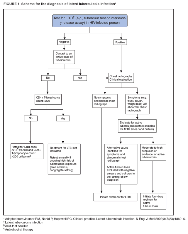
![TABLE 2. Drug therapy for treatment and chronic maintenance therapy of AIDS-associated opportunistic infections in adults and adolescents
Opportunistic infection
Preferred therapy, duration of therapy, chronic maintenance
Alternative therapy
Other options/issues
Pneumocystis pneumonia (PCP)
Preferred treatment for moderate to severe PCP
Trimethoprim-sulfamethoxazole (TMP-SMX): § [15–20 mg TMP and 75–100 mg SMX]/kg/day IV administered q6h or q8h (AI), may switch to PO after clinical improvement (AI)
Duration of therapy: 21 days§ (AII)
Preferred treatment for mild to moderate PCP
Same daily dose of TMP-SMX as above, § administered PO in 3 divided doses (AI); or
TMP-SMX (160 mg/800 mg or DS) 2 tablets § tid (AI)
Duration of therapy: 21 days § (AII)
Preferred secondary prophylaxis
TMP-SMX (160 mg/800 mg or DS) tablet PO § daily (AI); or
TMP-SMX (80 mg/400 mg or SS) tablet PO § daily (AI)
Alternative therapy for moderate to severe PCP
Pentamidine 4 mg/kg IV daily infused § over ≥60 minutes (AI), certain specialists reduce dose to 3 mg/kg IV daily because of toxicities (BI); or
Primaquine 15–30 mg (base) PO daily § plus clindamycin 600–900 mg IV q6h to q8h or clindamycin 300–450 mg PO q6h to q8h (AI)
Alternative therapy for mild-to-moderate PCP
Dapsone 100 mg PO daily and TMP 15 § mg/kg/day PO (3 divided dose) (BI); or
Primaquine 15–30 mg (base) PO daily § plus clindamycin 300–450 mg PO q6h to q8h (BI); or
Atovaquone 750 mg PO bid with food§ (BI)
Alternative secondary prophylaxis
TMP-SMX (160 mg/800 mg or DS) PO tiw § (BI)
Dapsone 50 mg PO bid or 100 mg PO § daily (BI); or
Dapsone 50 mg PO daily plus § pyrimethamine 50 mg PO weekly plus leucovorin 25 mg PO weekly (BI); or
Dapsone 200 mg PO plus pyrimethamine § 75 mg PO plus leucovorin 25 mg PO weekly (BI); or
Aerosolized pentamidine 300 mg every § month via Respirgard II™ nebulizer (BI); or
Atovaquone 1,500 mg PO daily § (BI); or
Atovaquone 1,500 mg + pyrimethamine § 25 mg + leucovorin 10 mg PO daily (CIII)
Indications for corticosteroids (AI)
PaO2 <70 mmHg at room air or alveolar-arterial O2 gradient >35 mmHg
Prednisone doses (beginning as early as possible and within 72 hours of PCP therapy) (AI):
Days 1–5 40 mg PO bid
Days 6–10 40 mg PO daily
Days 11–21 20 mg PO daily
IV methylprednisolone can be administered as 75% of prednisone dose
Benefits of corticosteroid if started after 72 hours of treatment is unknown, but a majority
of clinicians will use it in patients with moderate to severe PCP (BIII)
Whenever possible, patients should be tested for G6PD deficiency before use of primaquine](figures/r804a1t2a.gif)
![TABLE 2. (Continued) Drug therapy for treatment and chronic maintenance therapy of AIDS-associated opportunistic infections in adults and adolescents
Opportunistic infection
Preferred therapy, duration of therapy, chronic maintenance
Alternative therapy
Other options/issues
Toxoplasma gondii encephalitis (TE)
Preferred therapy
Pyrimethamine 200 mg PO x 1, then 50 mg § (<60 kg) to 75 mg (≥60 kg) PO daily plus sulfadiazine 1,000 mg (<60 kg) to 1,500 mg (≥60 kg) PO q6h plus leucovorin 10–25 mg PO daily (can increase 50 mg) (AI)
Duration for acute therapy
At least 6 weeks § (BII); longer duration if clinical or radiologic disease is extensive or response is incomplete at 6 weeks
Preferred chronic maintenance therapy
Pyrimethamine 25–50 mg PO daily plus sul
§ fadiazine 2,000–4,000 mg PO daily (in two to four divided doses) plus leucovorin 10–25 mg PO daily (AI)
Alternative therapy regimens
Pyrimethamine (leucovorin)* plus clin•
damycin 600 mg IV or PO q6h (AI); or
TMP-SMX (5 mg/kg TMP and 25 mg/kg • SMX) IV or PO bid (BI); or
Atovaquone 1,500 mg PO bid with • food (or nutritional supplement) plus pyrimethamine (leucovorin)* (BII); or
Atovaquone 1,500 mg PO bid with food • (or nutritional supplement) plus sulfadiazine
1,000–1,500 mg PO q6h (BII); or
Atovaquone 1,500 mg PO bid with food • (BII); or
Pyrimethamine (leucovorin)* plus • Azithromycin 900–1200 mg PO daily (BII)
Alternative chronic maintenance therapy/secondary prophylaxis
Clindamycin 600 mg PO every 8 hours • plus pyrimethamine 25–50 mg PO daily plus leucovorin 10–25 PO daily (BI) [should add additional agent to prevent PCP (AII)]; or
Atovaquone 750 mg PO every 6–12 hours • +/- [(pyrimethamine 25 mg PO daily plus leucovorin 10 mg PO daily) or sulfadiazine
2,000–4,000 mg PO] daily (BII)
Adjunctive corticosteroids (e.g., dexamethasone)
should be administered when clinically indicated only for treatment of mass effect attributed to focal lesions or associated edema (BIII); discontinue as soon as clinically feasible
Anticonvulsants should be administered to patients with a history of seizures (AIII) and continued through the acute treatment; but should not be used prophylactically (DIII)
Cryptosporidiosis
Preferred therapy
Initiate or optimize ART for immune § restoration (AII)
Symptomatic treatment of diarrhea § (AIII)
Aggressive oral or IV rehydration & § replacement of electrolyte loss (AIII)
Alternative therapy for cryptosporidiosis
A trial of nitazoxanide 500–1,000 mg PO § bid with food for 14 days (CIII) + optimized
ART, symptomatic treatment and rehydration & electrolyte replacement
Use of antimotility agents such as loperamide
or tincture of opium might palliate symptoms (BIII)](figures/r804a1t2b.gif)
![TABLE 2. (Continued) Drug therapy for treatment and chronic maintenance therapy of AIDS-associated opportunistic infections in adults and adolescents
Opportunistic infection
Preferred therapy, duration of therapy, chronic maintenance
Alternative therapy
Other options/issues
Mycobacterium tuberculosis (TB)
Empiric treatment should be initiated and continued in HIV-infected persons in whom TB is suspected until all diagnostic work-up is complete (AII)
Treatment of drug-susceptible active TB disease
(refer to Table 3 for dosing recommendations)
Initial phase (2 months) (AI)
Isoniazid (INH)† + [rifampin (RIF) or rifabutin (RFB)] + pyrazinamide (PZA) + ethambutol (EMB); if drug susceptibility report shows sensitivity to INH & RIF and PZA, then EMB may be discontinued before 2 months of treatment is completed (AI)
Continuation phase
INH + (RIF or RFB) daily or tiw § (AIII) or biw (if CD4+ count >100/μL) (CIII)
Duration of therapy:
Pulmonary TB – 6 months (AI)
Pulmonary TB w/ cavitary lung lesions & (+) culture after 2 months of TB treatment (AII) – 9 months
Extrapulmonary TB w/ CNS, bone, or joint infections – 9 to 12 months (AII);
Extrapulmonary TB in other sites – 6 to 9 months (AII)
Treatment for drug-resistant active TB
Resistant to INH
Discontinue INH (and streptomycin, if § used)
(RIF or RFB) + EMB + PZA for 6 months § (BII); or
(RIF or RFB) + EMB for 12 months (pref
§ erably with PZA during at least the first 2 months) (BII)
A fluoroquinolone may strengthen the § regimen for patients with extensive disease (CIII)
Resistant to rifamycins
INH + PZA + EMB + a fluoroquinolone for § 2 months, followed by 10–16 additional months with INH + EMB + fluoroquinolone
(BIII)
Amikacin or capreomycin may be § included in the first 2–3 months for patients with rifamycin resistance & severe disease (CIII)
Multidrug resistant (MDR, i.e., INH & RIF resistant) or extensively drug resistant (XDR, i.e., resistance to INH & RIF, fluoroquinolone
& at least 1 injectable agent) TB
Therapy should be individualized based § on resistance pattern and with close consultation
with experienced specialist (AIII)
Directly observed therapy (DOT) is recommended
for all HIV patients undergoing treatment for active TB (AII)
Initial phase of TB treatment may also be administered 5 days weekly (40 doses) (AII), or tiw (24 doses) (BII) by DOT
For CNS disease, corticosteroid should be initiated as early as possible and continued for 6–8 weeks (AII)
RIF is not recommended for patients receiving
HIV protease inhibitors (PI) because of its induction of PI metabolism (EII)
RFB is a less potent CYP 3A4 inducer than RIF and is preferred in patients receiving PIs
Rifapentine administered once weekly can result in development of resistance in HIV-infected patients and is not recommended (EI)
Therapeutic drug monitoring should be considered in patients receiving rifamycin and interacting ART
Paradoxical reaction that is not severe may be treated with nonsteroidal anti-inflammatory
drugs (NSAIDs) without a change in anti-TB or anti-HIV therapy (BIII)
For severe paradoxical reaction, may consider prednisone or methylprednisolone 1 mg/kg of body weight, gradually reduced after 1–2 weeks (BIII)](figures/r804a1t2d.gif)
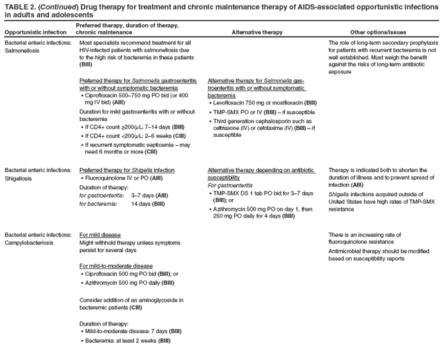
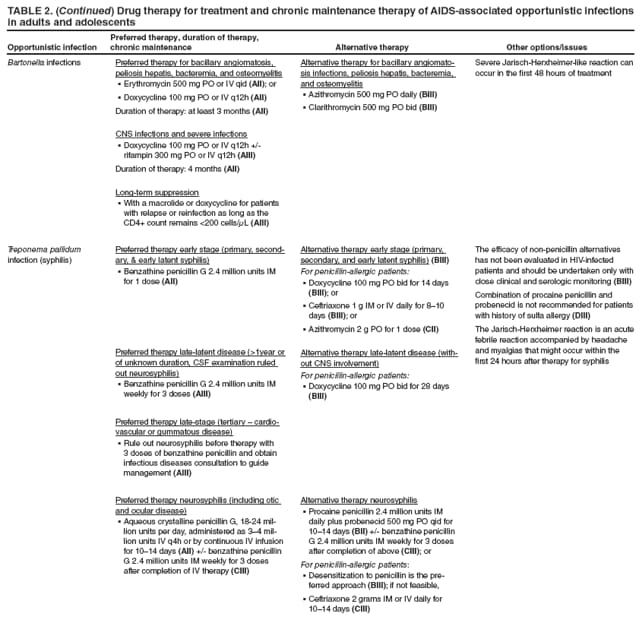
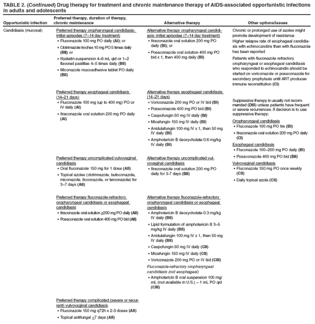
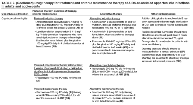
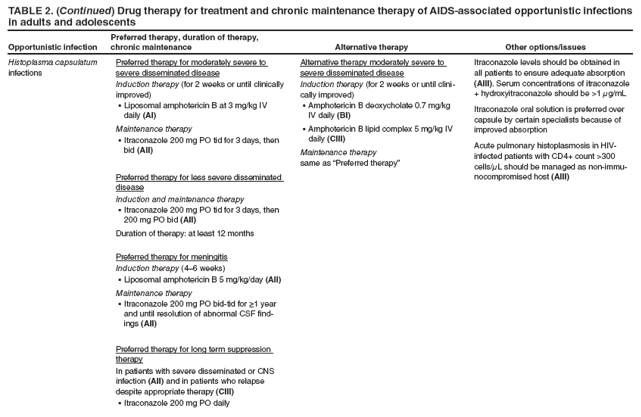
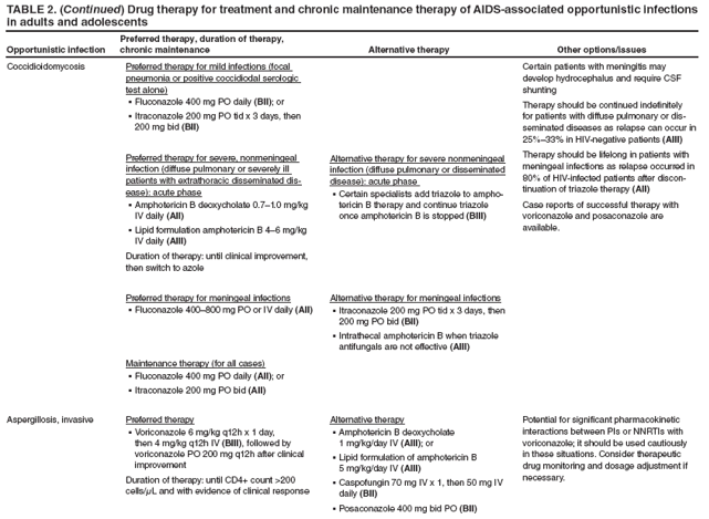
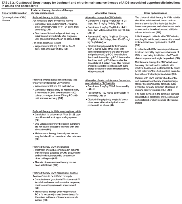
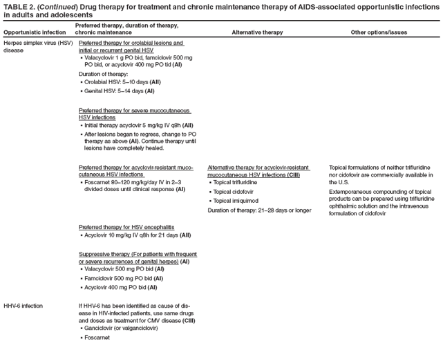
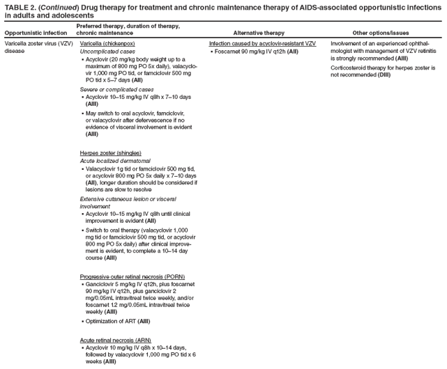
![TABLE 2. (Continued) Drug therapy for treatment and chronic maintenance therapy of AIDS-associated opportunistic infections in adults and adolescents
Opportunistic infection
Preferred therapy, duration of therapy, chronic maintenance
Alternative therapy
Other options/issues
HHV-8 diseases
(Kaposi’s Sarcoma [KS], primary effusion lymphoma [PEL], multicentric Castleman’s disease [MCD],)
Initiation or optimization of ART should be done for all patients with KS, PEL, or MCD (BII)
Preferred therapy for visceral KS (BII), disseminated
cutaneous KS (CIII), and PEL (BIII)
Chemotherapy + ART § (BII)
Oral valganciclovir or IV ganciclovir might be
§ useful as adjunctive therapy in PEL (BII)
Preferred therapy for MCD
Valganciclovir 900 mg PO bid § (BII); or
Ganciclovir 5 mg/kg IV q12h § (BII)
Alternative therapy for MCD
Rituximab, 375 mg/m2 given weekly x 4–8 weeks, may be an alternative to antiviral therapy (BII)
Human papillomavirus disease
Treatment of condyloma acuminata (genital warts)
Intralesional interferon-alpha is usually not recommended because of high cost, difficult administration, and potential for systemic side effects (DIII)
The rate of recurrence of genital warts is high, despite treatment
Patient-applied therapy
Podofilox 0.5% solution or 0.5% gel – apply § to all lesions bid x 3 consecutive days, followed
by 4 days of no therapy, repeat weekly for up to 4 cycles (BIII); or
Imiquimod 5% cream – apply to lesion at § bedtime and remove in the morning on 3 nonconsecutive nights weekly for up to 16 weeks. Each treatment should be washed with soap and water 6–10 hours after application
(BII)
Provider-applied therapy
Cryotherapy (liquid nitrogen or cryoprobe) § – apply until each lesion is thoroughly frozen; repeat every 1–2 weeks. Some providers allow the lesion to thaw, then freeze a 2nd time in each session (BIII).
Trichloroacetic acid or bicloroacetic acid § cauterization – 80%–90% aqueous solution,
apply to each lesion, repeat weekly for 3–6 weeks (BIII)
Surgical excision § (BIII) or laser surgery (CIII)
Podophyllin resin 10%–25% suspen
§ sion in tincture of benzoin – apply to all lesions, then wash off a few hours later, repeat weekly for 3–6 weeks (CIII)](figures/r804a1t2p.gif)
![TABLE 2. (Continued) Drug therapy for treatment and chronic maintenance therapy of AIDS-associated opportunistic infections in adults and adolescents
Opportunistic infection
Preferred therapy, duration of therapy, chronic maintenance
Alternative therapy
Other options/issues
Hepatitis B virus (HBV) disease
Therapy for patients who require ART
Patients should be treated with agents active § against both HIV and HBV or with agents with independent activity against each virus (CIII)
Consider tenofovir + emtricitabine as part of § HIV and HBV regimen (CIII)
Lamivudine or emtricitabine-naïve patients
[Lamivudine 150 mg PO bid (or 300 mg PO § daily) or emtricitabine 200 mg PO daily] + tenofovir (TDF) 300 mg PO daily (CIII) (+ additional agent[s] active against HIV)
Lamivudine or emtricitabine-experienced patients with detectable HBV DNA (assume lamivudine-resistance)
If not on TDF§ : Add TDF 300 mg PO daily as part of an ART regimen + lamivudine or emtricitabine (CIII); or
Adefovir 10 mg PO daily + lamivudine or § emtricitabine + other combination ART (BII); or
Entecavir 1 mg PO daily can be considered § in patients with complete HIV suppression (while on ART) who do not demonstrate YMDD (M204V/I) motif mutations in HBV DNA (CIII)
Duration of therapy: Because of the high rates of relapse, certain specialists recommend continuing therapy indefinitely (CIII)
Treatment for patients who do not require ART
Use agents with sole activity against HBV § and with the least potential of selecting HIV resistance mutations (BIII)
Consider early initiation of ART, especially § for patients with high HBV DNA
For patients with CD4+ count >350 cells/μL, HBeAg (-), HBV DNA >2,000 IU/mL (>20,000 copies/mL)
Adefovir 10 mg PO daily § (CIII)
For patients with CD4+ count >350 cells/μL, HBeAg (+), HBV DNA >20,000 IU/mL (>200,000 copies/mL), and elevated ALT
Peginterferon alfa-2a 180 § μg SQ weekly (CIII) x 48 weeks – with careful follow-up of HBeAg conversion
Emtricitabine, entecavir, lamivudine, or tenofovir should not be used for the treatment
of HBV infection in patients who are not receiving combination ART (EII)
Among patients coinfected with HIV, HBV, and HCV, consideration of starting ART should be the first priority. If ART is not required, an interferon-based regimen, which suppresses both HCV & HBV, should be considered (CIII)
If IFN-based treatment for HCV has failed, treatment of chronic HBV with nucleoside or nucleotide analogs is recommended (CIII)
Cross-resistance to emtricitabine or telbivudine
should be assumed in patients with suspected or proven 3TC resistance
When changing ART regimens, continue agents with anti-HBV activity because of the risk of IRIS
If anti-HBV therapy is discontinued and a flare occurs, therapy should be reinstituted, as it can be potentially life saving (BIII)
Hepatitis C virus (HCV) disease
Genotype 1, 4, 5, or 6 (AI)
Peginterferon alfa-2a 180 § μg SQ weekly, or
Peginterferon alfa-2b 1.5 mg/kg SQ weekly§
+
Ribavirin PO (wt-based dosing) § (AII)
<75 kg: 600 mg qAM and 400 mg qPM;
≥75 kg: 600 mg qAM and 600 mg qPM
Genotype 2 or 3 (AI)
Peginterferon alfa-2a 180§ μg SQ weekly, or
Peginterferon alfa-2b 1.5 mg/kg SQ weekly§
+
Ribavirin (fixed dose) PO 400 mg qAM and § 400 mg qPM
Duration of therapy:
48 weeks – genotypes 1 or 4, 5 or 6 § (AI) and genotypes 2 and 3 (BII)
At least 24 weeks – treatment of acute HCV § infection (<6 months from HCV exposure) (BIII)
In patients for whom ribavirin is contraindicated
(e.g. unstable cardiopulmonary disease, pre-existing anemia unresponsive
to erythropoietin, renal failure, or hemoglobinopathy)
Peginterferon alfa-2a 180 § μg SQ weekly (AII), or
Peginterferon alfa-2b 1.5 § μg/kg SQ weekly (AII)
In patients with decompensated liver disease
Liver transplantation if feasible § (CIII)
For patients with CD4+ count <200 cells/μL, initiation of ART may be considered before HCV treatment (CIII)
Didanosine + ribavirin may lead to increased mitochondrial toxicities; concomitant use is contraindicated (EI).
HCV therapy is not recommended in patients with hepatic decompensation. Liver transplantation, if feasible, should be the primary treatment option (CIII).
Interferon is abortifacient in high doses and ribavirin is teratogenic. HCV treatment is not recommended in pregnant women or women who are not willing to use birth control (EIII).](figures/r804a1t2q.gif)
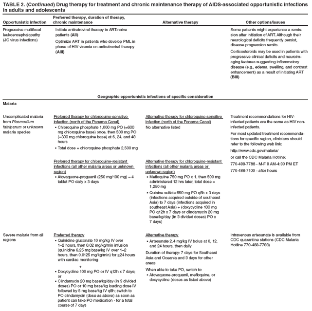
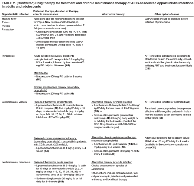
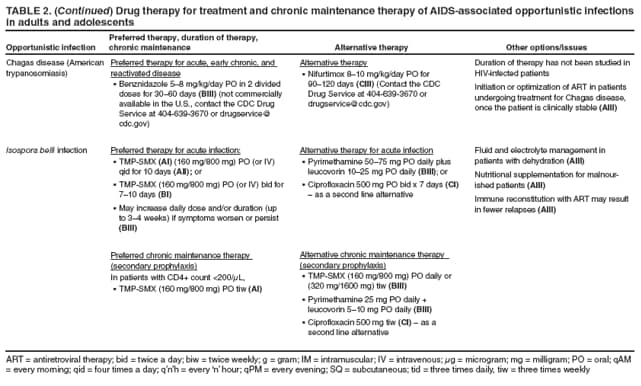
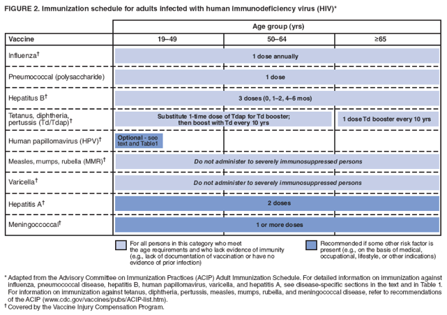
![TABLE 3. Recommended doses of first-line antituberculosis drugs for adults*
Drug
Daily
2x/wk
(not recommended for patients with CD4+ count <100 cells/μL)
3x/wk
Isoniazid
5 mg/kg (usual dose 300 mg)
15 mg/kg (usual dose 900 mg)
15 mg/kg (usual dose 900 mg)
Rifampin [NOTE: rifampin is not
recommended in patients receiving
HIV protease inhibitor(s) (PI) or the
CCR5 antagonist maraviroc]
10 mg/kg (usual dose 600 mg)
10 mg/kg (usual dose 600 mg)
10 mg/kg (usual dose 600 mg)
Rifabutin (w/o HIV PIs or NNRTI§)
5 mg/kg (usual dose 300 mg)
5 mg/kg (usual dose 300 mg)
5 mg/kg (usual dose 300 mg)
w/ HIV protease inhibitors
150 mg†
Not recommended†
150 mg†
w/ efavirenz
450–600 mg
450–600 mg
450–600 mg
Streptomycin/amikacin/ kanamycin
15 mg/kg
25 mg/kg
25 mg/kg
Pyrazinamide
40–55 kg body weight
1,000 mg (18.2–25.0 mg/kg)
2,000 mg (36.4–50.0 mg/kg)
1,500 mg (27.3–37.5 mg/kg)
56–75 kg body weight
1,500 mg (20.0–26.8 mg/kg)
3,000 mg (40.0–53.6 mg/kg)
2,500 mg (33.3–44.6 mg/kg)
>75 kg body weight
2,000 mg (22.2–26.3 mg/kg)
4,000 mg (44.4–52.6 mg/kg)
3,000 mg (33.3–44.6 mg/kg)
Ethambutol
40-55 kg body weight
800 mg (14.5–20.0 mg/kg)
2,000 mg (36.4–50.0 mg/kg)
1,200 mg/kg (21.8–30.0 mg/kg)
56-75 kg body weight
1,200 mg (16.0–21.0 mg/kg)
2,800 mg (37.3–50.0 mg/kg)
2,000 mg/kg (26.7–35.7 mg/kg)
>75 kg body weight
1,600 mg (17.8–21.0 mg/kg)
4,000 mg (44.4–52.6 mg/kg)
2,400 mg/kg (26.7–31.6 mg/kg)
* Some experts recommend that for severe tuberculosis infection (e.g., CNS, pericardial disease) considerations should be given to the use of adjuvant corticosteroids.
The dosing and taper schedule should be individualized according to severity of disease and adjusted, as needed, in response to treatment.
† Acquired rifamycin resistance has been reported in patients with inadequate rifabutin levels while on 150 mg twice weekly dosing together with ritonavir-boosted PIs. May consider therapeutic drug monitoring when rifabutin is used with a ritonavir-boosted PI and adjust dose accordingly.
§ Non-nucleoside reverse transcriptase inhibitor.](figures/r804a1t3.gif)