Key points
- Respirable crystalline silica (RCS) is a hazard for millions of U.S. workers in different industries.
- Dust containing RCS can enter in the air when working.
- Workers exposed to RCS can develop serious lung disease.
Overview
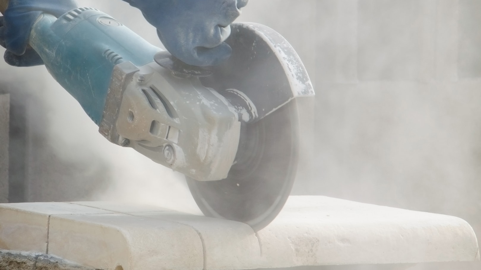
Silica is the compound silicon dioxide (SiO2). Crystalline silica forms when molecules of SiO2 arrange into crystalline form. Respirable crystalline silica (RCS) refers to particles containing crystalline silica and are small enough to inhale deep into the lungs (respirable). Work activities such as cutting, grinding, polishing, and drilling can generate dust from materials with RCS and then can become airborne.
Silicosis is a lung disease caused by inhaling excessive amounts of respirable crystalline silica. It is not reversible, but it is preventable. Excessive exposure to RCS can also cause other serious diseases, including lung cancer, chronic obstructive pulmonary disease (COPD), certain autoimmune diseases, and chronic renal failure.
Where it's found
Crystalline silica is typically found in:
- Soil
- Sand
- Concrete, brick, and cement
- Mortar and plaster
- Granite and other types of rock and stone
- Engineered (artificial) stone
The most common form of crystalline silica found in nature is quartz. Less commonly, it can occur in nature in the form of cristobalite and tridymite, typically in volcanic rock formations. Exposure to cristobalite also occurs in foundries where intense heat of molten metal converts other forms of silica in clay molds to cristobalite. Heating silica-containing materials during kiln and ceramic manufacturing can also form cristobalite and tridymite.
Risks
Silica exposure in countertop work
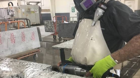
Countertop work is a primary concern for exposure risks to workers. Stone products used for countertops (like granite, quartz, or engineered stone) can contain have varying amounts of crystalline silica. Some engineered stone products are made of up to 95% crystalline silica, making dust control critically important.
The 2026 NIOSH and OSHA Hazard Alert compiles research on the health risks of RCS to workers exposed during countertop manufacturing, finishing, and installation. The document recommends a layered approach to protection, starting with the most effective options:
- Elimination or Substitution (best approach): Choose stone materials that contain little or no crystalline silica. This eliminates or lessens the hazard at its origin, provided the substitute material is not itself dangerous.
- Engineering Controls: Seven specific strategies are outlined (such as ventilation or wet-cutting methods) to reduce dust in the work environment.
- Administrative Controls: Nine strategies are described to limit workers' exposure through training and changes in procedures or scheduling.
- Personal Protective Equipment (PPE): Guidance on proper respirator and PPE use is included as an additional layer of protection.
The alert also covers requirements for medical screening and health monitoring of workers who are regularly exposed to RCS.
Other exposures
Exposures can also occur during activities like:
- Glass, pottery, ceramics, bricks, concrete, natural and engineered (artificial) stone manufacturing
- Abrasive blasting
- Foundry work
- Hydraulic fracturing
- Stonecutting
- Rock drilling
- Quarry work
- Tunneling
Workers in various industries can have jobs at risk for RCS exposure. Examples include:
- Construction
- Mining
- Oil and gas extraction
- Engineered (artificial) and natural stone countertop fabrication
- Foundries and other manufacturing settings
- Dentistry
Keep reading: Safe work practices
Resources
Key publications
More information
- Engineered Stone and Silicosis (NIOSH blog)
- Mining and Silicosis (webpage)
- Inhaling Silica Dust Can Cause Deadly Lung Disease - SPANISH, CHINESE (infographic)
Searchable database
NIOSHTIC-2 is a searchable bibliographic database of occupational safety and health publications, documents, grant reports, and other communication products supported in whole or in part by NIOSH. Search "silica" for more publications.