
Taeniasis
[Taenia asiatica] [Taenia saginata] [Taenia solium]
Causal Agents
The cestodes Taenia saginata (beef tapeworm), T. solium (pork tapeworm) and T. asiatica (Asian tapeworm). Taenia solium can also cause cysticercosis.
Life Cycle

Taeniasis is the infection of humans with the adult tapeworm of Taenia saginata, T. solium or T. asiatica. Humans are the only definitive hosts for these three species. Eggs or gravid proglottids are passed with feces  ; the eggs can survive for days to months in the environment. Cattle (T. saginata) and pigs (T. solium and T. asiatica) become infected by ingesting vegetation contaminated with eggs or gravid proglottids
  . In the animal’s intestine, the oncospheres hatch
  , invade the intestinal wall, and migrate to the striated muscles, where they develop into cysticerci. A cysticercus can survive for several years in the animal. Humans become infected by ingesting raw or undercooked infected meat
  . In the human intestine, the cysticercus develops over 2 months into an adult tapeworm, which can survive for years. The adult tapeworms attach to the small intestine by their scolex
  and reside in the small intestine
  . Length of adult worms is usually 5 m or less for T. saginata (however it may reach up to 25 m) and 2 to 7 m for T. solium. The adults produce proglottids which mature, become gravid, detach from the tapeworm, and migrate to the anus or are passed in the stool (approximately 6 per day). T. saginata adults usually have 1,000 to 2,000 proglottids, while T. solium adults have an average of 1,000 proglottids. The eggs contained in the gravid proglottids are released after the proglottids are passed with the feces. T. saginata may produce up to 100,000 and T. solium may produce 50,000 eggs per proglottid respectively.
Geographic Distribution
Taenia saginata and T. solium are worldwide in distribution. Taenia solium is more prevalent in poorer communities where humans live in close contact with pigs and eat undercooked pork. Taenia asiatica is limited to Asia and is seen mostly in the Republic of Korea, China, Taiwan, Indonesia, and Thailand.
Clinical Presentation
Taenia saginata taeniasis produces only mild abdominal symptoms. The most striking feature consists of the passage (active and passive) of proglottids. Occasionally, appendicitis or cholangitis can result from migrating proglottids. Taenia solium taeniasis is less frequently symptomatic than Taenia saginata taeniasis. The main symptom is often the passage (passive) of proglottids. The most important feature of Taenia solium taeniasis is the risk of development of cysticercosis.
Taenia spp. eggs.


Taenia spp. scoleces.


Taenia spp. proglottids.


Taenia spp. adults.


Laboratory Diagnosis
Microscopy
Microscopic identification of eggs and proglottids in feces is diagnostic for taeniasis, but is not possible during the first 3 months following infection, prior to development of adult tapeworms. Repeated examination and concentration techniques will increase the likelihood of detecting light infections. Nevertheless, identification of Taenia is not possible if solely based on microscopic examination of eggs, because all Taenia species produce eggs that are morphologically identical. Eggs of Taenia spp. are also indistinguishable from those produced by cestodes of the genus Echinococcus (tapeworms of dogs and other canid hosts). Microscopic identification of gravid proglottids (or, more rarely, examination of the scolex) allows species determination.
TAKE EXTREME CARE IN PROCESSING THE SPECIMENS! INGESTION OF EGGS CAN RESULT IN CYSTICERCOSIS!
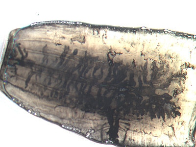
Figure B: Mature proglottid of T. saginata, stained with India ink. Note the number of primary uterine branches (>12). Image courtesy of the Orange County Public Health Laboratory, Santa Ana, CA.
Separation of T. saginata and T. solium is best accomplished by examination of mature proglottids. Taenia saginata has 12-30 primary lateral uterine branches, while T. solium has 7-13 primary lateral uterine branches. Visualization of the branches can be improved by clearing the specimen in lactophenol followed by India ink injection into the lateral genital pore. The procedure is as follows:
- Clear the formalin-fixed proglottids in lactophenol (50/50 liquefied phenol crystals in lactic acid) for at least 30 minutes (thicker specimen may take a few hours to overnight).
- Gently sandwich the proglottids between two glass microscope slides, with the genital pore exposed along the edge of the two slides.
- Using a small gauge (25 or 27 g) tuberculin syringe, slowly inject India ink into the genital pore.
- Allow the ink to flow down the uterine stem and into the primary uterine branches.
- Count the number of primary uterine branches to determine the species (7-13 for T. solium and 12-30 for T. saginata).
Antibody detection
May prove useful especially in the early invasive stages, when the eggs and proglottids are not yet apparent in the stools.
More on: Morphologic comparison with other intestinal parasites
Treatment Information
Treatment information for taeniasis can be found at: Clinical Treatment of Taeniasis
DPDx is an educational resource designed for health professionals and laboratory scientists. For an overview including prevention, control, and treatment visit www.cdc.gov/parasites/.