Metagonimiasis
[Metagonimus yokogawai]
Causal Agents
Metagonimus yokogawai, a minute intestinal fluke (and the smallest human fluke).
Life Cycle

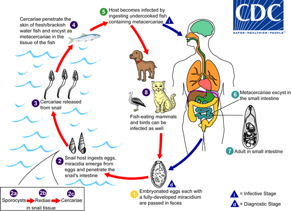
Adults release fully embryonated eggs each with a fully-developed miracidium, and eggs are passed in the host’s feces  . After ingestion by a suitable snail (first intermediate host), the eggs hatch and release miracidia which penetrate the snail’s intestine
  . Snails of the genus Semisulcospira are the most frequent intermediate host for Metagonimus yokogawai. The miracidia undergo several developmental stages in the snail, i.e. sporocysts
  , rediae
  , and cercariae
  . Many cercariae are produced from each redia. The cercariae are released from the snail
  and encyst as metacercariae in the tissues of a suitable fresh/brackish water fish (second intermediate host)
  . The definitive host becomes infected by ingesting undercooked or salted fish containing metacercariae
  . After ingestion, the metacercariae excyst, attach to the mucosa of the small intestine
  and mature into adults (measuring 1.0 mm to 2.5 mm by 0.4 mm to 0.75 mm)
  . In addition to humans, fish-eating mammals (e.g., cats and dogs) and birds can also be infected by M. yokogawai
  .
.
Geographic Distribution
Mostly the Far East, as well as Siberia, Manchuria, the Balkan states, Israel, and Spain.
Clinical Presentation
The main symptoms are diarrhea and colicky abdominal pain. Migration of the eggs to extraintestinal sites (heart, brain) can occur, with resulting symptoms.
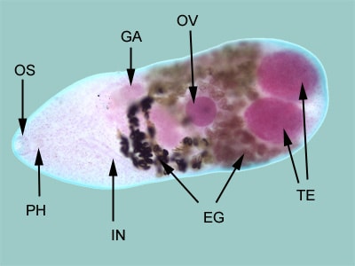
Metagonimus yokogawai, adult fluke.

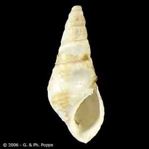
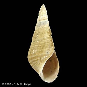
Snail intermediate hosts of M. yokogawai.


Laboratory Diagnosis
The diagnosis is based on the microscopic identification of eggs in the stool. However, the eggs are indistinguishable from those of Heterophyes heterophyes and resemble those of Clonorchis and Opisthorchis. Specific diagnosis is based on identification of the adult fluke evacuated after antihelminthic therapy, or found at autopsy.
Treatment Information
For information about treatment please contact CDC-INFO.
DPDx is an educational resource designed for health professionals and laboratory scientists. For an overview including prevention, control, and treatment visit www.cdc.gov/parasites/.