bannermenu

Babesiosis

[Babesia divergens] [Babesia duncani] [Babesia microti] [Babesia MO-1]

Causal Agents

Babesiosis is caused by apicomplexan parasites of the genus, Babesia. While more than 100 species have been reported, only a few have been identified as causing human infections, including B. microti, B. divergens, B. duncani, and a currently un-named strain designated MO-1.

Life Cycle

lifecycle image

The Babesia microti life cycle involves two hosts, which includes a rodent, primarily the white-footed mouse, Peromyscus leucopus, and a tick in the genus, Ixodes. During a blood meal, a Babesia-infected tick introduces sporozoites into the mouse host The Number 1. Sporozoites enter erythrocytes and undergo asexual reproduction (budding) The Number 2. In the blood, some parasites differentiate into male and female gametes although these cannot be distinguished at the light microscope level The Number 3. The definitive host is the tick. Once ingested by an appropriate tick The Number 4, gametes unite and undergo a sporogonic cycle resulting in sporozoites The Number 5. Transovarial transmission (also known as vertical, or hereditary, transmission) has been documented for “large” Babesia spp. but not for the “small” babesiae, such as B. microti The Letter A.

Humans enter the cycle when bitten by infected ticks. During a blood meal, a Babesia-infected tick introduces sporozoites into the human host The Number 6. Sporozoites enter erythrocytes The Letter B and undergo asexual replication (budding) The Number 7. Multiplication of the blood stage parasites is responsible for the clinical manifestations of the disease. Humans are, for all practical purposes, dead-end hosts and there is probably little, if any, subsequent transmission that occurs from ticks feeding on infected persons. However, human to human transmission is well recognized to occur through blood transfusions The Number 8.

Geographic Distribution

Worldwide, but little is known about the prevalence of Babesia in malaria-endemic countries, where misidentification as Plasmodium probably occurs. In Europe, most reported cases are due to B. divergens and occur in splenectomized patients. In the United States, B. microti is the agent most frequently identified (Northeast and Midwest), and can occur in nonsplenectomized individuals. Babesia duncani has been isolated in patients in Washington and California. MO-1 has been isolated from patients in Missouri.

Clinical Presentation

Most infections are probably asymptomatic, as indicated by serologic surveys. Manifestations of disease include fever, chills, sweating, myalgias, fatigue, hepatosplenomegaly, and hemolytic anemia. Symptoms typically occur after an incubation period of 1 to 4 weeks, and can last several weeks. The disease is more severe in patients who are immunosuppressed, splenectomized, and/or elderly. Infections caused by B. divergens tend to be more severe (frequently fatal if not appropriately treated) than those due to B. microti, where clinical recovery usually occurs.

Babesia sp. in thick blood smears stained with Giemsa.
Babesia parasites resemble Plasmodium falciparum, however Babesia has several distinguishing features: the parasites are pleomorphic (vary in shape and size), can be vacuolated, and do not produce pigment.
Figure A: <em>Babesia</em> sp. in thick blood smears stained with Giemsa.
Figure A: Babesia sp. in thick blood smears stained with Giemsa.
Figure B: <em>Babesia</em> sp. in thick blood smears stained with Giemsa.
Figure B: Babesia sp. in thick blood smears stained with Giemsa.
Babesia sp. in thin blood smears stained with Giemsa.
Figure A: <em>Babesia</em> sp. in a thin blood smear stained with Giemsa.
Figure A: Babesia sp. in a thin blood smear stained with Giemsa.
Figure E: <em>Babesia</em> sp. in a thin blood smear stained with Giemsa. Image contributed by the Arizona State Health Laboratory.
Figure E: Babesia sp. in a thin blood smear stained with Giemsa. Image contributed by the Arizona State Health Laboratory.
Figure B: <em>Babesia</em> sp. in a thin blood smear stained with Giemsa.
Figure B: Babesia sp. in a thin blood smear stained with Giemsa.
Figure F: <em>Babesia</em> sp. in a thin blood smear stained with Giemsa. Image contributed by the Arizona State Health Laboratory.
Figure F: Babesia sp. in a thin blood smear stained with Giemsa. Image contributed by the Arizona State Health Laboratory.
Figure C: <em>Babesia</em> sp. in a thin blood smear stained with Giemsa.
Figure C: Babesia sp. in a thin blood smear stained with Giemsa.
Figure D: <em>Babesia</em> sp. in a thin blood smear stained with Giemsa.
Figure D: Babesia sp. in a thin blood smear stained with Giemsa.
Babesia sp. (tetrad forms) in thin blood smears stained with Giemsa.
Figure A: <em>Babesia</em> sp. in a thin blood smear stained with Giemsa. Note the tetrads, a dividing form characteristic for <em>Babesia</em>.
Figure A: Babesia sp. in a thin blood smear stained with Giemsa. Note the tetrads, a dividing form characteristic for Babesia.
Figure B: <em>Babesia</em> sp. in a thin blood smear stained with Giemsa. Note the tetrads, a dividing form characteristic for <em>Babesia</em>.
Figure B: Babesia sp. in a thin blood smear stained with Giemsa. Note the tetrads, a dividing form characteristic for Babesia.
Figure C: <em>Babesia</em> sp. in a thin blood smear; note the tetrad form and ameboid trophozoite.
Figure C: Babesia sp. in a thin blood smear; note the tetrad form and ameboid trophozoite.
Figure D: <em>Babesia</em> sp. in a thin blood smear; tetrad form, pairs aligned.
Figure D: Babesia sp. in a thin blood smear; tetrad form, pairs aligned.
Babesia sp. (extracellular forms) in a thin blood smear stained with Giemsa.
Figure A: <em>Babesia</em> sp. in a thin blood smear stained with Giemsa. Note the clumped extracellular forms indicative of <em>Babesia</em>.
Figure A: Babesia sp. in a thin blood smear stained with Giemsa. Note the clumped extracellular forms indicative of Babesia.
Figure B: <em>Babesia</em> sp. in a thin blood smear stained with Giemsa. Note the extracellular forms as well as intra-erythrocytic forms, one of which is vacuolated.
Figure B: Babesia sp. in a thin blood smear stained with Giemsa. Note the extracellular forms as well as intra-erythrocytic forms, one of which is vacuolated.
Figure C: <em>Babesia</em> sp. in a thin blood smear stained with Giemsa, showing extracellular forms. Image was courtesy of the Connecticut Department of Public Health Laboratory.
Figure C: Babesia sp. in a thin blood smear stained with Giemsa, showing extracellular forms. Image was courtesy of the Connecticut Department of Public Health Laboratory.
Babesia microti in thin blood smears stained with Giemsa.
Figure A: <em>Babesia microti</em> in a thin blood smear stained with Giemsa. Babesia sp. cannot be identified to the species level by morphology alone; additional testing, such as PCR, is always recommended.
Figure A: Babesia microti in a thin blood smear stained with Giemsa. Babesia sp. cannot be identified to the species level by morphology alone; additional testing, such as PCR, is always recommended.
Figure B: <em>Babesia</em> microti in a thin blood smear stained with Giemsa. <em>Babesia</em> sp. cannot be identified to the species level by morphology alone; additional testing, such as PCR, is always recommended. Note the tetrad form in this image.
Figure B: Babesia microti in a thin blood smear stained with Giemsa. Babesia sp. cannot be identified to the species level by morphology alone; additional testing, such as PCR, is always recommended. Note the tetrad form in this image.
Figure C: <em>Babesia microti</em> in a thin blood smear stained with Giemsa. Note the intra-erythrocytic vacuolated forms indicated by the black arrows.
Figure C: Babesia microti in a thin blood smear stained with Giemsa. Note the intra-erythrocytic vacuolated forms indicated by the black arrows.
Babesia MO-1 in thin blood smears stained with Giemsa.
Figure A: <em>Babesia</em> MO-1 in a thin blood smear stained with Giemsa. Babesia sp. cannot be identified to the species level by morphology alone; additional testing, such as PCR, is always recommended. Note the vacuolated parasites (black arrows) in the image.
Figure A: Babesia MO-1 in a thin blood smear stained with Giemsa. Babesia sp. cannot be identified to the species level by morphology alone; additional testing, such as PCR, is always recommended. Note the vacuolated parasites (black arrows) in the image.
Figure E: <em>Babesia</em> MO-1 in a thin blood smear stained with Giemsa.
Figure E: Babesia MO-1 in a thin blood smear stained with Giemsa.
Figure I: <em>Babesia</em> MO-1 in a thin blood smear stained with Giemsa.
Figure I: Babesia MO-1 in a thin blood smear stained with Giemsa.
Figure B: <em>Babesia</em> MO-1 in a thin blood smear stained with Giemsa. Babesia sp. cannot be identified to the species level by morphology alone; additional testing, such as PCR, is always recommended. Note the vacuolated parasites (black arrows) in the image.
Figure B: Babesia MO-1 in a thin blood smear stained with Giemsa. Babesia sp. cannot be identified to the species level by morphology alone; additional testing, such as PCR, is always recommended. Note the vacuolated parasites (black arrows) in the image.
Figure F: <em>Babesia</em> MO-1 in a thin blood smear stained with Giemsa.
Figure F: Babesia MO-1 in a thin blood smear stained with Giemsa.
Figure J: <em>Babesia</em> MO-1 in a thin blood smear stained with Giemsa. Note the tetrad (black arrow).
Figure J: Babesia MO-1 in a thin blood smear stained with Giemsa. Note the tetrad (black arrow).
Figure C: <em>Babesia</em> MO-1 in a thin blood smear stained with Giemsa.
Figure C: Babesia MO-1 in a thin blood smear stained with Giemsa.
Figure G: <em>Babesia</em> MO-1 in a thin blood smear stained with Giemsa. Babesia sp. cannot be identified to the species level by morphology alone; additional testing, such as PCR, is always recommended.
Figure G: Babesia MO-1 in a thin blood smear stained with Giemsa. Babesia sp. cannot be identified to the species level by morphology alone; additional testing, such as PCR, is always recommended.
Figure D: <em>Babesia</em> MO-1 in a thin blood smear stained with Giemsa.
Figure D: Babesia MO-1 in a thin blood smear stained with Giemsa.
 Figure H: <em>Babesia</em> MO-1 in a thin blood smear stained with Giemsa. Babesia sp. cannot be identified to the species level by morphology alone; additional testing, such as PCR, is always recommended.
Figure H: Babesia MO-1 in a thin blood smear stained with Giemsa. Babesia sp. cannot be identified to the species level by morphology alone; additional testing, such as PCR, is always recommended.
Babesia duncani in a thin blood smear stained with Giemsa.
Babesia parasites resemble Plasmodium falciparum, however Babesia has several distinguishing features: the parasites vary more in shape and in size (pleomorphic), and they do not produce pigment.
Figure A: <em>Babesia duncani</em> in a thin blood smear stained with Giemsa. Babesia sp. cannot be identified to the species level by morphology alone; additional testing, such as PCR, is always recommended.
Figure A: Babesia duncani in a thin blood smear stained with Giemsa. Babesia sp. cannot be identified to the species level by morphology alone; additional testing, such as PCR, is always recommended.
Ixodes spp., vectors of babesiosis.


Babesia spp. are transmitted by ticks, primarily of the genus Ixodes. In Europe, the primary vector for babesiosis is I. ricinus; in North America it is I. scapularis. Adults of Ixodes spp. are characterized by having mouthparts longer than the basis capituli, a lack of festoons, an inornate dorsal shield without eyes, and an inverted, U-shaped anal groove.
Figure A: Larva (A), nymph (B), adult male (C), adult female (D), and engorged female with eggs (E) of <em>Ixodes scapularis</em>. Image courtesy of James Occi.
Figure A: Larva (A), nymph (B), adult male (C), adult female (D), and engorged female with eggs (E) of Ixodes scapularis. Image courtesy of James Occi.
Figure B: Close-up of the head of a nymph of <em>Ixodes</em>. Notice the palps (PA) and hypostome (HY) are long, in comparison to the basis capituli (BC). Image courtesy of the Washington State Public Health Laboratories.
Figure B: Close-up of the head of a nymph of Ixodes. Notice the palps (PA) and hypostome (HY) are long, in comparison to the basis capituli (BC). Image courtesy of the Washington State Public Health Laboratories.
Figure C: Ventral view of the specimen in Figure B. Notice the inverted, U-shaped anal groove (AG). Also shown is one of the spiracular plates (SP).

Laboratory Diagnosis

Diagnosis can be made by microscopic examination of thick and thin blood smears stained with Giemsa. Repeated smears may be needed.
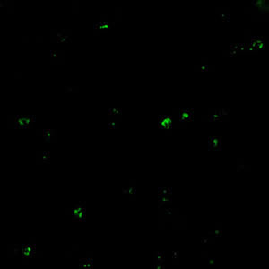
Antibody Detection

Diagnosis of Babesia infection should be made by detection of parasites in patients’ blood smears.  However, antibody detection tests are useful for detecting infected individuals with very low levels of parasitemia (such as asymptomatic blood donors in transfusion-associated cases), for diagnosis after infection is cleared by therapy, and for discrimination between Plasmodium falciparum and Babesia infection in patients whose blood smear examinations are inconclusive and whose travel histories cannot exclude either parasite.

The indirect fluorescent antibody test (IFA) using B. microti parasites as antigen detects antibodies in 88-96% of patients with B. microti infection. IFA antigen slides are prepared using washed, parasitized erythrocytes produced in hamsters. Patients’ titers generally rise to ≥1:1024 during the first weeks of illness and decline gradually over 6 months to titers of 1:16 to 1:256 but may remain detectable at low levels for a year or more. Specificity is 100% in patients with other tick-borne diseases or persons not exposed to the parasite.  Cross-reactions may occur in serum specimens from patients with malaria infections, but generally titers are highest with the homologous antigen.

The extent of cross-reactivity between Babesia species is variable. A negative result with B. microti antigen for a patient exposed on the West Coast may be a false-negative reaction for Babesia infection. Individuals whose exposure could have occurred on the West Coast should be tested also for antibodies to the Babesia duncani, because of the lack of cross-reactivity with B. microti.

Reference:

Krause PJ, Telford S RI, Ryan R, et al. Diagnosis of babesiosis: Evaluation of a serologic test for the detection of Babesia microti antibody. J Infect Dis 1994;169:923-926.

Positive IFA result with B. microti antigen.

Molecular diagnosis

In some infections with intraerythrocytic parasites, the morphologic characteristics observed on microscopic examination of blood smears do not allow an unambiguous differentiation between Babesia and Plasmodium. Moreover, potential blood donors may have subclinical symptoms and very low parasitemia, undetectable in blood smears. In such cases, the diagnosis can be derived from molecular techniques, such as PCR. In addition, molecular approaches are very valuable in investigations of new Babesia variants (or species) observed in recent human infections in the United States and in Europe.

References:

1. Hojgaard A, Lukacik G, Piesman J. Detection of Borrelia burgdorferi, Anaplasma phagocytophilum and Babesia microti, with two different multiplex PCR assays. Ticks and Tick-borne Diseases 2014 (5):349–351.

2. Bonnet S, Jouglin M, Malandrin L, Becker C, A. Agoulon A, L’Hostis M, Chauvin A. Transstadial and transovarial persistence of Babesia divergens DNA in Ixodes ricinus ticks fed on infected blood in a new skin-feeding technique. Parasitol 2007;134:197–207.

Treatment Information

Treatment information for babesiosis can be found at: https://www.cdc.gov/parasites/babesiosis/health_professionals/index.html#tx

DPDx is an educational resource designed for health professionals and laboratory scientists. For an overview including prevention, control, and treatment visit www.cdc.gov/parasites/.

Page last reviewed: October 30, 2017