 ShareCompartir

Monthy Case Studies - 2003
Case #116 - September, 2003
An HIV positive, middle-aged man with severe weight loss was being managed at a local hospital. The attending physician requested that the patient be tested for microsporidia and that a routine ova and parasites (O & P) examination be performed. The lab prepared a fecal smear from a specimen preserved with 10% formalin and stained the smear using the Chromotrope 2R staining method. Figure A shows what was observed at 1000× magnification. What is your diagnosis? Based on what criteria?
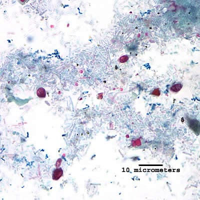
Figure A
Answer to Case #116
This was a case of microsporidiosis. The diagnostic morphologic features observed were:
- the presence of round to oval spores of microsporidia (black arrows, Figure A). They will stain pinkish red when using the Chromotrope 2R staining technique. Often, an equatorial belt-like stripe can be seen in the spores.
- spores within the size range of microsporidia. Spores of Encephalitozoon spp. measure 1.0 to 1.5 by 2.5 to 3.0 micrometers, consistently appearing larger than Enterocytozoon bieneusi spores (0.8 to 1.4 micrometers). E. bieneusi is the most commonly recognized species causing diarrhea in immunocompromised patients.
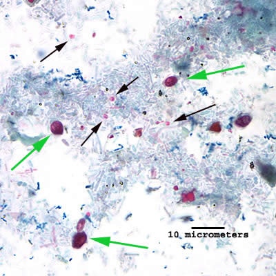
Figure A
The difference in size between the spores allows a tentative genus-level diagnosis, but definitive confirmation depends on laboratory testing using one or more of the techniques listed above. Green arrows in Figure A indicate yeast cells present in the specimen, and not a causal agent of disease.
More on: Microsporidiosis
Images presented in the monthly case studies are from specimens submitted for diagnosis or archiving. On rare occasions, clinical histories given may be partly fictitious.
