 ShareCompartir

Monthy Case Studies - 2003
Case #108 - May, 2003
A local laboratory received a fecal specimen for ova and parasites (O & P) as part of an investigation to identify clusters of cyclosporiasis cases. The specimen was concentrated using the formalin-ethyl acetate (FEA) method. The lab responsible for the diagnostic tests did not have all reagents to perform acid-fast staining. In addition, the light bulb of their microscope had recently burned out, so they could not perform any bright-field-based microscopic examination. Nevertheless, the lab did have a microscope equipped with UV light to perform fluorescence microscopy, which allowed them to examine the concentrate. Figure A shows an object observed in the specimen, which was present in very low numbers.
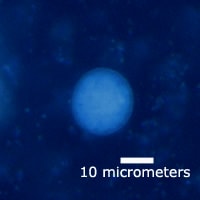
Figure A
Answer to Case #108
The correct response is that the object was not Cyclospora cayetanensis. The object was about 20 micrometers in diameter. This is too large to be a C. cayetanensis oocyst, which measure 8 to 10 micrometers in diameter. Also, the autofluorescence of a Cyclospora oocyst wall is much more intense than the autofluorescence of the object in Figure A due to the thickness of the Cyclospora oocyst wall.
When observed using conventional bright-field microscopy (Figure B), it can be determined that this is actually a cyst of Entamoeba coli based on the number of nuclei inside the cyst (greater than 4). The objective of this case was to demonstrate that other organisms autofluoresce when exposed to UV light. With experience, many of these can be readily recognized, but when a suspicious object is observed, follow-up examination using bright-field and/or stained smears should be performed.
Figure A
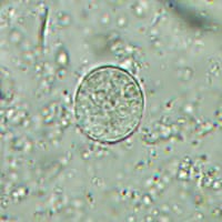
Figure B
More on: Cyclosporiasis
More on: Intestinal Amoebae
Images presented in the monthly case studies are from specimens submitted for diagnosis or archiving. On rare occasions, clinical histories given may be partly fictitious.
