 ShareCompartir

Monthy Case Studies - 2002
Case #86 - June, 2002
Revised 6/20/2002
A 45-year-old man, who recently returned from an extended trip to Ethiopia, sought treatment for a 3 to 5 week history of abdominal pain, diarrhea, rash, and a 20-pound weight loss. He was admitted to the hospital for dehydration. Several procedures were performed, including bronchoalveolar lavage (BAL). The image below is from a Giemsa stained BAL smear (Figure A). What is your diagnosis? Based on what criteria?
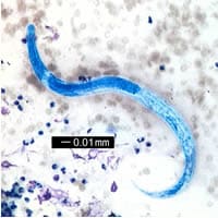
Figure A
Acknowledgement: This case was kindly provided by Drs. C. Touchie, A. McCarthy, and M. Desjardins of Ottawa Hospital, Ontario, Canada. Please contact Dr. Marc Desjardins with questions or comments.
Marc Desjardins Ph.D.
Clinical Microbiologist
Ottawa Hospital
501 Smyth Rd
Ottawa, Ontario,
Canada
K1H-8L6
Phone: 613-737-8899 ext 72242
Fax: 613-737-8324
e-mail: madesjardins@ottawahospital.on.ca
Answer to Case #86
The image shows an L3 larva (= third-stage or infective-stage larva), of Strongyloides stercoralis. L3 larvae are the stage responsible for internal autoinfection, and also the stage most commonly seen in sputum, BAL, or other tissues in cases of disseminated strongyloidiasis.
These larvae measure slightly less than 20 micrometers in diameter by 500 to 600 micrometers in length, and have the same typical features seen in third-stage larvae recovered from culture or the environment, including size, an esophagus that is approximately 1/2 the length of the body and not of the rhabditoid type, and a notched tail. Two of these features, overall size and length and shape of the esophagus, were visible in the image. The scale bar on the image should have been labeled 0.01 mm instead of 0.1 mm.
There are few other nematode larvae besides Strongyloides stercoralis that one would encounter in such a preparation. The morphologic features seen here would allow one to make a diagnosis of Strongyloides.
More on: Strongyloidiasis
Images presented in the monthly case studies are from specimens submitted for diagnosis or archiving. On rare occasions, clinical histories given may be partly fictitious.
