 ShareCompartir

Monthy Case Studies - 2001
Case #68 - September, 2001
An adult man passed a segmented worm and submitted it to his local state health department for identification. The worm was placed in 10% formalin and sent to CDC (Figure A). In addition to gross examination, a segment was removed from the distal end and compressed between two glass slides, producing the objects shown in Figure B. What is your diagnosis? Based on what criteria?
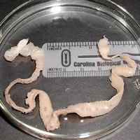
Figure A
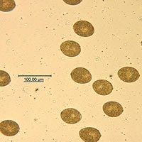
Figure B
Answer to Case #68
This was a case of diphyllobothriasis caused by Diphyllobothrium sp., most likely D. latum. The diagnostic features observed included:
- the presence of a stroblia made up of proglottids, indicative of a tapeworm (cestode). The size of this specimen excluded small tapeworms, such as Hymenolepis nana.
- proglottids that were wider than they were long. This feature should be carefully considered since immature proglottids of Taenia spp. can also have this appearance. The more distal proglottids are longer than they are wide in these species.
- eggs that were an average of 66 micrometers in length, with an abopercular knob.
Often, the scolex is not present on worms passed in the stool, so other methods of species determination must be used. In Diphyllobothrium spp., the uterus is in the form of a rosette, occupying the middle field of the proglottid and can sometimes be seen as a dark dot in more mature proglottids when examining the whole worm. These dark dots were seen in some proglottids in Figure A. In addition, a segment was removed to see if it had a gravid proglottid (containing eggs) by pressing it between two glass slides. The eggs were oval and operculated, consistent with those of Diphyllobothrium spp. Eggs from cestodes belonging to the families Taeniidae and Hymenolepididae are round and do not have an operculum.
More on: Diphyllobothriasis
Images presented in the monthly case studies are from specimens submitted for diagnosis or archiving. On rare occasions, clinical histories given may be partly fictitious.
