 ShareCompartir

Monthy Case Studies - 1999
Case #5 - February, 1999
A single proglottid was sent to the Oregon State Public Health Laboratory for identification. It came from a 9-month-old boy. Figure A is a picture of the proglottid. It was 3 mm by 15 mm and preserved in formalin. In the formalin, the proglottid became opaque and tough, making it difficult to observe the location of the pore or pores. It was longer than it was wide and both ends were slightly pointed. It was impossible to observe the number and location of pores; however, several small granules extruding from the proglottid were observed when pressure was applied to the organism. Some of the granules were placed on a microscope slide and examined at 100× and 400× (Figure B). The objects measured 230 micrometers by 170 micrometers. Individual granules measured approximately 40 micrometers in diameter. What is your diagnosis? Based on what criteria? After treating the child, what else should the family do? If mature eggs had not been available, how would you examine the proglottid structure for species identification?
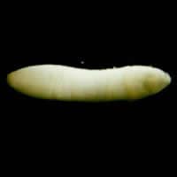
Figure A
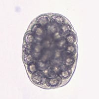
Figure B
Acknowledgement: This case was prepared by Dustin Brown of the Oregon State Public Health Laboratory.
Answer to Case #5
This was a tapeworm infection caused by Dipylidium caninum. Morphologic diagnostic features included:
- a proglottid in Figure A and an egg capsule in Figure B (case studies page). Figure A below shows an egg from the same case at a higher magnification to demonstrate the hooklets.
While the child is being treated, the family should also treat the pets in the family for tapeworms and for fleas. This will disrupt the life cycle from mammal (dog?) to flea and back to mammal so that the child will not get reinfected.
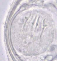
Figure A
More on: Dipylidium
Images presented in the monthly case studies are from specimens submitted for diagnosis or archiving. On rare occasions, clinical histories given may be partly fictitious.
