
Case #164 – September, 2005
A 42-year-old animal trapper sought medical attention regarding an ulcerative lesion on his arm (Figure A). He reported that he had been in Bolivia two months ago and that the lesion on his arm had grown worse since his return. A biopsy of the lesion on his arm was performed and a touch prep smear prepared and stained with Giemsa. The tissue was sent to pathology to be sectioned and stained with H & E. Figures B and C show objects observed on the Giemsa stained touch prep smears, and Figures D and E show objects observed on one of the H & E slides. What is your diagnosis? Based on what criteria?
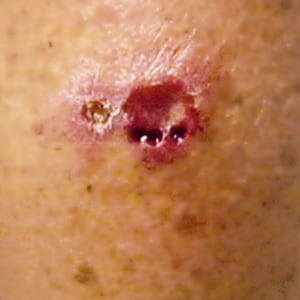
Figure A
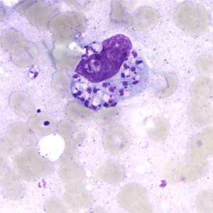
Figure B
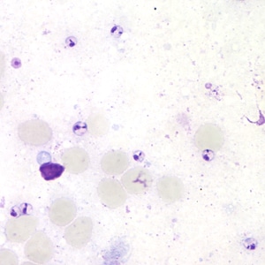
Figure C
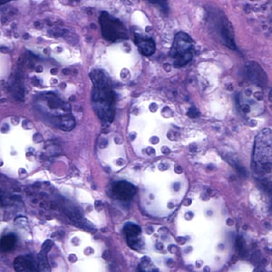
Figure D
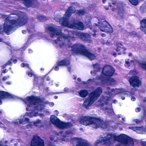
Figure E
Images presented in the DPDx case studies are from specimens submitted for diagnosis or archiving. On rare occasions, clinical histories given may be partly fictitious.
DPDx is an educational resource designed for health professionals and laboratory scientists. For an overview including prevention, control, and treatment visit www.cdc.gov/parasites/.