Skip directly to site content Skip directly to page options Skip directly to A-Z link Skip directly to A-Z link Skip directly to A-Z link
Volume 30, Number 4—April 2024
Research

Divergent Pathogenesis and Transmission of Highly Pathogenic Avian Influenza A(H5N1) in Swine

Bailey ArrudaComments to Author , Amy L. Vincent Baker, Alexandra Buckley, Tavis K. Anderson, Mia Torchetti, Nichole Hines Bergeson, Mary Lea Killian, and Kristina Lantz
Author affiliations: Agriculture Research Service, Ames, Iowa, USA (B. Arruda, A.L. Vincent Baker, A. Buckley, T.K. Andersen); Animal and Plant Health Inspection Service, Ames (M. Torchetti, N. Hines Bergeson, M.L. Killian, K. Lantz)

Main Article

Figure 5

Microscopic lung lesions of swine infected with highly pathogenic avian influenza A(H5N1) belonging to the goose/Guangdong 2.3.4.4b hemagglutinin phylogenetic clade. A) Perivascular mononuclear inflammatory infiltrate (arrowheads) and suppurative bronchiolitis (arrows) in the lung of pig 777 infected with A/turkey/MN/22 necropsied at 3 days postinoculation (dpi). B) Peribronchiolar mononuclear inflammatory infiltrate (arrowhead), suppurative bronchiolitis (arrow), necrotizing bronchiolitis and bronchitis (chevrons), and alveolar luminal accumulation of cellular debris (asterisk) in the lung of pig 796 infected with A/bald eagle/FL/22 necropsied at 5 dpi. C) Peribronchiolar mononuclear inflammatory infiltrate (arrowheads), suppurative bronchiolitis (arrows), and necrotizing bronchiolitis and bronchitis (chevrons) in the lung of pig 58 infected with A/raccoon/WA/22 necropsied at 3 dpi. D) Peribronchiolar and peribronchial mononuclear inflammatory infiltrate (arrowheads), suppurative bronchiolitis (arrow), and necrotizing bronchiolitis and bronchitis (chevrons) in the lung of pig 78 infected with A/redfox/MI/22 necropsied at 3 dpi. Hematoxylin & eosin stain; original magnification ×40.

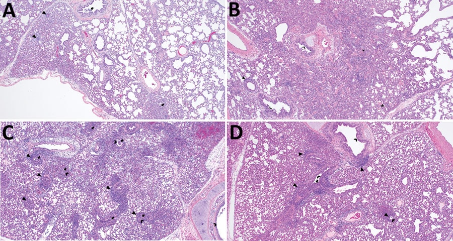
Figure 5. Microscopic lung lesions of swine infected with highly pathogenic avian influenza A(H5N1) belonging to the goose/Guangdong 2.3.4.4b hemagglutinin phylogenetic clade. A) Perivascular mononuclear inflammatory infiltrate (arrowheads) and suppurative bronchiolitis (arrows) in the lung of pig 777 infected with A/turkey/MN/22 necropsied at 3 days postinoculation (dpi). B) Peribronchiolar mononuclear inflammatory infiltrate (arrowhead), suppurative bronchiolitis (arrow), necrotizing bronchiolitis and bronchitis (chevrons), and alveolar luminal accumulation of cellular debris (asterisk) in the lung of pig 796 infected with A/bald eagle/FL/22 necropsied at 5 dpi. C) Peribronchiolar mononuclear inflammatory infiltrate (arrowheads), suppurative bronchiolitis (arrows), and necrotizing bronchiolitis and bronchitis (chevrons) in the lung of pig 58 infected with A/raccoon/WA/22 necropsied at 3 dpi. D) Peribronchiolar and peribronchial mononuclear inflammatory infiltrate (arrowheads), suppurative bronchiolitis (arrow), and necrotizing bronchiolitis and bronchitis (chevrons) in the lung of pig 78 infected with A/redfox/MI/22 necropsied at 3 dpi. Hematoxylin & eosin stain; original magnification ×40.

Main Article

Page created: March 07, 2024
Page updated: March 20, 2024
Page reviewed: March 20, 2024
The conclusions, findings, and opinions expressed by authors contributing to this journal do not necessarily reflect the official position of the U.S. Department of Health and Human Services, the Public Health Service, the Centers for Disease Control and Prevention, or the authors' affiliated institutions. Use of trade names is for identification only and does not imply endorsement by any of the groups named above.
file_external