 ShareCompartir

Monthy Case Studies - 2003
Case #109 - June, 2003
A 29-year-old man was seen by his primary care physician for intermittent abdominal pain and traces of blood in his stool. The man had traveled to Mexico on a camping vacation 1 week prior to the onset of symptoms. He reported that he was careful about what he ate and drank during the trip. The man submitted a stool specimen for ova and parasites (O & P) examination. The images below show what was seen in large numbers on a trichrome stained smear of his PVA preserved stool (Figures A and B, 1000× and Figure C, 500×). What is your diagnosis? Based on what criteria?
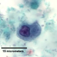
Figure A
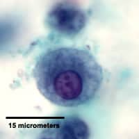
Figure B
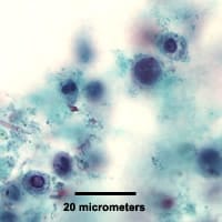
Figure C
Answer to Case #109
The objects seen in the images are leukocytes (white blood cells). Large numbers of white blood cells (WBCs) are often found in patients with bacterial infections and therefore other tests should be considered. Figures A and B were magnified views of macrophages, which can resemble trophozoites of Entamoeba species such as E. histolytica/dispar or E. coli. Figure C showed both macrophages and some polymorphonuclear leuckocytes (PMN). This image should have been used to evaluate the case along with Figures A and B.
The following composites show the macrophages and PCR confirmed E. histolytica trophozoites side by side. Note the difference in the ratio of the nucleus to the cytoplasm.
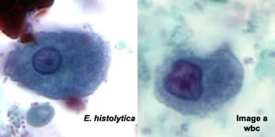
Figure A (Composite)
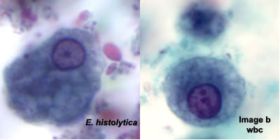
Figure B (Composite)
More on: Artifacts
More on: Amebiasis
Images presented in the monthly case studies are from specimens submitted for diagnosis or archiving. On rare occasions, clinical histories given may be partly fictitious.
