 ShareCompartir

Monthy Case Studies - 2001
Case #71 - November, 2001
A 30-year-old woman discovered a worm in her stool (Figure A). She took the specimen to her doctor, reporting no symptoms or international travel. The specimen was preserved in 10% formalin and sent to CDC for identification. The worm was identified using a dissecting microscope. The laboratorians at CDC were also interested in determining the gender of the worm. The anterior and posterior ends of the worm were dissected and placed in lacto-phenol solution to clear the worm so that morphologic/diagnostic features could be seen (posterior end—Figure B and Figure C; anterior end—Figure D and Figure E). A frontal view of the anterior tip is pictured in Figure F. What is your diagnosis? Based on what criteria? For bonus points, what is the worm's gender?
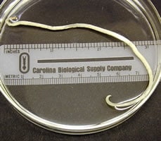
Figure A
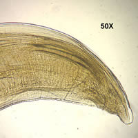
Figure B

Figure C
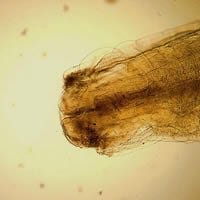
Figure D
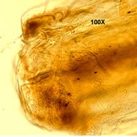
Figure E
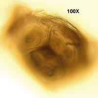
Figure F
Answer to Case #71
This was a case of ascariasis caused by Ascaris lumbricoides. The worm shown in the images was an adult male. Diagnostic morphologic features observed included:
- the presence of three prominent lips around the mouth, an important characteristic of ascarids.
- a size range which was within that for male Ascaris worms (15 to 32 cm). These worms are the largest nematodes found in the intestinal tract of humans.
- the presence of a curved tail with two spicules, indicating that this worm was a male.
More on: Ascariasis
Images presented in the monthly case studies are from specimens submitted for diagnosis or archiving. On rare occasions, clinical histories given may be partly fictitious.
