 ShareCompartir

Monthy Case Studies - 2001
Case #62 - June, 2001
An adult female was diagnosed with malaria. Her travel history included trips to Ghana and Sierra Leone. The hospital laboratory consulted CDC after getting clearance from the state health department for assistance in identifying the species responsible for this case. The original blood smears were sent to CDC along with whole blood in EDTA for confirmation. What is your diagnosis? Based on what criteria?
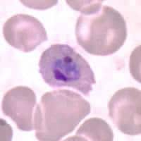
Figure A
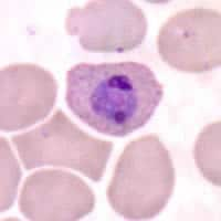
Figure B
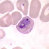
Figure C
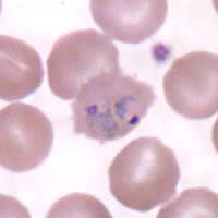
Figure D

Figure E
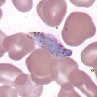
Figure F
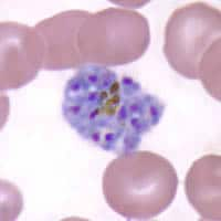
Figure G
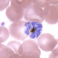
Figure H
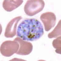
Figure I
Answer to Case #62
This was a case of malaria caused by Plasmodium ovale. Diagnostic features observed included:
- the presence of Schüffner's dots (Figures A, B, and C).
- trophozoites that were slightly ameboid (Figures C and D) or compact; most with a large nucleus (chromatin dot). Most parasites were in slightly enlarged and/or oval red blood cells with some appearing fimbriated (Figures A and D).
- gametocytes (Figures E and F) that were greatly distorted, almost resembling those of P. falciparum. Closer scrutiny, however, revealed that the pigment was too dispersed and that the gametocyte was, in fact, just abnormally shaped. A few gametocytes were seen on the slide, several of which were deformed.
- schizonts (Figures G, H, and I) containing 9 to 14 merozoites found in slightly enlarged red blood cells. Some schizonts had centrally located or clumped coarse pigment. This feature, sometimes seen in P. ovale infections, should not be confused with P. malariae schizonts, which typically have the rosette configuration, but fewer nuclei.
PCR analysis was performed and confirmed the species of Plasmodium to be P. ovale.
More on: Malaria
Images presented in the monthly case studies are from specimens submitted for diagnosis or archiving. On rare occasions, clinical histories given may be partly fictitious.
