 ShareCompartir

Monthy Case Studies - 2000
Case #36 - May, 2000
A 24-year-old thrill seeker from Montana went on a three week Central American excursion to Guatemala, Honduras, Nicaragua, and Panama. He traveled as much as possible on his trail bicycle. Though recommended, he did not take malaria prophylaxis. Two days before his return to the United States, he developed a low-grade fever. The next day his fever was accompanied by chills and muscle aches. After arriving home, he went to the hospital emergency room at the request of his mother. A thin blood smear was made and stained with Giemsa. The following images show the objects seen in his blood smears (Figures A through G). What is your diagnosis? Based on what criteria?
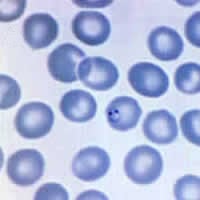
Figure A
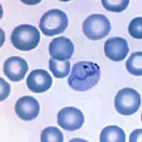
Figure B
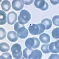
Figure C
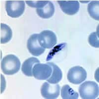
Figure D
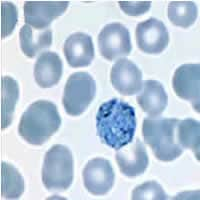
Figure E
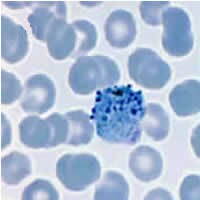
Figure F
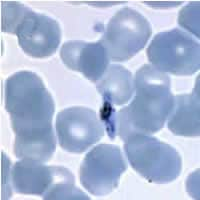
Figure G
Answer to Case #36
This was a case of malaria caused by a mixed infection with Plasmodium falciparum and P. vivax. Diagnostic features observed were:
Plasmodium falciparum:
- a ring with double chromatin dots, thin and delicate, in a normal sized red blood cell (Figure A).
- a thin, appliqué ring in a normal sized red blood cell (Figure C).
- typical banana-shaped P. falciparum gametocytes (Figures D and G), the former of which shows the outline of the red blood cell, sometimes referred to as Laveran's bib.
Plasmodium vivax:
- amoeboid trophozoite in an enlarged red blood cell (Figure B).
- a gametocyte in an enlarged (1.5×) red blood cell (Figure E).
- a super-enlarged red blood cell (>2×) with what appears to contain both an immature schizont and a gametocyte (Figure F).
- a sturdy ring with a large chromatin dot in a red blood cell which is slightly enlarged and is showing what appears to be coarse stippling (Figure H). There is also a large gametocyte in an enlarged (1.5×) red blood cell.
Note the color of the red blood cells. Since the red blood cells are very blue in these images, we believe that the pH of the Giemsa stain was higher than what is recommended (about 7.2). This is how the smear will appear under basic conditions. As such, diagnostic features such as Schüffner's dots may not be seen. Usually, the red blood cells have a pinkish hue when stained with Giemsa at the recommended pH.
More on: Malaria
Images presented in the monthly case studies are from specimens submitted for diagnosis or archiving. On rare occasions, clinical histories given may be partly fictitious.
