 ShareCompartir

Monthy Case Studies - 1999
Case #20 - September, 1999
A 27-year-old serviceman had a routine blood smear examined for parasites at a Georgia Army hospital laboratory after returning from active duty in the Philippines. Several objects were noted in the Giemsa stained blood film, of which two are shown in the following images. What is your diagnosis? Based on what criteria?
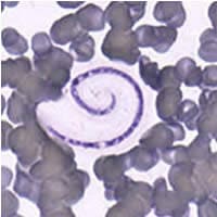
Figure A
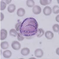
Figure B
Answer to Case #20
The objects were fungal spores of Helicosporium. This is an artifact that contaminated the blood smear, probably during preparation or drying. We chose not to include a scale bar since the erythrocytes can easily be used as a reference of size (about six to seven micrometers in diameter). Helicosporium is sometimes misdiagnosed as some type of microfilaria. However, its smaller size (length and diameter) is enough to rule out microfilaria. We have attached a composite slide containing the original images of Helicosporium (Figures B and C) and two images of true microfilaria. Figures A (Wuchereria) and D (Mansonella), illustrate the larger overall size of the microfilaria. Even the smallest of the human microfilaria (Figure D, M. ozzardi) is clearly larger than Helicosporium. Mansonella ozzardi is usually 160 to 200 micrometers in length and three to five micrometers in width on blood films. Microfilariae of Wuchereria (Figure A) are much larger, typically 250 to 300 micrometers in length and about 7 to 10 micrometers in width. In the larger Wuchereria, you can also see the individual nuclei as well as the sheath, both of which are diagnostic features of microfilaria (although not all microfilariae are sheathed). In Figure D, the nuclei are so dense that they appear to blend together as a solid mass, but there are clear spaces which correspond to anatomic landmarks. The small dots in the hyphae in the Helicosporium also resemble nuclei, but they are not the right size. Please note that measurement of microfilariae on thin films is the least accurate indication of size due to the wide variation in their shrinkage. Measurement of microfilariae in thick blood films is a bit more representative of the true size, but for the most accurate indication of size, measurement of microfilariae preserved in 2% formalin (Knott's preparation) is recommended.
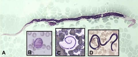
Figure A
More on: Artifacts
Images presented in the monthly case studies are from specimens submitted for diagnosis or archiving. On rare occasions, clinical histories given may be partly fictitious.
