Anthrax Photos
Vaccines Recommended for Travel and Some Specific Groups
WARNING: Some of these photos might be unsuitable for children. Viewing discretion is advised.
Photos of the Disease
A few example photos from the Public Health Image Library
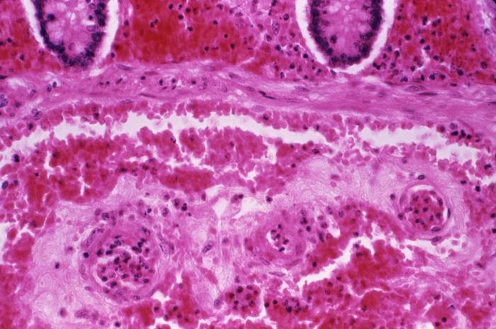
This micrograph reveals submucosal hemorrhage in the small intestine, in a case of fatal human anthrax; H&E stain; Mg. 240X.
Source: PHIL Photo ID# 4629
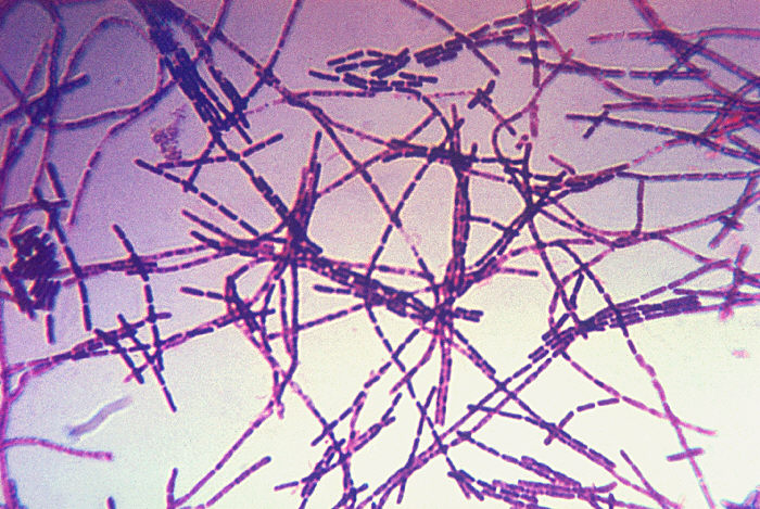
Bacillus anthracis.
Source: PHIL Photo ID# 1811
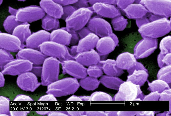
Electron micrograph (SEM) depicted spores from the Sterne strain of Bacillus anthracis bacteria.
Source: PHIL Photo ID# 10123
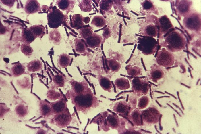
A photomicrograph of Bacillus anthracis bacteria using Gram’s stain technique.
Source: PHIL Photo ID# 2226
Images of People Affected by the Disease
A few example photos from the Public Health Image Library
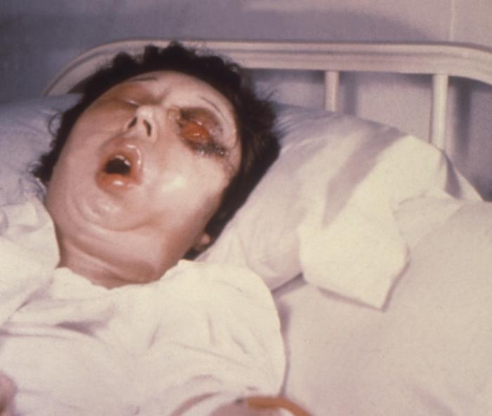
This female patient is shown here on the 5th day of a Bacillus anthracis infection involving her left eye.
Source: PHIL Photo ID# 4504
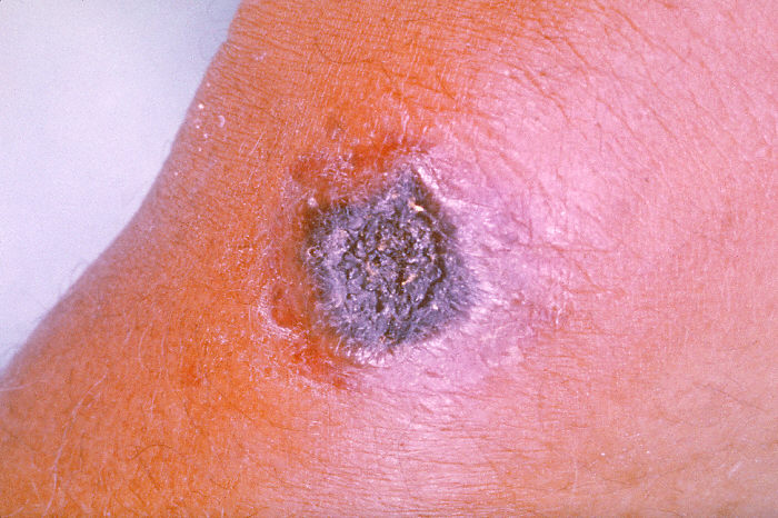
Anthrax lesion on the skin of the forearm caused by the bacterium Bacillus anthracis.
Source: PHIL Photo ID# 2033
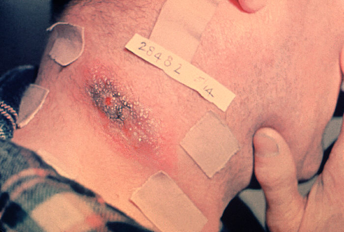
Anthrax lesion on the neck.
Source: PHIL Photo ID# 1794
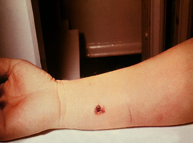
Anthrax, skin of right forearm, 7th day.
Source: PHIL Photo ID# 1801
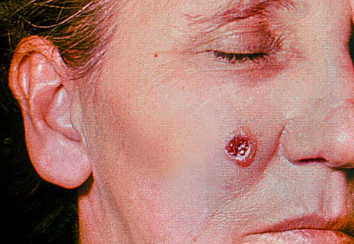
Anthrax, skin of face, 6th day.
Source: PHIL Photo ID# 1804
Additional Images & Regulations
Be sure to see the regulations and copyright rules for these sites offering more images/photos.