Case #283 – September, 2010
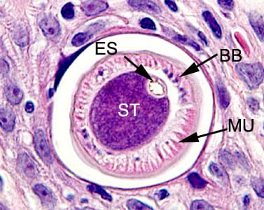
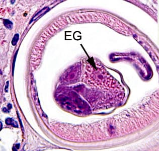
A 49-year-old man presented to his primary care physician with a lump in his throat which had developed over the past three months. He was ultimately referred to an oral surgeon with multiple oral ulcers and submucosal nodules. The patient was originally from Mexico, but has lived in Iowa for nearly 20 years, with recent visits back to Mexico in the past two years. The patient currently works as a landscaper, but has also worked in a foundry and on a pig farm. Biopsy specimens were taken from the submucosal nodules and sent to a pathology laboratory for routine histologic sectioning. Figures A–F show what was observed in sections of the nodule, stained with hematoxylin and eosin (H&E). Figure A was taken at 100x magnification. Figures B and C were taken at 400x magnification. Figures D–F were taken at 1000x magnification. What is your diagnosis? Based on what criteria?
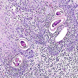
Figure A
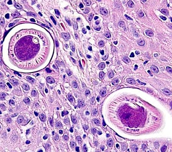
Figure B
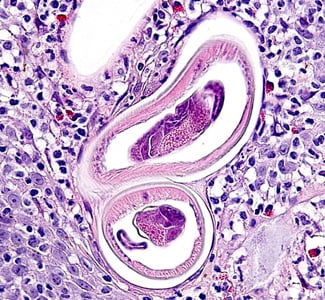
Figure C
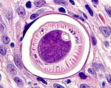
Figure D

Figure E
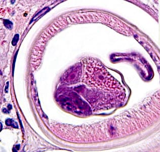
Figure F
This case and images were courtesy of Dr. John Hellstein, Oral and Maxillofacial Pathology, University of Iowa.
Images presented in the DPDx case studies are from specimens submitted for diagnosis or archiving. On rare occasions, clinical histories given may be partly fictitious.
DPDx is an educational resource designed for health professionals and laboratory scientists. For an overview including prevention, control, and treatment visit www.cdc.gov/parasites/.
