
Case #259 – September, 2009
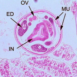
A four-year-old child was admitted to the hospital for sever abdominal pain mimicking appendicitis. A small section of bowel was removed and sent to the Pathology Department for work-up. A section of tissue was preserved in formalin, sectioned, and stained with hematoxylin and eosin (H&E). Images A–C show what was observed at 100x magnification of slides made from the tissue specimen. In addition to the biopsy, stool was collected for routine ova and parasite (O&P) examination. The object is Figure D, which measured on average 73 micrometers long by 37 micrometers wide, was seen in low numbers in a concentrated wet mount from a formalin-preserved aliquot of the stool. What is your diagnosis? Based on what criteria?

Figure A

Figure B

Figure C

Figure D

Images presented in the DPDx case studies are from specimens submitted for diagnosis or archiving. On rare occasions, clinical histories given may be partly fictitious.
DPDx is an educational resource designed for health professionals and laboratory scientists. For an overview including prevention, control, and treatment visit www.cdc.gov/parasites/.