 ShareCompartir

Monthy Case Studies - 2003
Case #114 - August, 2003
A 32-year-old freelance photographer went on a field assignment in the Amazon rain forest. Upon his return, he complained of intermittent abdominal discomfort such as mild cramping and occasional loose stools. The company he had traveled for instructed him to report to their health clinic for a check-up. He provided a stool specimen for an O & P (ova and parasites) examination. Figures A-E are images of organisms that were seen on a LV-PVA smear, stained with Wheatley's trichrome and examined at 1000× magnification. What is your diagnosis? Based on what criteria?
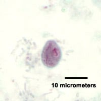
Figure A

Figure B
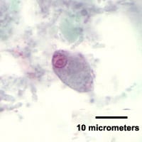
Figure C
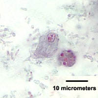
Figure D

Figure E
Answer to Case #114
The organisms shown were Chilomastix mesnili (cysts and trophozoites) and Blastocystis hominis. The pathogenecity of Blastocystis is still a topic of debate and therefore may or may not be the cause of this individual's intestinal symptoms; C. mesnili is considered nonpathogenic. Diagnostic features included:
C. mesnili (Figures A, C, D, and E):
- lemon-shaped cysts within the size range for C. mesnili (7–10 micrometers) with a large nucleus containing a large karyosome, and fibrils along the cytostome
- pyriform trophozoites within the size range for C. mesnili (10–24 micrometers) with a large nucleus at the anterior end with granular peripheral chromatin (may be evenly or irregular distributed); a cytostome can sometimes be seen bordered with fibrils.
B. hominis (Figures B and D)
- organisms within the size range for B. hominis (usually 5–15 micrometers but can range from 4–35 micrometers).
- spherical to ellipsoidal cyst-like bodies with a large central vacuole and a narrow rim of cytoplasm that contains multiple nuclei and inclusion bodies. In trichrome stained smears, the nuclei and inclusion bodies typically stain bright to dark red and the central vacuole will stain green to gray.
More on: Chilomastix mesnili
More on: Blastocystis hominis
Images presented in the monthly case studies are from specimens submitted for diagnosis or archiving. On rare occasions, clinical histories given may be partly fictitious.
