Volume 13, Number 6—June 2007
Dispatch
Loop-Mediated Isothermal Amplification for Influenza A (H5N1) Virus
Abstract
We describe a 1-step reverse-transcription loop-mediated isothermal amplification assay for detection of highly pathogenic avian influenza A (H5N1) viruses. The assay was tested by using a panel of highly pathogenic H5N1 subtypes isolated over the past 10 years and clinical specimens. The assay produced negative results for all non-H5N1 subtypes.
Appendix Figure 1
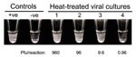
Appendix Figure 1. Visual inspection of the positivity of LAMPreactions. Heated, treated viral culture was serially diluted and tested by the LAMP assay. Reactions were visually inspected after the incubation. Positive reactions would produce...
Highly pathogenic avian influenza A (H5N1) virus has had a significant global effect on the poultry industry, human healthcare, and many other sectors (1). Several molecular tests have been developed for the rapid detection of influenza (H5) virus subtypes (2), but they often require sophisticated equipment (e.g., PCR machine) and are difficult for researchers and clinicians to perform in resource-limited settings. Loop-mediated isothermal amplification (LAMP) provides a molecular testing option for this scenario (3). The LAMP mechanism has been described (3,4). Using this approach, nucleic acids are amplified under isothermal conditions (e.g., in a water bath) with high specificity, efficiency, and speed (3). The assay is highly specific due to recognition of target DNA by 6 independent sequences. An attractive feature of LAMP is its ability to generate large amounts of white magnesium pyrophosphate precipitate in positive reactions (4). Examples of positive and negative reactions are shown in Appendix Figure 1. LAMP enables easy visual identification of positive reactions (4,5) and avoids additional cost and labor for postamplification analysis. This closed-tube method can also minimize the problem of carryover contamination in less controlled environments.
We aimed to develop a reverse transcription (RT) LAMP assay that could detect a variety of highly pathogenic influenza (H5N1) viruses. For primer design, we used highly pathogenic influenza (H5N1) sequences publicly available from the Influenza Sequence Database (www.flu.lanl.gov/index.html) in August 2006. We used hemagglutinin (HA) from an influenza (H5N1) prototype vaccine strain (A/Vietnam/1203/2004, GenBank accession no. AY651334) as the reference for this study. The sequences included highly pathogenic influenza (H5N1) viruses identified in the past 2 years. In addition, sequences from all the other HA subtypes (e.g., 1,127 for H1 and 1,472 for H3) in the above database were used for the analysis. Because highly pathogenic influenza (H5N1) gene sequences are genetically diverse, all studied H5 sequences (n = 711) were aligned, and a highly conserved region at the 5′ end of HA2 encoding sequence (corresponding to nt 1,065–1,298 of the reference sequence) was selected as the target (Appendix Figure2A, and Appendix Figure 3). Of all the downloaded highly pathogenic influenza (H5N1) sequences, 115 did not contain the target site and were excluded from analysis. The GenBank accession numbers for the highly pathogenic influenza (H5N1) sequences used for the primer design are listed in the Appendix Table.
Figure
![Thumbnail of Detection of influenza (H5) virus by loop-mediated isothermal amplification (LAMP). A) Serially diluted RNA from A/Vietnam/1203/2004 was tested by the reverse transcription (RT)–LAMP (upper panel) and RT-PCR (lower panel) assays. The viral titers used in these reactions are indicated. Viral RNA was extracted by using the QIAamp Viral RNA Mini Kit (QIAGEN, Valencia, CA, USA) according to the manufacturer’s instructions. For a typical 25-μL reaction, 2 μL of sample was mixed with 2× in-house reaction buffer (40 mmol/L Tris-HCl, pH 8.8; 20 mmol/L KCl; 16 mmol/L MgSO4; 20 mmol/L [NH4]2SO4; 0.2% Tween 20 [v/v]; 1.6 mol/L betaine; 2.8 mmol/L each dNTP), 50 U Bst DNA polymerase (New England Biolabs, Ipswitch, MA, USA), 8 U avian myeloblastosis virus reverse transcriptase (Invitrogen, Gaithersburg, MD, USA), 40 pmol/L primers FIP and BIP, 20 pmol/L primers LPF and LPR, and 5 pmol/L primers F3 and B3. Reaction mixtures were incubated at 60ºC for 120 min, and the turbidity of these reactions was examined by use of a turbidity meter (LA-200, Treamecs; Kyoto, Japan) in real time. The turbidities of these reactions 5–20 min after incubation were taken as the baseline. The threshold value for a positive reaction was set to be 10× above the standard deviation of the baseline. For the H5-specific RT-PCR assay, primers H5-1 (5′-GCCATTCCACAACATACACCC-3′) and H5-3 (5′-CTCCCCTGCTCATTGCTATG-3′) were used according to the protocol optimized by the World Health Organization H5 Reference Laboratory Network (7). Positive (219 bp) and nonspecific products from the PCR reaction are highlighted by the arrow and arrowhead, respectively. B) Detection of H5 virus in postmortem lung tissues from a patient with influenza (H5). Signals from the tested samples, positive control, and water control are indicated. C) Direct detection of influenza (H5) viruses from culture supernatants. Heat-treated supernatant from cells infected with A/Vietnam/1203/2004 were serially diluted and directly used as input in the LAMP assay. The plaque-forming units (pfu) of influenza (H5) virus in these reactions are shown.](/eid/images/06-1572-F1-tn.jpg)
Figure. Detection of influenza (H5) virus by loop-mediated isothermal amplification (LAMP). A) Serially diluted RNA from A/Vietnam/1203/2004 was tested by the reverse transcription (RT)–LAMP (upper panel) and RT-PCR (lower panel) assays. The...
Previous demonstration that degenerate primers could be used in LAMP assays (6) led us to use the same approach for designing a set of degenerate primers for the targeted region (Appendix Figure 2B). Our HA sequence analysis also showed that HA sequences from influenza (H5N2) viruses of North American lineage and other viruses with other HA subtypes contain extensive mismatches to the primers used in the LAMP assay (Appendix Figure 2A, B). In the initial phase of the study, we used the reference strain (A/Vietnam/1203/2004) to determine the sensitivity of the assay. Purified RNA from a viral supernatant with a known titer was serially diluted and subjected to the LAMP assay. As shown in the Figure panel A, the detection limit of the assay was found to be 2×10⁻³ plaque-forming units (pfu) per reaction. The sensitivity of this assay was also compared with that of an optimized RT-PCR assay recommended by the World Health Organization (7). As shown in the Figure panel A, the detection limit of the RT-PCR assay was identical to that from the LAMP assay. We also spiked various amounts of influenza (H5) viruses into non-H5 nasopharyngeal aspirate samples and tested the purified RNA from these clinical specimens by the LAMP and RT-PCR assays. The results from these assays were identical (data not shown). These findings are in accordance with previous findings that the sensitivity of LAMP assays is comparable to that of conventional PCR methods (5,6,8). In addition, all positive reactions were visually examined for the presence of white precipitate at the end of the reaction incubation. As expected, all results correlated exactly with those deduced by the real-time turbidity meter (data not shown).
We also tested the feasibility of using this assay to detect influenza (H5) viruses in clinical specimens from a patient with influenza (H5). RNA from 2 different postmortem lung tissue samples from a patient infected with influenza A/HK/212/03 was subjected to the LAMP assay. As shown in the Figure panel B, both RNA samples had positive results for the influenza (H5) viruses.
To evaluate the specificity of the LAMP assay for influenza (H5) viruses, a comparative study including all 16 HA subtypes and a variety of H5N1 strains was performed (Table). All 14 H5N1 strains isolated from different geographic regions during the past 10 years were positive, whereas none of the non-H5 samples were positive (Table). All the reactions were visually examined, and the results corresponded with those generated from the real-time turbidity meter. Influenza A/chicken/Wajo/BBVM/2005, A/duck/Vietnam/568/2005, and A/bar-headed goose/Qinghai/5/2005 are clade 2 influenza (H5N1) viruses that have recently emerged from different geographic regions (9), and these 3 viruses are phylogenetically distinct (10,11). Our results demonstrate that this assay is applicable to a wide variety of highly pathogenic influenza (H5N1) viruses. In addition, other virus reassortants with the HA of this lineage are expected to be positive in the assay.
We previously demonstrated that heat-treated blood samples could be directly tested by a LAMP assay specific for DNA of a bloodborne pathogen (5). These modifications could avoid the need for nucleic acid purification, thereby reducing the cost and turnaround time for molecular diagnosis. We tested the feasibility of detecting the H5 sequence from viral cultures without RNA extraction. In a Biosafety Level 3 facility, 50 μL of viral culture of the prototype virus was heat inactivated (99°C for 10 min), the heat-treated sample was serially diluted with standard viral culture medium, and 2 μL of serially diluted samples was added to the LAMP assay. As shown in the Figure panel C, and in Appendix Figure 1, H5N1 subtype could still be detected by the assay. However, the detection limit (9.6 pfu/reaction) was ≈1,000× less sensitive than the limit with purified RNA as an input.
The RT-LAMP assay is highly specific, and its sensitivity is comparable to that of an optimized RT-PCR assay for influenza (H5N1) viruses. The detection limit is equivalent to that of a similar LAMP assay (12), but our assay was extensively tested by using a wide variety of influenza (H5) viruses, including recent clade 2 influenza (H5) viruses (9). LAMP does not require thermocyclers and gel electrophoresis. Reactions can simply be incubated in a water bath or heating block, and the results can be confirmed by direct visual inspection. Because early identification of influenza (H5) is crucial for the containment of the disease, this novel assay can provide an efficient option for the preliminary molecular detection of highly pathogenic influenza (H5) viruses in basic laboratory or clinical settings. Combined with the use of a turbidity meter, which costs much less than a real-time RT-PCR system, RT-LAMP can provide quantitative data for viral load studies. In conclusion, the LAMP assay is a promising tool for the detection of influenza (H5) viruses.
Ms Jayawardena is a PhD student in the Department of Microbiology of The University of Hong Kong. Her research interests mainly relate to the molecular biology of influenza viruses.
Acknowledgments
We thank Bonnie W.Y. Wong for her technical assistance.
We acknowledge research funding from Public Health Research Grant A195357 from the US National Institutes of Health, the Research Grant Council of Hong Kong (HKU 7530/06M), and the Seed Funding Programme for Basic Research from HKU.
References
- Perez DR, Sorrell EM, Donis RO. Avian influenza: an omnipresent pandemic threat. Pediatr Infect Dis J. 2005;24:S208–16. DOIPubMedGoogle Scholar
- Brown IH. Advances in molecular diagnostics for avian influenza. Dev Biol (Basel). 2006;124:93–7.PubMedGoogle Scholar
- Notomi T, Okayama H, Masubuchi H, Yonekawa T, Watanabe K, Amino N, Loop-mediated isothermal amplification of DNA. Nucleic Acids Res. 2000;28:e63. DOIPubMedGoogle Scholar
- Mori Y, Nagamine K, Tomita N, Notomi T. Detection of loop-mediated isothermal amplification reaction by turbidity derived from magnesium pyrophosphate formation. Biochem Biophys Res Commun. 2001;289:150–4. DOIPubMedGoogle Scholar
- Poon LLM, Wong BWY, Ma EHT, Chan KH, Chow LMC, Abeyewickreme W, Sensitive and inexpensive molecular test for falciparum malaria: detecting Plasmodium falciparum DNA directly from heat-treated blood by loop-mediated isothermal amplification. Clin Chem. 2006;52:303–6. DOIPubMedGoogle Scholar
- Poon LLM, Leung CSW, Chan KH, Lee JHC, Yuen KY, Guan Y, Detection of human influenza A viruses by loop-mediated isothermal amplification. J Clin Microbiol. 2005;43:427–30. DOIPubMedGoogle Scholar
- World Health Organization. Recommended laboratory tests to identify avian influenza A virus in specimens from humans. 2005 [cited 2006 September 24]. Available from http://www.who.int/csr/disease/avian_influenza/guidelines/avian_labtests2.pdf
- Parida M, Posadas G, Inoue S, Hasebe F, Morita K. Real-time reverse transcription loop-mediated isothermal amplification for rapid detection of West Nile virus. J Clin Microbiol. 2004;42:257–63. DOIPubMedGoogle Scholar
- Webster RG, Govorkova EA. H5N1 influenza–continuing evolution and spread. N Engl J Med. 2006;355:2174–7. DOIPubMedGoogle Scholar
- Chen H, Smith GJD, Li KS, Wang J, Fan XH, Rayner JM, Establishment of multiple sublineages of H5N1 influenza virus in Asia: implications for pandemic control. Proc Natl Acad Sci U S A. 2006;103:2845–50. DOIPubMedGoogle Scholar
- Smith GJ, Naipospos TS, Nguyen TD, de Jong MD, Vijaykrishna D, Usman TB, Evolution and adaptation of H5N1 influenza virus in avian and human hosts in Indonesia and Vietnam. Virology. 2006;350:258–68. DOIPubMedGoogle Scholar
- Imai M, Ninomiya A, Minekawa H, Notomi T, Ishizaki T, Tashiro M, Development of H5-RT-LAMP (loop-mediated isothermal amplification) system for rapid diagnosis of H5 avian influenza virus infection. Vaccine. 2006;24:6679–82. DOIPubMedGoogle Scholar
Figures
Table
Cite This ArticleTable of Contents – Volume 13, Number 6—June 2007
| EID Search Options |
|---|
|
|
|
|
|
|
Please use the form below to submit correspondence to the authors or contact them at the following address:
Leo L.M. Poon, Department of Microbiology, The University of Hong Kong, Queen Mary Hospital, Pokfulam, Hong Kong Special Administrative Region, People’s Republic of China;
Top