 |
|
|
|
|
|
|
| ||||||||||
|
|
|
|
|
|
|
||||
| ||||||||||
|
|
|
|
|
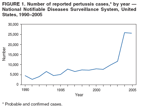
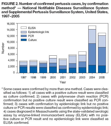
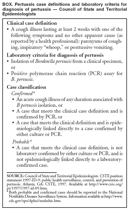
Persons using assistive technology might not be able to fully access information in this file. For assistance, please send e-mail to: mmwrq@cdc.gov. Type 508 Accommodation and the title of the report in the subject line of e-mail. Outbreaks of Respiratory Illness Mistakenly Attributed to Pertussis --- New Hampshire, Massachusetts, and Tennessee, 2004--2006Pertussis, or whooping cough, is a highly infectious, nationally notifiable* respiratory disease associated with prolonged cough illness and paroxysms of coughing, inspiratory "whoop," or posttussive vomiting. Reported pertussis cases have tripled in the United States since 2001, with 25,616 probable or confirmed cases reported in 2005 (Figure 1). This increase has been attributed to increased circulation of Bordetella pertussis, waning vaccine-induced immunity among adults and adolescents, heightened awareness of pertussis among health-care providers, increased public health reporting, and increased use of polymerase chain reaction (PCR) testing for diagnosis (1). To minimize the spread of pertussis, control measures must be implemented early in the course of illness when the risk for transmission is highest. However, diagnosis of pertussis is complicated by nonspecific signs and symptoms, particularly in the early catarrhal stage of disease. In addition, the lack of rapid, sensitive, and specific laboratory tests makes early and accurate identification of pertussis challenging. This report describes two hospital outbreaks and one community outbreak of respiratory illness during 2004--2006 in New Hampshire, Massachusetts, and Tennessee that were attributed initially to pertussis. However, subsequent investigations revealed negative or equivocal laboratory results and epidemiologic and clinical features atypical of pertussis, suggesting that pertussis was not the cause of these outbreaks. The findings in this report underscore the need for thorough epidemiologic and laboratory investigation of suspected pertussis outbreaks when considering extensive control measures. New Hampshire. In March 2006, a laboratory worker from a 396-bed hospital visited the occupational medicine clinic with a 3-week history of paroxysmal cough and posttussive vomiting. The laboratory worker tested positive with the hospital's single-target PCR assay for pertussis (IS481).† The worker subsequently was treated with azithromycin and furloughed for 5 days. Postexposure prophylaxis (PEP) with azithromycin was administered to all close contacts. Case investigation from mid-March to early April identified 15 additional health-care personnel (HCP) in the same laboratory with respiratory illness and either a positive or equivocal PCR test result for pertussis, leading hospital investigators to suspect an outbreak. Suspected pertussis in HCP was defined as either 1) cough of any duration and at least one classic pertussis symptom (i.e., paroxysms of coughing, whoop, or posttussive vomiting) or 2) a positive or equivocal PCR test result. In April, to control the spread of the outbreak, the hospital's infection-control and occupational-medicine staff members offered PEP and vaccination with the newly licensed tetanus toxoid, reduced diphtheria toxoid, acellular pertussis vaccine (Tdap) to all personnel in the hospital's clinical laboratories. Despite these interventions, from late April to early May, 18 additional ill HCP with suspected pertussis were identified through passive surveillance in other parts of the hospital, including patient-care areas. In May, the hospital began screening all HCP for signs and symptoms of upper respiratory tract infection and began PCR testing for pertussis on symptomatic HCP. By June, 134 suspected pertussis cases had been identified: 98 (73%) by positive or equivocal PCR results and 36 (27%) by clinical symptoms alone. A total of 192 nasopharyngeal swabs or aspirates from symptomatic HCP, including specimens from 27 (20%) of the 134 HCP with suspected pertussis, were submitted for isolation of B. pertussis by culture throughout the course of the outbreak; none yielded B. pertussis. Review of surveillance data revealed no increased pertussis activity in the surrounding community. No pertussis cases were identified among vaccinated or unvaccinated infants, either in the hospital or surrounding community. Retrospective interviews of 120 (90%) HCP with suspected pertussis indicated that 25 (21%) of those interviewed never had cough, a hallmark symptom of pertussis. Among the 95 (79%) HCP with cough, 33 (35%) reported never having a classic pertussis symptom (i.e., paroxysms, whoop, or posttussive vomiting). Myalgia, not typically associated with pertussis, was reported by 32 (34%) of 93 HCP who were asked whether they had this symptom. Additional laboratory evaluation included retesting of initial DNA extracts at CDC using a two-target PCR assay (IS481 and ptxS1). Among 111 extracts available for testing, one was positive for both targets and interpreted as B. pertussis, and 24 extracts were positive by single target alone (IS481) and interpreted as indeterminate. Sera from 39 HCP who had not been vaccinated during the outbreak with Tdap and who met the hospital's definition for suspected pertussis were collected and tested at Vanderbilt University Medical Center in Nashville, Tennessee, for antipertussis toxin immunoglobulin G (IgG) by enzyme-linked immunosorbent assay (ELISA); one sample had a positive IgG level, one was intermediate, and 37 were negative. Samples of aspirates and DNA extracts were tested at the hospital and CDC for a panel of viral pathogens, other Bordetella species, Chlamydia pneumoniae, and Mycoplasma pneumoniae. PCR testing yielded two specimens with results consistent with Bordetella holmesii. Substantial resources were invested to control this outbreak. During March--May 2006, approximately 1,700 visits by HCP to the occupational medicine clinic for respiratory illness were reported. Among 6,289 hospital HCP, 978 (16%) ill HCP were tested by PCR, treated, and furloughed pending negative PCR results. An additional 1,311 contacts of HCP with suspected pertussis received PEP. Other control measures included a 1-week Tdap vaccination campaign in May, during which 4,524 (72%) HCP were vaccinated. Massachusetts. A child aged 20 months was admitted to a 347-bed pediatric hospital on September 21, 2006, with respiratory symptoms; the child had not received all age-appropriate doses of diphtheria and tetanus toxoids and acellular pertussis (DTaP) vaccine. Initial tests on September 24 were positive for respiratory syncytial virus. Subsequent testing for pertussis by two-target PCR assays (IS481 and ptxS1) at the Massachusetts State Laboratory Institute (MSLI) were positive for both targets on October 2, 2006. In October, the hospital initiated enhanced screening of symptomatic HCP with suspected pertussis and other HCP who had been in contact with the child. A total of 507 HCP with upper respiratory symptoms were identified during the course of the investigation. Nasopharyngeal specimens from symptomatic HCP were tested by culture, PCR, or both during October 1--November 14. By December 2006, 36 specimens from HCP had tested positive for pertussis by PCR (33 at MSLI and three at a commercial laboratory). Twenty-eight of the 36 (78%) HCP had reported cough of fewer than 2 weeks and 33 (92%) had reported no classic pertussis symptoms. Of the 33 PCR-positive specimens tested for two targets at MSLI, 29 (88%) were positive by a single target (IS481) and four (12%) were positive by both targets (IS481 and ptxS1). Of the 32 PCR-positive specimens submitted for culture, none yielded B. pertussis. Sera were collected from 23 HCP who had positive PCR test results and were not vaccinated during the outbreak; all were negative for antipertussis toxin IgG by ELISA at MSLI. Because a number of HCP had atypical symptoms and no culture or serologic confirmation of pertussis, repeat PCR testing was conducted at CDC and the Provincial Laboratory for Public Health in Alberta, Canada. Twenty-five initial DNA extracts with positive PCR test results were retested at CDC using two-target PCR assays (IS481 and ptxS1). One sample was positive by both targets (IS481 and ptxS1) and interpreted as positive for B. pertussis, and 24 were positive by a single target only (IS481) and interpreted as indeterminate. Six of the 25 initial DNA extracts also were retested by the Canadian laboratory; two extracts were positive by IS481 and ptxS1 (interpreted as positive for B. pertussis), three were positive by IS481 only (interpreted as possibly B. pertussis), and one result was uninterpretable. Overall, only one of six specimens tested by MSLI, CDC, and the Canadian laboratory was interpreted as positive for B. pertussis by all three laboratories. Six DNA extracts were tested for M. pneumoniae by PCR, and none were positive. Tennessee. In April 2004, pertussis in an infant aged 5 weeks was confirmed by isolation of B. pertussis from a nasopharyngeal specimen. Before diagnosis, the infant had been taken to the local health department and two other medical facilities. Aggressive contact tracing and testing of symptomatic contacts was undertaken by the local health department. For this investigation, a laboratory-confirmed case was defined as a PCR-positive case in a symptomatic contact, using a single-target repeating sequence found in B. pertussis (RSBP1). A clinical case was defined as either cough illness of at least 2 weeks' duration or cough of any duration with paroxysms of coughing, whoop, or posttussive vomiting and an epidemiologic link to a laboratory-confirmed case. Antimicrobial treatment was offered to all patients, and PEP was offered to all asymptomatic close contacts. Further contact tracing and control measures were implemented for all patients with laboratory-confirmed or clinical diagnoses of pertussis. During a 2-month period, 1,459 persons in the community who visited health-care providers with pertussis symptoms were evaluated for pertussis and offered treatment or PEP with erythromycin or azithromycin. A total of 317 symptomatic persons were tested by PCR; 43 (14%) were positive. Of these, only two (5%) had cough of at least 2 weeks' duration. Among 284 samples submitted for culture, only the specimen from the infant yielded B. pertussis. Because of the lack of culture confirmation, serologic testing for antipertussis toxin IgG by ELISA was performed at Vanderbilt University Medical Center on 21 patients and contacts. Four of 11 patients who were positive by PCR also had serologic evidence of recent pertussis infection. Testing for alternate pathogens was not performed. Reported by: KB Kirkland, MD, EA Talbot, MD, RA Lasky, RK McLellan, MD, Dartmouth-Hitchcock Medical Center, Lebanon, New Hampshire; JT Montero, MD, New Hampshire Dept of Health. MA Barry, MD, TA McCarthy, MD, JE Gunn, MPH, JL Pendarvis, MPH, Boston Public Health Commission, Massachusetts; LL Han, MD, Massachusetts Dept of Public Health. RA Devasia, MD, KM Edwards, MD, Vanderbilt Univ Medical Center, Nashville, Tennessee; TF Jones, MD, Tennessee Dept of Public Health. K Kretsinger, MD, ML Tondella, PhD, KM Tatti, PhD, KH Brown, MPH, BA Slade, MD, K-H Wu, PhD, Div of Bacterial Diseases, X Lu, MS, Div of Viral Diseases, National Center for Immunization and Respiratory Diseases; M Patel, MD, EIS Officer, CDC. Editorial Note:Although the respiratory outbreaks in New Hampshire, Massachusetts, and Tennessee initially were considered caused by pertussis, retrospective investigations demonstrated that pertussis was unlikely to have been the primary etiology. The results of these investigations underscore the importance of confirming pertussis as the etiology of respiratory outbreaks when control measures are being implemented, particularly when laboratory results are inconsistent and supporting clinical and epidemiologic data are lacking. Several laboratory methods, including culture, serology, and PCR, are available for pertussis diagnosis. Culture is a reference standard and 100% specific. Its sensitivity can be as high as 56% early in the course of illness but decreases with delays in specimen collection or in patients who have received antimicrobial treatment or previous vaccination (1--3). Other factors that can affect the yield of culture include technical methods for obtaining specimens, availability of appropriate media, transport of specimens, and experience with isolation of B. pertussis (2,4). Isolating B. pertussis in culture can take 7--14 days and might not be timely for acute case management. However, confirming the etiology with culture in the early stages of a suspected pertussis outbreak will help guide the public health response (3,4), and continued isolation of B. pertussis from a subset of clinical samples will provide laboratory evidence of ongoing transmission. Serology using paired acute- and convalescent-phase sera requires at least a 4-week interval between specimen collections and is not useful for immediate diagnosis (4). Single-sample serology tests for antipertussis toxin IgG have been developed for research purposes but must be collected at least 2 weeks after symptom onset (5). Pertussis serology assays using commercially available reagents also are available, but these assays are not clinically validated and might not differentiate between recent and remote infection or vaccination. In 1997, introduction of PCR test results into the pertussis case definition of the Council of State and Territorial Epidemiologists (CSTE) (Box) facilitated laboratory diagnosis of disease, particularly among adults and adolescents, who often visit health-care providers late in the course of illness when the yield of culture is lower (2). The use of PCR, a rapid and sensitive diagnostic test, has become widespread. Among confirmed pertussis cases reported to NNDSS, the percentage of cases confirmed by PCR increased from 12% in 1997 to 44% in 2005, and the percentage of cases confirmed by culture decreased from 52% in 1997 to 20% in 2005. Overall, during 1997--2005, the number of PCR-confirmed cases increased while the number of culture-confirmed cases remained stable (Figure 2; CDC, unpublished data, 2007). During the same period, the percentage of pertussis cases confirmed both by PCR and culture ranged from 1.1%--3.1% annually (mean: 2.3%). Presumed false-positive PCR test results in persons with nonspecific clinical features, such as rhinorrhea, sneezing, and sore throat, have raised concerns regarding the widespread application of PCR in an outbreak setting (6). No standardized PCR protocols for pertussis testing exist; approximately 100 different assays that use the IS481 target sequence have been documented (7). Laboratories vary in DNA purification techniques, primers and probes used in testing, and quality assurance procedures (1,4). Although these assays might undergo analytic sensitivity testing for technical performance standards (e.g., detection limits and reproducibility), a limited number of laboratories have established the accuracy of their PCR test (1). In addition, as illustrated in the Massachusetts outbreak, interpretation of PCR results can vary among laboratories. Use of standardized rapid and reliable laboratory tests to improve the specificity of the CSTE case definition is a public health priority. CDC, the Food and Drug Administration, and state and local public health partners have implemented a clinical validation study to evaluate several PCR and serologic assays. The results from that study should provide the basis for future validated laboratory assays to diagnose and manage pertussis cases and outbreaks. The outbreaks described in this report illustrate the limitations of relying solely on PCR assays to confirm pertussis. PCR is an important tool for diagnosing individual cases of pertussis in persons for whom a high index of suspicion exists and for whom timely treatment and PEP are essential. However, the positive predictive value can be lower if PCR is used as a screening tool without culture confirmation during a suspected pertussis outbreak (3). Overreliance on the results of PCR assays can lead to implementation of unnecessary and resource-intensive control measures (e.g., case identification, antimicrobial treatment, furlough of ill persons, and administration of PEP) (8). In outbreak settings, positive PCR results should be interpreted in conjunction with epidemiologic investigation, evaluation of clinical symptoms, and confirmation by culture. CDC recommends timely collection and testing (early in the course of illness and during the initial stages of the outbreak) of nasopharyngeal specimens for culture in at least a subset of persons who are symptomatic to confirm pertussis as the etiology of the outbreak (3). Absent or inconsistent supporting data and negative pertussis cultures in appropriately collected specimens should prompt testing for alternate pathogens. Cocirculation of other pathogens can cause respiratory illness with symptoms similar to pertussis. Circulation of B. pertussis in communities is common and occurs in a background of other causes of respiratory illness. In retrospect, the culture-confirmed pertussis in the infant in Tennessee might have reflected sporadic disease rather than the beginning of an outbreak. Because confirmation of pertussis outbreaks by culture can take several weeks, simultaneous testing of acutely symptomatic persons for other pathogens (e.g., viruses or atypical bacteria) might be appropriate. Guidance on appropriate approaches to respiratory outbreaks of unknown etiology is available to state and local health departments through consultation with CDC at telephone 770-488-7100. Considering the challenges of diagnosing pertussis and controlling outbreaks, prevention of pertussis outbreaks through widespread vaccination is an important strategy. The Advisory Committee on Immunization Practices recommends vaccination of persons aged 11--64 years with the newly licensed Tdap vaccines (1,9), which have been estimated 85%--92% effective (1,9). Achieving high coverage is expected to prevent disease and decrease the likelihood of future pertussis outbreaks. Although the effectiveness of vaccination with Tdap in interrupting transmission of pertussis during an outbreak has not been established, persons previously vaccinated with Tdap should have a lower risk for acquiring and transmitting pertussis, thereby preventing the outbreak from expanding. Investigation of suspected pertussis outbreaks should include timely consideration of clinical, laboratory, and epidemiologic data, including vaccination status of the population affected, to help health officials implement appropriate control measures. References
* Information available at http://www.cdc.gov/epo/dphsi/nndsshis.htm. † The assays identified in this report have not been approved by the Food and Drug Administration.
Figure 1
Disclaimer All MMWR HTML versions of articles are electronic conversions from ASCII text into HTML. This conversion may have resulted in character translation or format errors in the HTML version. Users should not rely on this HTML document, but are referred to the electronic PDF version and/or the original MMWR paper copy for the official text, figures, and tables. An original paper copy of this issue can be obtained from the Superintendent of Documents, U.S. Government Printing Office (GPO), Washington, DC 20402-9371; telephone: (202) 512-1800. Contact GPO for current prices. **Questions or messages regarding errors in formatting should be addressed to mmwrq@cdc.gov.Date last reviewed: 8/23/2007 |
|||||||||
|