 ShareCompartir

Monthy Case Studies - 2003
Case #117 - October, 2003
A survey of intestinal parasites in school-aged children was conducted in a village in Kenya. Specimens were sent to a reference laboratory for processing and parasite identification. A microscope capable of UV fluorescence microscopy was available to screen for Cyclospora cayetanensis after completing a standard FEA concentration. Figures A and B show what was found in one of the samples. What is your diagnosis? Based on what criteria?
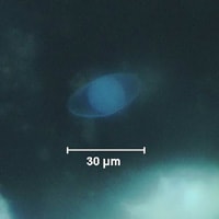
Figure A
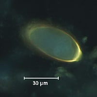
Figure B
Answer to Case #117
This case demonstrated a mixed infection of cystoisosporiasis, caused by Cystoisospora belli, and trichuriasis, caused by Trichuris trichiura. Diagnostic morphologic features included:
Cystoisospora belli
- oocysts within the size range for C. belli (25 to 30 micrometers in length).
- the presence of a single sporoblast in the Cystoisospora oocyst, indicating that it is immature.
Trichuris trichiura
- eggs within the size range for T. trichiura (50 to 54 micrometers in length).
- the presence of polar plugs, although one end is out of focus in Figure B.
When using UV fluorescence microscopy, one important diagnostic feature includes the fluorescent color of the oocyst wall of Cystoisospora and the shell of the Trichuris egg. To confirm these diagnoses, one can switch to bright-field microscopy and view the same organisms (shown in the images below).
Cystoisospora—UV
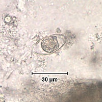
Cystoisospora—bright-field
Trichuris—UV

Trichuris—bright-field
More on: Cystoisosporiasis
More on: Trichuriasis
Images presented in the monthly case studies are from specimens submitted for diagnosis or archiving. On rare occasions, clinical histories given may be partly fictitious.
