 ShareCompartir

Monthy Case Studies - 2003
Case #100 - January, 2003
A 35-year-old woman, who wears contact lenses, was seen by a physician for conjunctivitis she had experienced for one year. She had been camping in the high Sierras and Yosemite Park area about six weeks prior to being seen by her doctor. A worm, approximately 12 to 14 mm in length, was found under the patient's contact lens. The doctor removed the worm and sent it to a reference laboratory for identification. The specimen was forwarded to CDC for confirmatory identification. A more detailed microscopic examination revealed the presence of several structures shown in the images below (Figures A, B, and C). Figures A and B show details of the worm's morphology, including the buccal capsule, esophagus, and striations, as indicated on the images. Figure C shows thin-shelled eggs seen in the worm; the eggs measured approximately 47 µm by 30 µm. What is your diagnosis? Based on what criteria?
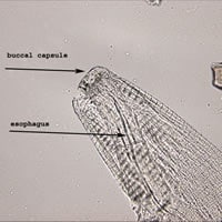
Figure A
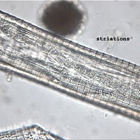
Figure B
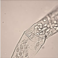
Figure C
Answer to Case #100
This was a case of thelaziasis caused by a nematode in the genus, Thelazia. Diagnostic features observed included:
- an adult female worm within the size range for the genus (females range from 8 to 18 mm in length; males are smaller, ranging from 7 to 12 mm in length).
- the presence of short cuticular buccal capsule.
- a cuticle with transverse striations that appeared like slightly overlapping shingles.
- eggs within the size range for Thelazia sp. (45 to 53 µm by 27 to 33 µm).
More on: Thelaziasis
Images presented in the monthly case studies are from specimens submitted for diagnosis or archiving. On rare occasions, clinical histories given may be partly fictitious.
