 ShareCompartir

Monthy Case Studies - 2002
Case #94 - October, 2002
A 50-year-old scholar was seen in a hospital in Connecticut for severe diarrhea. The patient’s travel history in the last six months included India and Mexico. The following images show an object from trichrome-stained smears of the patient’s stool specimen. The size of the object was 13 micrometers. The three images show the same object in three different focal planes. What is your diagnosis? Based on what criteria? Would you recommend a confirmatory test(s) for this case? What test(s) and for what reason(s)?
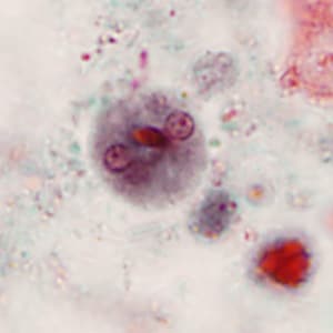
Figure A

Figure B
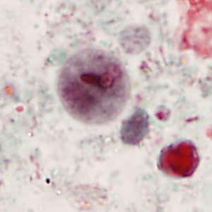
Figure C
Answer to Case #94
This was a case of amebiasis caused by Entamoeba histolytica/Entamoeba dispar. Diagnostic features observed included:
- the shape and size of the cyst, which was compatible with E. histolytica/dispar. Cysts of E. histolytica/dispar range in size from 12 to 15 micrometers.
- the presence of 4 nuclei (observed in the different focal planes), compatible with E. histolytica/dispar.
- the presence of chromatoid bodies with blunt ends.
Entamoeba histolytica and E. dispar are morphologically-identical species except when RBC’s are observed within the cytoplasm of trophozoites, which is confirmatory for E. histolytica. Diagnostic tests should be used to confirm the presence of E. histolytica since it may cause severe extraintestinal infections (e.g., hepatic or pulmonary) that can lead to death. Specific E. histolytica antigen detection by ELISA and PCR using specific primers that differentiate E. histolytica from E. dispar can be used for this purpose. Serology is not useful for the detection of extra-intestinal or active intestinal invasion.
More on: Amebiasis
Images presented in the monthly case studies are from specimens submitted for diagnosis or archiving. On rare occasions, clinical histories given may be partly fictitious.
