 ShareCompartir

Monthy Case Studies - 2001
Case #72 - November, 2001
A sixteen-year-old teenage boy spent a month in the Amazon basin in Brazil. After returning to the United States, he noticed a small indurated nodule on hisforehead. He treated the nodule with topical antibiotics. During the nextthree months, the lesion did not heal, but became ulcerated and continued togrow. The teenager visited his doctor who photographed the lesion and performeda biopsy. Figure A is a photograph of the lesion on the young man'sforehead. Figures B, C, and D are Giemsa stained sectionsof the tissue that was sent to CDC for identification/diagnosis. What is yourdiagnosis? Based on what criteria?

Figure A
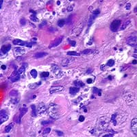
Figure B
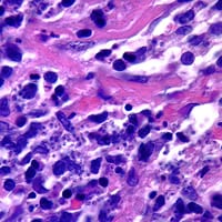
Figure C
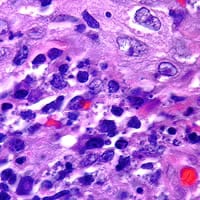
Figure D
Answer to Case #72
This was a case of leishmaniasis. The organisms shown in the Figures B-D were amastigotes, the rounded morphological stageof Leishmania sp. that has no external flagellum. A large nucleus and asmaller kinetoplast could be seen in a few of these amastigotes. Both of thesefeatures are not always seen on all organisms due to the thickness of histologyspecimens. Also, both of these features may not be in the same focal plane onthe slide.
One may speculateas to the species of Leishmania based on epidemiology and symptoms. However, more precise diagnostic procedures, such as PCR or isoenzyme analysis,should be implemented for an accurate diagnosis and subsequent propertreatment.
More on: Leishmaniasis
Images presented in the monthly case studies are from specimens submitted for diagnosis or archiving. On rare occasions, clinical histories given may be partly fictitious.
