 ShareCompartir

Monthy Case Studies - 2001
Case #66 - August, 2001
This case does not involve a human. However, we thought that the information was interesting and worthy of sharing.
A biologist at a university in the state of Tennessee made a Giemsa stained smear of urine obtained from a single insect, similar to the one shown in Figure A. The insect was found by a physician from Mexico in the outdoor hallway of an apartment complex in Tennessee. Several similar insects that ranged in size from 13 to 25 micrometers in length were found in the hallway. What kind of insect is shown in Figure A? What are the organisms shown in Figures B, C, and D? Do your findings have any public health significance?
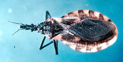
Figure A
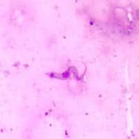
Figure B
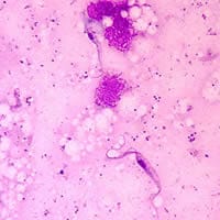
Figure C
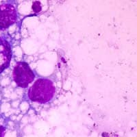
Figure D
Answer to Case #66
The insect was Triatoma sanguisuga. The organisms in the insect were epimastigote and trypomastigote forms of Trypanosoma cruzi, the organism responsible for Chagas Disease. Diagnostic features observed were:
- the size of the organisms, which was within the range for T. cruzi.
- the presence of a kinetoplast (Figure B), which was very large and located at the posterior end. This feature is used to distinguish the infective (to humans) trypomastigote stage from the epimastigote stage (Figures C and D) where the kinetoplast is located next to the nuclei of the organism.
Trypanosoma cruzi in enzootic in a number of states in the United States, including Tennessee, but this is not usually how T. cruzi is identified. Instead, various public health agencies survey a given animal population, such as raccoons, and screen for T. cruzi organisms in the animals' blood or conduct serologic assays for anti-T. cruzi antibodies.
More on: American Trypanosomiasis
Images presented in the monthly case studies are from specimens submitted for diagnosis or archiving. On rare occasions, clinical histories given may be partly fictitious.
