 ShareCompartir

Monthy Case Studies - 2001
Case #57 - April, 2001
A 32-year-old woman from Sudan submitted a stool specimen as part of a general physical examination. She was a refugee from Sudan and had been living in a holding camp in the Middle East before relocating to Texas. The woman's health was compromised because of poor living conditions, including inadequate sanitary facilities and exposure to animals. The parasitology lab at the Texas Department of Health, Bureau of Laboratories identified the organism shown in Figures A and B. Figure A was captured from a direct wet mount prepared from a formalin preserved specimen and stained with Lugol's iodine. Figure B, which simulates focusing through four different planes, is a series of four individual images of the same organism from a PVA stool smear stained with the trichrome stain. The objects ranged in size from 10 to 18 micrometers in diameter. What is your diagnosis? Based on what criteria?
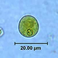
Figure A
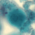
Figure B
Acknowledgement: This case was kindly contributed by the Texas Department of Health, Bureau of Laboratories.
Answer to Case #57
The organisms in this case were cysts of Entamoeba polecki. Diagnostic features observed were:
- a uninucleate cyst with the nucleus being one third or less the diameter of the cyst.
- the presence of a pleomorphic karyosome which can be minute or large, compact or diffuse. The position of the karyosome can be central or eccentric. In the image from the formalin specimen, small granules can also be seen.
- many pleomorphic chromatoid bodies, seen in the composite image as rods with round ends, throughout the cyst. These bodies can be highly variable, appearing as small particles, small or large rods with rounded or splintered ends.
This organism is primarily a parasite of monkeys and pigs with a worldwide distribution.
More on: E. polecki
Images presented in the monthly case studies are from specimens submitted for diagnosis or archiving. On rare occasions, clinical histories given may be partly fictitious.
