 ShareCompartir

Monthy Case Studies - 2000
Case #42 - August, 2000
A reference laboratory received one trichrome stained slide from a local health care facility requesting an O & P (ova and parasites) examination on a 25-year-old man whose symptoms included abdominal cramping and intermittent diarrhea. The images below show the objects seen on the submitted slide. The objects averaged 4.5 to 5.5 micrometers in diameter. What is your diagnosis? Based on what criteria? What else would you recommend?
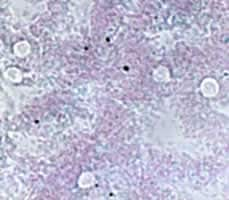
Figure A
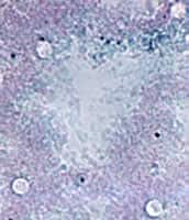
Figure B
Answer to Case #42
This was a case of cryptosporidiosis caused by Cryptosporidium sp. On rare occasions, one may not receive the optimal specimen to make an accurate diagnosis. It is important to be able to use what you have to make a tentative diagnosis on the specimen. Then, you can request additional specimens to correctly confirm or dismiss your suspicions. Trichrome stained smears are normally not used to diagnose Cryptosporidium since the oocysts usually will not stain, as shown in Figures A and B (on the case studies page). Key features included the shape and size of the objects, which were consistent for Cryptosporidium oocysts. On a few of the objects, you could almost make out the faint outline of the sporozoites. The clear area surrounding the objects suggests a cellular wall is present. Based on the findings of this case, requesting a formalin preserved specimen or acid-fast stained fecal smear would be the appropriate follow-up. We have included an image (Figure A below) illustrating the same specimen stained using a modified acid-fast.
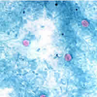
Figure C
More on: Cryptosporidiosis
Images presented in the monthly case studies are from specimens submitted for diagnosis or archiving. On rare occasions, clinical histories given may be partly fictitious.
