 ShareCompartir

Monthy Case Studies - 1999
Case #23 - November, 1999
A 45-year-old Korean man was assigned to perform construction work in the Philippines. He went to a local hospital in the Philippines for persistent diarrhea. A stool examination was performed on a formalin fixed stool concentrate of his specimen. The objects in the following images were seen at 400×. What is your diagnosis? Based on what criteria?
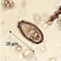
Figure A
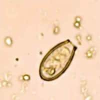
Figure B
Acknowledgement: This case was kindly contributed by the College of Public Health, University of the Philippines, Manila.
Answer to Case #23
This was a case of clonorchiasis caused by Clonorchis sinensis, a liver fluke. Opisthorchis species are also acceptable answers since the eggs of these three species are virtually indistinguishable and both are found in Asia (Korea). The diagnostic features noted were size, obvious presence of an operculum, and the small abopercular knob (indicated by the arrow in Figure B below). The images are from a formalin fixed stool concentrate (400×).
These eggs are sometimes confused with those of intestinal flukes such as heterophyids, which also have small, operculate eggs. Usually, the eggs of intestinal flukes are smaller, ranging from 20 to 30 micrometers by 15 to 17 micrometers whereas eggs of the liver flukes are generally larger (27 to 5 micrometers by 12 to 9 micrometers) and have a more conspicuous, or seated, operculum and have a small knob at the abopercular end. Geographic location of where the infection was acquired is helpful in making an accurate diagnosis.
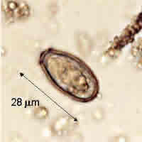
Figure A
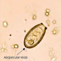
Figure B
More on: Clonorchiasis
Images presented in the monthly case studies are from specimens submitted for diagnosis or archiving. On rare occasions, clinical histories given may be partly fictitious.
