 ShareCompartir

Monthy Case Studies - 1999
Case #10 - April, 1999
The Wisconsin State Laboratory of Hygiene (Tim Monson and Linda Kelly) sent this image to CDC, with the following message:
"We are wondering if you can help us diagnose/rule out a body consistent with a Fasciola/Fasciolopsis egg. It measures around 140 micrometers in length and there is no apparent operculum present (not that we can detect anyway). Two stools were submitted in 10% formalin, and we could only find the one "egg" in question here. The patient history that we obtained is as follows: 26 year old male; travel to Cancun, Mexico 3/6 through 3/13; onset of symptoms on 3/13 (stomach pain, diarrhea [4/day], intestinal discomfort); stools collected on 3/18; symptoms resolved by 3/26."
Is this a parasite egg? Based on what criteria? If not a parasite egg, what is it? Based on what criteria? What other examination(s) would you recommend?
Our colleagues from Wisconsin added: "The image was taken in black and white… We just used a Polaroid camera that fits onto one of the scope's oculars."
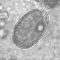
Figure A
Answer to Case #10
The object shown in this case was an artifact and the proper diagnosis was No Parasites Found (NPF). There was no visible operculum in the image, and careful observation by our colleagues from Wisconsin did not detect an operculum either, eliminating Fasciola hepatica and Fasciolopsis buski, whose eggs would have been approximately this size. The object resembled a mite egg (e.g., from cheese mites), that are known contaminants of stool specimens. The object was the right size, and the bubble globule (arrow on Figure A below) is sometimes seen in mite eggs. As an added illustration, Figure B below shows a mite egg (size 155 micrometers by 77 micrometers) in a stool specimen with many such eggs, sent by the Georgia Public Health Laboratory.
To obtain more certain identification, we would suggest repeat stool specimens to detect any additional objects. If found, such objects should be investigated for the presence of an operculum (including tapping the coverslip to pop the operculum open).
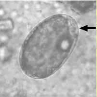
Figure A
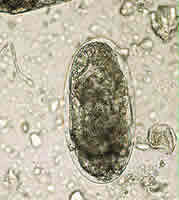
Figure B
More on: Artifacts
Images presented in the monthly case studies are from specimens submitted for diagnosis or archiving. On rare occasions, clinical histories given may be partly fictitious.
