 ShareCompartir

Monthy Case Studies - 1999
Case #8 - March, 1999
The Michigan Department of Health received a worm that was passed rectally by a woman. She reported abdominal pain and that she had passed 15 similar worms. She had no other symptoms. The patient had three cats and one parrot. After a preliminary diagnosis at the Michigan Department of Health, the worm was sent to CDC for confirmation. The worm was 5.5 cm long and is shown in Figures A (tail) and B (head). What is your diagnosis? Based on what criteria? Would you make any other inquiries or perform any additional tests?
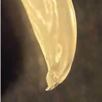
Figure A
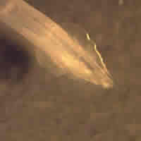
Figure B
Answer to Case #8
The worm was an adult male of the feline ascarid, Toxocara cati. The diagnostic features observed were:
- the worm's length, which at 5.5 cm was compatible with Toxocara (length of mature males: four to six cm); this length excludes human ascarids that are much longer (males 15 to 31 cm) and pinworms that are much shorter (males 0.2 to 0.5 cm).
- the caudal digitiform process (green arrow, Figure A below), a diagnostic feature of a male Toxocara.
- cervical alae (yellow arrows on Figure B below) that were striated, broad, rounded posteriorly, and tapered toward the anterior end, thus giving a broad arrowhead shape. This is diagnostic for compatible with Toxocara cati; in T. canis, they would be narrower and longer.
- three characteristic ascarid lips, one dorsal, and two subventral (blue arrow on Figure B below).
The fact that the patient had three cats also speaks in favor of T. cati.
The report of this passing per rectum of an adult worm of T. cati is interesting because it is such a rare occurrence, and because it is difficult to explain. In a recent article (Adult Toxocara cati infections in US children: report of four cases. Am J Trop Med Hyg 1998;59:404-406.), Eberhard and Alfano reported on four cases, all in children, who passed adult T. cati either per rectum (two cases) or by vomiting (two cases). These patients were serologically negative for Toxocara, which indicated that the worms had in all likelihood been acquired by ingestion of immature larvae which then matured in the digestive tract of the patients (and, in two cases, went through that tract). If these adult worms had derived from infective eggs ingested by the children, this would have necessitated a worm migration outside the digestive tract into the patient's body, resulting in a positive serology and possibly symptoms of visceral/ocular larva migrans; one last possibility, that ingested eggs could develop into adults without worm migration outside the digestive tract, has not, to our knowledge, been observed in humans.
In this case, the serology for Toxocara was negative and there were no symptoms suggestive of visceral/ocular larva migrans. Thus, if the patient did actually pass the 15 adult worms, as reported, she would have acquired them by ingesting at least 15 immature larvae!
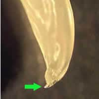
Figure A
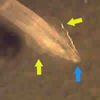
Figure B
More on: Toxocariasis
Images presented in the monthly case studies are from specimens submitted for diagnosis or archiving. On rare occasions, clinical histories given may be partly fictitious.
