 ShareCompartir

Monthy Case Studies - 1999
Case #6 - February, 1999
A 52-year-old man developed malaria 13 months after returning from a visit to Kenya and the island of Zanzibar (Tanzania). Some images from his blood smears are shown in Figures A through D. What is your diagnosis? Based on what criteria?
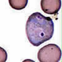
Figure A

Figure B

Figure C
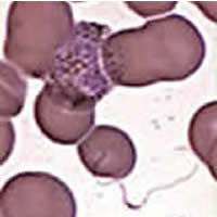
Figure D
Acknowledgement: Smears provided by a health facility in Utah and by the Utah Public Health Laboratory. This case was prepared by Dustin Brown of the Oregon State Public Health Laboratory.
Answer to Case #6
This was a case of malaria caused by Plasmodium vivax. Diagnostic morphologic features included:
- infected erythrocytes that were markedly enlarged and deformed (Figures A and D); Schüffner's dots were visible in Figures A, C, and D.
- an amoeboid trophozoite, typical of P. vivax (Figures A).
- a schizont with 16 merozoites (Figures B), a number compatible with P. vivax (P. ovale, would have only 6 to 14 merozoites).
- a rounded gametocyte (Figures C).
Figures D showed another gametocyte (red arrow). In addition, there is an exflagellated microgamete (Figures D below, blue arrow), recognizable by its filamentous shape and the presence of nuclear material (near the tip of the blue arrow). Exflagellation usually occurs in the mosquito midgut. The microgametocyte (male gametocyte) releases eight microgametes that move around very actively. If a microgamete encounters a macrogamete (female gamete—each macrogametocyte produces only one macrogamete), fertilization occurs. Exflagellated microgametes are occasionally seen in blood smears, especially when the blood is collected in a tube and left to sit for a while. The environmental conditions (temperature, pH) change, approaching those found in the mosquito's midgut, inducing the microgametocytes to exflagellate. Charles Louis Alphonse Laveran, a French military physician, discovered the malaria parasite on November 6, 1880 in Constantine (Algeria) while examining an unstained wetprep of the blood of a febrile soldier. He saw very actively motile filaments as well as pigment granules and round and crescent shaped bodies, which were exflagellating Plasmodium falciparum. He first named the parasite Oscillaria malariae, probably in homage to the motility of the gametes.
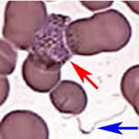
Figure D
More on: Malaria
Images presented in the monthly case studies are from specimens submitted for diagnosis or archiving. On rare occasions, clinical histories given may be partly fictitious.
