 ShareCompartir

Monthy Case Studies - 1998
Case #2 - December, 1998
A concentrate (formalin-ethyl acetate technique) of a stool specimen was preserved in formalin. Figures A and B show a bright-field examination of the wet mount. The objects measured approximately 300 micrometers in length. What is your diagnosis? Based on what criteria?
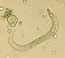
Figure A
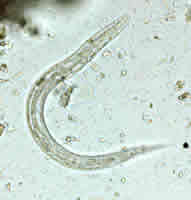
Figure B
Acknowledgement: Specimen contributed by Ray Kaplan, SmithKline Beecham Laboratories, Atlanta, GA.
Answer to Case #2
The organisms were rhabditoid larvae of Strongyloides stercoralis. Diagnostic features observed were:
- the size (up to 380 micrometers in length)
- a short buccal canal (red arrow in Figure B)
- a prominent genital primordium (blue arrows, especially in Figure A)
The buccal canal and genital primordium are useful for differentiating Strongyloides rhabditoid larvae from those of hookworms. The latter have a long buccal canal and an inconspicuous genital primordium. In Strongyloides, the rhabditoid larvae hatch in the small intestine and are passed in the feces, constituting a diagnostic stage; in hookworm, the eggs are passed in the feces and larvae are found only when the unpreserved stool specimen has been stored for one to two days under conditions that allow the eggs to hatch.
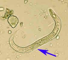
Figure A
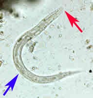
Figure B
More on: Strongyloidiasis
Images presented in the monthly case studies are from specimens submitted for diagnosis or archiving. On rare occasions, clinical histories given may be partly fictitious.
